Treatment for Flashes of Light in Peripheral Vision: How It Works, Results and What to Expect

Flashes often occur when the gel inside the eye pulls on the retina. New flashes with many floaters, a curtain-like shadow, or vision loss need same-day urgent eye care.
Key Takeaways
- Flashes often occur when the gel inside the eye pulls on the retina.
- New flashes with many floaters, a curtain-like shadow, or vision loss need same-day urgent eye care.
- A retinal tear may be sealed with laser treatment or cryotherapy to help prevent retinal detachment.
- Not all flashes need a procedure; treatment is directed at the underlying cause.
- Even after a reassuring examination, new or worsening symptoms should be checked promptly.
Treatment for flashes of light in peripheral vision depends on why they are happening. Many flashes are caused by age-related changes in the eye and need monitoring, but sudden new flashes—especially with floaters, a shadow, or reduced vision—require urgent dilated eye examination to rule out a retinal tear or detachment.
Overview: Treatment for Flashes of Light in Peripheral Vision
Treatment for flashes of light in peripheral vision is not one single treatment. Flashes are a symptom, often described as brief sparks, lightning streaks, flickering, or camera-flash effects at the edge of sight. The appropriate care depends on whether the flashes come from harmless vitreous changes, a retinal tear, retinal detachment, migraine-related visual symptoms, inflammation, or another eye or neurological condition.
The first and most important step is a prompt eye assessment when flashes are new, sudden, or increasing. An ophthalmologist can dilate the pupils and examine the retina at the back of the eye. If there is no retinal injury, observation and follow-up may be all that is needed. If a retinal tear or detachment is found, treatment is usually recommended urgently to protect vision.
Flashes can be unsettling, but they do not automatically mean permanent damage. Early assessment helps identify the people who need immediate treatment and reassures those whose symptoms are due to a non-dangerous cause.
What could be causing weird flashes of light in my peripheral vision?
A common cause is posterior vitreous detachment (PVD). The vitreous is a clear, gel-like substance inside the eye. With age, it naturally becomes more liquid and may separate from the retina. During this process, the vitreous can tug on the retina, creating flashes that are often most noticeable in dim light or when moving the eyes.
Sometimes vitreous pulling causes a retinal tear. Fluid can then pass through the tear and lift the retina from its normal position, causing retinal detachment. This is an eye emergency because the retina is essential for vision. Flashes may also follow an eye injury, occur with inflammation inside the eye, or arise after eye surgery.
Visual migraine or migraine aura can also cause flashing lights, shimmering zigzags, or moving patterns. These symptoms commonly affect both eyes and may last minutes to an hour, sometimes with no headache. However, a person should not assume new flashes are migraine without first considering an eye examination, particularly if symptoms affect one eye or are accompanied by floaters or a field-of-vision shadow.
- Age-related vitreous changes or posterior vitreous detachment
- Retinal tear or retinal detachment
- Eye trauma or recent eye surgery
- Inflammation affecting the eye
- Migraine aura or, less commonly, neurological causes
When should I worry about flashes in my peripheral vision?

New, sudden, frequent, or worsening flashes should be assessed promptly by an ophthalmologist or urgent eye service. The need for urgent care is greater if flashes are accompanied by a sudden increase in floaters, dark dots or cobweb-like shapes, blurred vision, a curtain or veil across vision, loss of side vision, or a dark shadow. These symptoms can indicate a retinal tear or detachment.
People at higher risk include those who are very short-sighted, have had previous retinal tears or detachment, have had cataract or other eye surgery, have sustained an eye injury, or have a family history of retinal detachment. Diabetes and certain inflammatory eye conditions may also increase the need for careful evaluation, depending on the individual situation.
A painless retinal problem can still be urgent. If a curtain-like shadow, sudden visual loss, or significant new floaters occurs, the person should seek same-day emergency eye care rather than waiting to see whether the symptoms settle.
How treatment works: examination, candidacy and procedure options
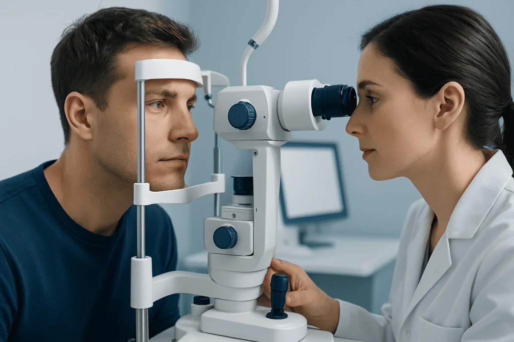
An ophthalmologist usually begins by reviewing the symptom pattern, medical history, medications, injury history, and previous eye conditions. Vision is tested, and eye drops are used to widen the pupils. The doctor then examines the vitreous and retina, often using a slit lamp, indirect ophthalmoscopy, and sometimes retinal imaging or ultrasound if the view is blocked by bleeding or cataract.
People with uncomplicated posterior vitreous detachment are commonly candidates for monitoring rather than a procedure. The flashes often reduce as vitreous traction settles. Follow-up may be advised because a retinal tear can occasionally develop after the initial symptoms begin. The clinician will explain which new symptoms should prompt earlier review.
If a retinal tear is identified before detachment develops, laser photocoagulation or cryotherapy may be used to create a controlled scar around the tear. This helps secure the retina to the underlying tissue and reduces the chance that fluid will spread beneath it. These treatments are generally performed by a retinal specialist and are directed at the retinal problem, not simply at the sensation of flashing.
If retinal detachment is present, surgery may be needed. Depending on the type and location of detachment, options can include a gas-bubble procedure, scleral buckle surgery, vitrectomy, or a combination of approaches. The choice is individual and depends on the retina findings, the person’s eye health, and the urgency of repair.
Step-by-step: what to expect from retinal tear or detachment treatment
For laser treatment of a retinal tear, dilating drops and numbing eye drops are usually given first. The person is seated at a laser machine, and the ophthalmologist applies laser spots around the tear. The procedure commonly takes a short time, although the exact duration varies with the number and position of tears. Bright lights and mild discomfort or pressure can occur, but severe pain is not expected.
Cryotherapy uses a small probe placed on the outside of the eye to freeze the tissue around a retinal tear. It may be selected when the tear is difficult to treat with laser. The eye is numbed, and additional sedation or anesthesia may be considered in selected situations.
Retinal detachment repair is more involved and may be performed in an operating room. In vitrectomy, the surgeon removes some or all of the vitreous gel, repairs retinal breaks, and may place a gas or silicone oil bubble to support the retina. A scleral buckle is a small band placed around the outside wall of the eye to relieve traction on the retina. Postoperative positioning instructions can be important, particularly when a gas bubble is used.
Acibadem International’s ophthalmology teams can assess flashes and coordinate retinal evaluation and treatment for international patients through its multidisciplinary specialists and JCI-accredited hospitals.
Recovery timeline, benefits and possible risks
After laser treatment or cryotherapy for a retinal tear, vision may be temporarily blurred from dilation and the procedure. Most people can return to many usual activities relatively soon, but the treating clinician may recommend avoiding strenuous exercise or heavy lifting for a period. The scar that seals the area takes time to develop, so follow-up examinations remain important.
After retinal detachment surgery, recovery can take weeks or longer. Vision often improves gradually, but the final result depends on factors such as how long the retina was detached, whether the central retina was involved, and the underlying cause. Some people notice persistent floaters, distortion, or reduced vision despite successful anatomical repair.
The main benefit of treating a retinal tear is lowering the risk of detachment. The goal of detachment surgery is to reattach the retina and preserve as much vision as possible. Possible risks include infection, bleeding, pressure changes in the eye, cataract development after vitrectomy, recurrence of detachment, and the need for further procedures. The ophthalmologist discusses the expected benefits and risks based on the individual diagnosis.
A gas bubble can temporarily blur vision and may require specific head positioning. Air travel and high-altitude travel are generally unsafe while an intraocular gas bubble remains because pressure changes can dangerously raise pressure inside the eye. The surgical team will advise when travel is safe.
How long do peripheral eye flashes last?
The duration varies with the cause. With posterior vitreous detachment, flashes may be most frequent over days or weeks and then gradually become less noticeable over several weeks to months. Some people may occasionally notice flashes for longer, particularly in darkness or with sudden eye movement.
Flashes from a retinal tear can continue until the traction changes or the tear is treated. Treatment may prevent further retinal damage, but it may not make every flash disappear immediately. Ongoing or newly increased flashes after an eye assessment should be reported, because symptoms can change over time.
Migraine-related visual symptoms typically develop gradually and resolve within a shorter period, often within an hour. A new pattern, symptoms in only one eye, or symptoms accompanied by loss of vision should still be assessed urgently rather than presumed to be migraine.
How to get eye flashes to go away?
There is no safe home method to make flashes stop, because treatment must address the cause. For uncomplicated vitreous changes, flashes often fade naturally as the eye adapts and the pulling on the retina decreases. Rest, hydration, eye exercises, or supplements have not been shown to repair a retinal tear or prevent retinal detachment.
If flashes are related to a retinal tear, laser treatment or cryotherapy may be used to reduce the risk of more serious retinal separation. If there is retinal detachment, surgery may be needed. For migraine-related visual symptoms, management may include identifying triggers and discussing headache or migraine care with a qualified clinician.
Until the eye has been assessed, it is sensible to avoid rubbing or pressing on the eye and to protect the eye from further injury. People should follow their ophthalmologist’s instructions about activity, follow-up, driving after dilation, and any warning signs that require urgent reassessment.
When to seek medical care
A person should arrange prompt eye care for any new flashes, particularly when they occur in one eye or begin suddenly. Same-day urgent assessment is recommended when flashes occur with a shower of new floaters, a dark curtain or shadow, blurred or reduced vision, eye injury, or recent eye surgery. These features may indicate a retinal tear or detachment that needs timely treatment.
Emergency medical care is also appropriate if visual symptoms occur with weakness, difficulty speaking, facial drooping, severe sudden headache, confusion, or other neurological symptoms. These signs may have causes outside the eye and need immediate evaluation.
Even after a normal dilated retinal examination, patients should contact their eye specialist if symptoms become more frequent, new floaters appear, or any area of vision becomes dark or missing. Regular eye examinations are especially valuable for people with high short-sightedness, diabetes, prior eye surgery, or a personal or family history of retinal disease.
Frequently asked questions
Are flashes in peripheral vision always a retinal detachment?
No. A common cause is posterior vitreous detachment, an age-related change in the gel inside the eye. However, retinal tears and detachment can cause similar symptoms, so new flashes should be checked with a dilated eye examination.
Can stress cause flashes of light in vision?
Stress can contribute to migraine in some people, and migraine aura may include flashing or shimmering visual effects. Stress should not be assumed to be the cause of sudden one-eye flashes, especially if floaters, a shadow, or reduced vision are also present.
Do flashes from posterior vitreous detachment need treatment?
Uncomplicated posterior vitreous detachment usually does not require a procedure. An ophthalmologist may recommend follow-up because retinal tears can occasionally occur during or after the vitreous separates.
What is the difference between floaters and flashes?
Floaters are small moving dots, lines, or cobweb-like shapes seen against a bright background. Flashes are brief sparks or streaks of light, often caused by traction on the retina. A sudden combination of both needs urgent eye assessment.
Will laser treatment stop eye flashes immediately?
Laser treatment seals a retinal tear to help prevent retinal detachment; it is not designed primarily to stop the flash sensation immediately. Flashes may lessen over time as vitreous traction settles, but follow-up is important if they persist or worsen.
Can a retinal tear heal on its own?
A retinal tear does not reliably heal in a way that prevents fluid from passing underneath the retina. When a clinically significant tear is found, an ophthalmologist may recommend laser or cryotherapy to create a protective seal around it.
References
- American Academy of Ophthalmology
- National Eye Institute
- Royal College of Ophthalmologists
- Mayo Clinic
This article is for general information only and is not a substitute for professional medical advice. Please consult a qualified doctor about your individual situation.
Explore treatments in Turkey — costs, top hospitals & a free quote
JCI-accredited · board-certified surgeons · reply within 24h
Add us as a Preferred Source to see more of our trusted health content across Google Search, AI Overviews and Discover.