Tubular Adenoma Colonoscopy Follow Up: Preparation, Procedure and Results

Tubular adenomas are common precancerous colon polyps, but they are not cancer. Complete removal during colonoscopy greatly lowers the chance that the removed polyp will cause future harm.
Key Takeaways
- Tubular adenomas are common precancerous colon polyps, but they are not cancer.
- Complete removal during colonoscopy greatly lowers the chance that the removed polyp will cause future harm.
- Most people with one or two small tubular adenomas need a repeat colonoscopy in 7 to 10 years.
- Three or more adenomas, larger polyps or advanced features may require surveillance in about 3 to 5 years.
- Only laboratory examination can confirm whether a removed polyp contains cancer cells.
- A clinician should individualize follow-up when there is a family history of colorectal cancer, hereditary risk or bowel symptoms.
Tubular adenoma colonoscopy follow up helps prevent colorectal cancer by checking for new polyps after an adenoma has been removed. The recommended interval depends mainly on the number, size and microscopic features of the polyps, as well as the quality of the initial examination.
Overview: tubular adenoma colonoscopy follow up
Tubular adenoma colonoscopy follow up means scheduling a future colonoscopy after a tubular adenoma has been found and removed. Tubular adenomas are growths that arise from the lining of the large intestine (colon) or rectum. They are considered precancerous because some can slowly develop into colorectal cancer if left in place, but the vast majority are not cancer at the time they are detected.
Follow-up colonoscopy is also called surveillance colonoscopy. Its purpose is not usually to recheck a successfully removed polyp immediately; rather, it looks for new polyps that may develop elsewhere in the colon. The timing is guided by the pathology report and procedure report, including the number of adenomas, their size, whether they were fully removed, and whether the bowel preparation allowed a clear examination.
For many people, the result is reassuring: one or two small tubular adenomas with low-grade dysplasia generally indicate a relatively low future risk when removed completely. A gastroenterologist can explain the individual plan and make sure it is aligned with current colorectal cancer surveillance guidance.
What is the recommended follow-up for tubular adenomas after a colonoscopy?
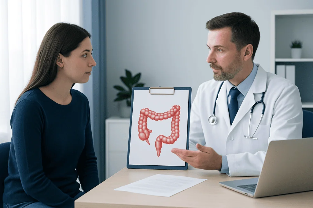
For adults whose colonoscopy was high quality and who had one or two tubular adenomas smaller than 10 millimeters removed completely, a repeat colonoscopy is commonly recommended in 7 to 10 years. This longer interval reflects the relatively low risk after complete removal of a small number of low-risk adenomas.
A shorter interval may be advised when there are three to four small tubular adenomas, often around 3 to 5 years, or when there are five to 10 adenomas, often around 3 years. A repeat examination at about 3 years is also commonly considered for an adenoma that is 10 millimeters or larger, has villous features, or has high-grade dysplasia. These findings are sometimes described as advanced adenoma features.
The exact recommendation may differ if a polyp was removed in pieces, if removal was uncertain, if bowel cleansing was inadequate, or if there is a personal or family history that changes colorectal cancer risk. People should retain both their colonoscopy and pathology reports, since these documents provide the details needed to set an appropriate surveillance interval.
- One or two small tubular adenomas: often 7 to 10 years.
- Three to four small tubular adenomas: often 3 to 5 years.
- Five to 10 adenomas or an advanced adenoma: often about 3 years.
- More than 10 adenomas: earlier follow-up and possible genetic assessment may be considered.
How the surveillance colonoscopy works
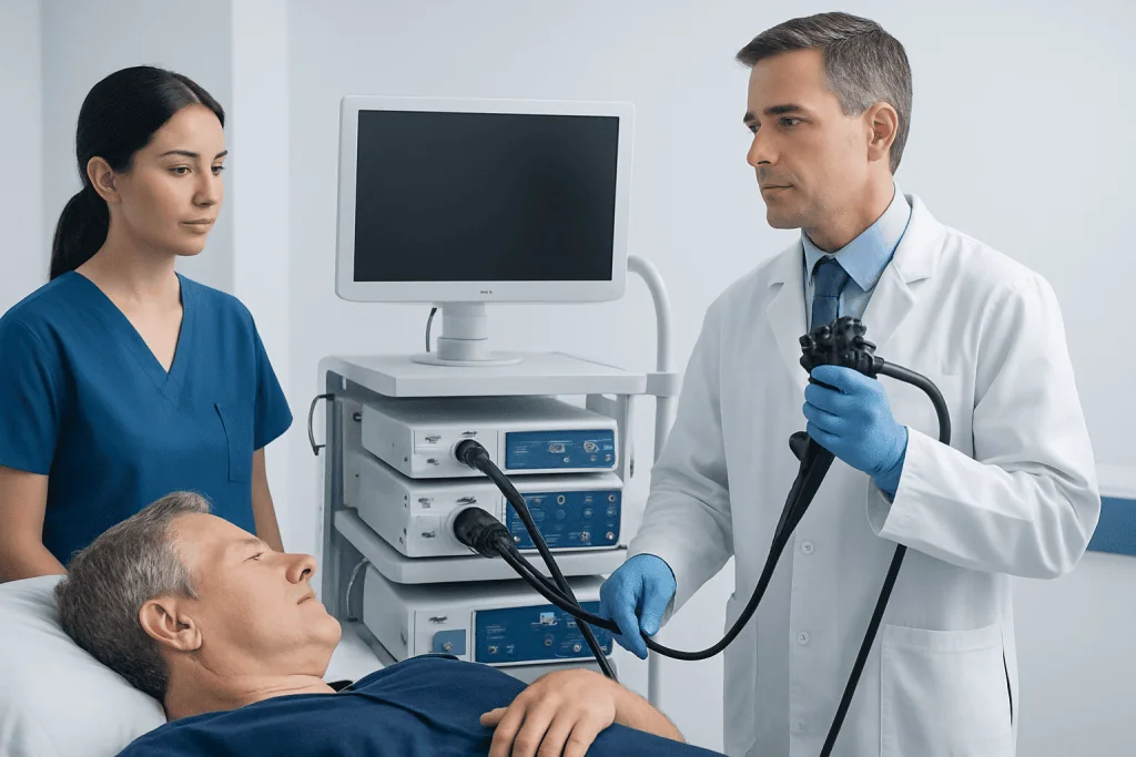
A surveillance colonoscopy is performed using a flexible, camera-equipped instrument called a colonoscope. The gastroenterologist gently passes it through the rectum and around the colon, viewing the lining on a monitor. Air or carbon dioxide is introduced to open the colon slightly and improve visibility; carbon dioxide is absorbed relatively quickly and may reduce discomfort after the test.
Most colonoscopies are performed with sedation or anesthesia so the patient is relaxed and usually does not remember much of the examination. Small polyps can often be removed during the same procedure using specialized instruments passed through the colonoscope. Removed tissue is sent to a pathology laboratory for microscopic review.
The main benefit of surveillance is prevention. Finding and removing adenomas before they become more advanced can reduce the likelihood of colorectal cancer. Colonoscopy also allows direct inspection of the bowel rather than relying only on symptoms, which often do not occur with early polyps.
Who needs follow-up and how to prepare
People who have had a tubular adenoma removed are candidates for surveillance, although the interval is individualized. Age, overall health, previous colonoscopy quality, family history, inflammatory bowel disease and known inherited cancer syndromes can all influence the plan. For older adults or those with serious health conditions, the expected benefit of another colonoscopy should be weighed thoughtfully against the burdens and risks of the procedure.
Preparation is essential because small polyps can be missed if stool remains in the bowel. The care team will provide a bowel-cleansing plan, usually involving dietary adjustments for a short period before the procedure and a prescribed laxative solution. Split-dose preparation, in which part of the solution is taken the evening before and part closer to the procedure, is commonly used because it can improve cleansing.
Patients should tell the clinical team about all medicines and supplements, particularly blood thinners, diabetes medicines, iron products and kidney disease treatments. Medication changes should only be made under the direction of the prescribing clinician or endoscopy team. Because sedation can impair judgment for the rest of the day, a responsible adult usually needs to accompany the patient home.
Step-by-step procedure and recovery timeline
On arrival, the patient is checked in and asked about medical history, allergies and medications. A clinician reviews consent, explains sedation and answers questions. During the procedure, which commonly lasts less than an hour but may take longer when multiple or complex polyps are removed, the patient is monitored for breathing, heart rate and blood pressure.
If a polyp is found, the clinician may remove it with a snare or another technique. Some larger lesions require advanced endoscopic removal, staged treatment or referral to a specialist center. The patient generally rests in a recovery area while the sedative wears off, and the endoscopist may share initial visual findings before discharge.
Mild bloating, gas or brief cramping can occur on the day of the test and generally settles quickly. Most people can eat and drink after recovery unless they are given specific instructions otherwise. Sedation means driving, operating machinery, drinking alcohol and making important legal or financial decisions should be avoided until the following day or as directed.
Pathology results can take several days. These results confirm whether the growth was a tubular adenoma, describe any higher-risk features and help determine the next surveillance date. It is important to attend the follow-up conversation or review the report through the care team’s recommended process.
How long does it take for a tubular adenoma to turn into cancer?
When an adenoma progresses to cancer, the process is usually slow and often takes many years. There is no fixed timeline for an individual polyp, and many tubular adenomas never become cancerous. Progression depends on factors such as size, cellular changes, genetic alterations and the polyp’s growth pattern.
Tubular adenomas are generally less likely to contain advanced changes than adenomas with substantial villous features, but risk rises with increasing size and with high-grade dysplasia. This is why pathology matters: it provides information that cannot be determined from symptoms alone and helps guide the timing of surveillance.
The gradual nature of adenoma development is a key reason screening and surveillance are effective. Removing adenomas interrupts a possible pathway toward cancer. Patients do not need to assume that a past tubular adenoma means cancer is inevitable; instead, they should follow the personalized monitoring schedule given by their clinician.
Can a doctor tell during a colonoscopy if a polyp is cancerous?
During colonoscopy, an experienced endoscopist can assess features that may suggest a polyp is more likely to be advanced, such as its size, shape, surface pattern, bleeding tendency or whether it appears to invade deeper tissue. High-definition imaging and specialized visual techniques can improve this assessment. However, appearance alone cannot definitively establish whether cancer cells are present.
Pathology is the definitive test. A pathologist examines removed tissue under a microscope to identify the polyp type, grade of dysplasia and, if present, invasive cancer. If cancer is identified within a polyp, additional tests or treatment may be needed to determine whether endoscopic removal was sufficient.
When a lesion looks complex or suspicious, the endoscopist may take biopsies, remove it using an advanced technique, refer the patient to an advanced endoscopist, or recommend surgical assessment. This careful approach supports accurate diagnosis while avoiding assumptions based only on the visual appearance of a polyp.
Risks, benefits and when to seek medical care
The benefits of surveillance colonoscopy include detecting new adenomas and removing them before they can progress. Colonoscopy is generally safe, but it is an invasive procedure. Possible complications include reactions to sedation, bleeding after polyp removal, infection and a tear in the colon wall (perforation). These events are uncommon, and the risk may be higher with larger or more complex polyp removal.
After colonoscopy, urgent medical advice is appropriate for severe or worsening abdominal pain, fever, persistent vomiting, dizziness or fainting, a swollen abdomen, or significant rectal bleeding. A small amount of blood may occur after some polyp removals, but heavy bleeding, blood clots or bleeding that does not stop should be assessed promptly. Patients should follow the discharge instructions supplied by their endoscopy team.
Medical review should also be arranged sooner than the planned surveillance date for new rectal bleeding, an ongoing change in bowel habits, unexplained iron-deficiency anemia, unintended weight loss or persistent abdominal symptoms. These symptoms often have causes other than cancer, but they deserve proper evaluation rather than waiting for the next routine colonoscopy.
Acibadem International’s multidisciplinary specialists and JCI-accredited hospitals support international patients needing evaluation and treatment for colorectal conditions, including surveillance planning after polyp removal.
Frequently asked questions
How often should you have a colonoscopy if a tubular adenoma is found?
The interval depends on the number, size and pathology features of the adenomas, as well as the quality of the initial colonoscopy. One or two small tubular adenomas are commonly followed with colonoscopy in 7 to 10 years, while multiple adenomas or advanced features often lead to follow-up in about 3 to 5 years. The treating gastroenterologist should provide the specific recommendation.
Is a tubular adenoma cancer?
No. A tubular adenoma is a precancerous polyp, meaning it has the potential to develop into cancer over time but is not itself cancer. Removal during colonoscopy is an important preventive step.
What does low-grade dysplasia in a tubular adenoma mean?
Low-grade dysplasia means that the cells show mild abnormal changes under the microscope. It is a common finding in adenomas and does not mean invasive cancer is present. The number and size of polyps still help determine the follow-up plan.
Do tubular adenomas cause symptoms?
Most tubular adenomas cause no symptoms and are found during screening or evaluation for another concern. Larger polyps may occasionally bleed or contribute to changes in bowel habits, but symptoms are not a reliable way to detect them. This is why recommended surveillance is important.
Can tubular adenomas come back after they are removed?
A completely removed individual adenoma does not usually regrow in the same location. However, a person who has developed an adenoma can develop new polyps elsewhere in the colon over time. Surveillance colonoscopy is designed to find and remove new growths early.
Should family members be screened sooner if someone has a tubular adenoma?
A family member’s screening plan depends on the patient’s age at diagnosis and whether the adenoma had advanced features, as well as the broader family history. Relatives should discuss this information with their own clinician. Earlier screening may be appropriate in some families, especially when there is colorectal cancer or advanced adenoma in a close relative.
References
- U.S. Multi-Society Task Force on Colorectal Cancer
- American College of Gastroenterology
- National Cancer Institute
- American Cancer Society
- World Health Organization
This article is for general information only and is not a substitute for professional medical advice. Please consult a qualified doctor about your individual situation.
Persistent digestive symptoms? Get evaluated in Turkey
JCI-accredited · board-certified surgeons · reply within 24h
Add us as a Preferred Source to see more of our trusted health content across Google Search, AI Overviews and Discover.