Ultrasound Anomaly: Preparation, Procedure and Results

The anomaly scan is a detailed, non-invasive ultrasound commonly offered around 18–22 weeks of pregnancy. It evaluates many fetal structures, including the brain, spine, face, heart, abdomen, kidneys, limbs and placenta.
Key Takeaways
- The anomaly scan is a detailed, non-invasive ultrasound commonly offered around 18–22 weeks of pregnancy.
- It evaluates many fetal structures, including the brain, spine, face, heart, abdomen, kidneys, limbs and placenta.
- Most people do not need extensive preparation, but the imaging centre may give individual instructions about bladder filling.
- The scan is usually painless and commonly takes about 20–45 minutes, though timing can vary.
- A report described as normal means no significant abnormality was seen during that examination; it does not guarantee that every condition has been excluded.
- An unexpected finding often needs repeat imaging or specialist assessment and does not always mean a serious problem.
An ultrasound anomaly scan, also called an anatomy scan, is a detailed pregnancy ultrasound that assesses the developing baby’s structures, growth and surrounding pregnancy tissues. It is usually performed between 18 and 22 weeks of pregnancy and provides important screening information, although it cannot detect every health condition.
Ultrasound anomaly scan: what it is and what it checks
An ultrasound anomaly scan is a detailed prenatal examination that uses sound waves to create images of a developing baby in the uterus. It is also known as a mid-pregnancy scan, morphology scan or anatomy ultrasound. The examination is generally offered between 18 and 22 weeks of pregnancy, when many body structures can be assessed clearly.
The purpose is to screen for structural ultrasound anomalies and to confirm key features of the pregnancy. A trained sonographer or doctor assesses the baby’s head and brain, face, spine, heart, stomach, abdominal wall, kidneys, bladder, arms and legs. The scan also reviews growth measurements, the placenta, umbilical cord and the amount of amniotic fluid.
This is a screening test rather than a diagnosis in itself. A reassuring scan result is valuable, but some conditions may be too small to see, develop later in pregnancy, or not have visible ultrasound signs. If an image is unclear or a possible difference is identified, the care team may recommend another scan or consultation with a fetal medicine specialist.
Who has an anomaly scan and when is it performed?
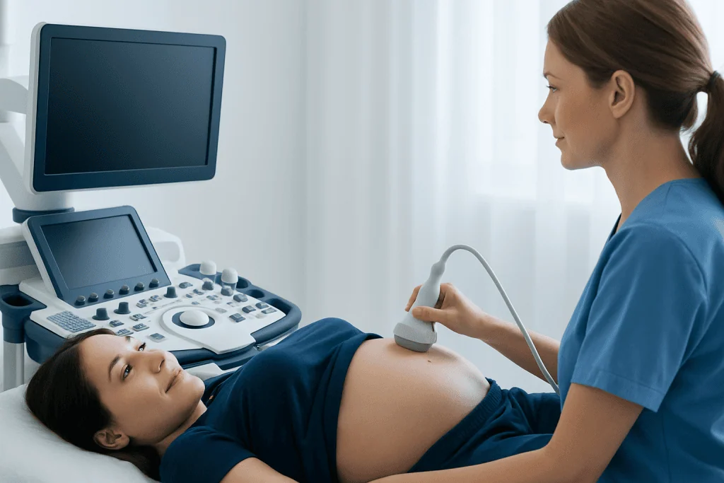
Most pregnant people are offered an anomaly scan as part of routine antenatal care. The ideal timing may differ slightly according to local guidance, the dating of the pregnancy and the imaging centre’s protocol. The appointment is commonly scheduled in the second trimester because the baby is large enough for detailed assessment while there is still sufficient amniotic fluid for imaging.
Some pregnancies need more tailored ultrasound assessment. This may apply when there is a family history of an inherited condition, a previous pregnancy affected by a birth difference, certain medical conditions, medication exposure, assisted conception, or an earlier screening result that needs clarification. Multiple pregnancies may also require additional views and follow-up.
Referral for a detailed scan does not automatically mean that there is a problem. It may simply allow the team to obtain better images or examine a particular area more closely. A clinician can explain why a scan is being recommended and what information it may provide in an individual pregnancy.
How the ultrasound anatomy scan protocol works
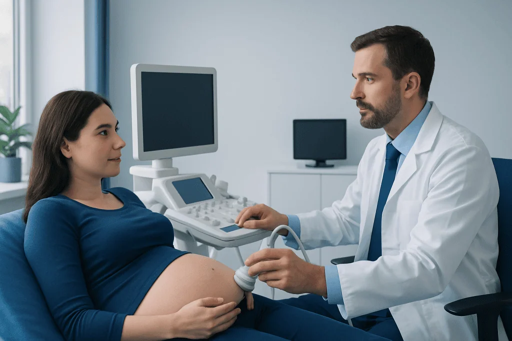
Ultrasound uses a small handheld device called a transducer. It sends high-frequency sound waves through the skin and receives echoes from tissues inside the body. A computer converts those echoes into moving images. Unlike X-rays and CT scans, routine obstetric ultrasound does not use ionising radiation.
During an ultrasound anatomy scan protocol, the operator records standard views and measurements in a systematic order. They may assess the baby’s position first, then obtain images of the head, brain, spine, chest, heart, abdomen, kidneys, limbs and other structures. The placenta’s location, the cervix when indicated, and the fluid around the baby may also be reviewed.
Image quality can be influenced by fetal position, the stage of pregnancy, maternal body shape, scar tissue and other ordinary factors. If the baby is facing an unfavourable direction or moving frequently, the sonographer may ask the pregnant person to change position, walk briefly, or return for another appointment. A repeat scan is often a practical way to complete missing views.
Anatomy ultrasound preparation: what to do before the appointment
Preparation before anomaly scan appointments is usually straightforward. The imaging centre should provide the most relevant instructions because requirements vary. Some centres ask patients to arrive with a comfortably full bladder, while others do not require this later in pregnancy. It is best to follow the instructions provided rather than drinking large amounts of water unnecessarily.
Patients should bring identification, referral paperwork if required, and any relevant previous ultrasound reports or pregnancy records. Wearing a comfortable two-piece outfit can make access to the abdomen easier. Eating normally is usually appropriate unless a clinician has given different instructions for a specific reason.
It can be helpful to arrive a little early, especially when completing forms or reviewing medical history. Patients may wish to ask whether a support person can attend, as visiting policies vary. It is also reasonable to prepare questions about how and when results will be discussed.
What not to do before your anatomy scan? Do not fast, restrict fluids, take medicines differently, or try unproven methods to make the baby move unless the care team has specifically instructed otherwise. Avoid overfilling the bladder, as discomfort can make the examination more difficult. Regular prescribed medicines should generally be continued, but any uncertainty should be discussed with the pregnancy care provider.
What happens during the procedure and how long does it take?
At the appointment, the pregnant person usually lies on an examination couch, often slightly tilted or with a pillow for comfort. Gel is placed on the abdomen to help the transducer make good contact with the skin. The sonographer moves the probe over different areas of the abdomen while viewing images on a monitor.
The examination may involve gentle pressure, particularly when the baby is positioned deep in the pelvis or when a specific view is needed. The sonographer may pause to take measurements, save images or ask the patient to change position. In selected situations, a transvaginal ultrasound may be offered to obtain clearer views of the cervix or placenta; this is a separate, explained procedure and requires consent.
How many minutes for an anomaly scan? Many scans take approximately 20 to 45 minutes. A longer appointment may be needed for twins or higher-order pregnancies, when the baby’s position limits the images, or when additional views are required. The time needed does not by itself indicate whether results are normal or abnormal.
Is an anomaly scan painful? An anomaly scan is not usually painful. The gel may feel cool, and pressure from the probe can be mildly uncomfortable for some people, but it should not cause significant pain. Patients should tell the sonographer promptly if they feel pain, dizziness, breathlessness or discomfort while lying down.
Results: how to understand a normal or unclear report
Results may be discussed immediately, at a later antenatal appointment, or once a doctor has reviewed the images, depending on the centre’s process. The report commonly lists the fetal structures that were examined, measurements that support gestational growth, the placenta location and any limitations of the study. Medical terminology in a report can sound concerning without context, so it is important to review it with a qualified clinician.
How do I know if my anomaly scan report is normal? A report may use wording such as “no structural abnormality detected,” “anatomy appears normal,” or “within normal limits” when the assessed structures look typical for the stage of pregnancy. It may also state that the scan was complete and that growth, fluid and placenta findings are appropriate. However, a normal scan cannot guarantee that every genetic, developmental or health condition has been ruled out.
An “incomplete” or “suboptimal” report often means that one or more views could not be obtained clearly, frequently because of fetal position. This commonly leads to a repeat examination. If a potential difference is seen, further assessment may include a targeted ultrasound, fetal echocardiography, genetic counselling, blood testing or referral to maternal-fetal medicine, depending on the finding.
In some cases, a follow-up scan provides reassurance. In others, it helps define an issue more precisely so that parents can receive clear information, supportive counselling and appropriate pregnancy planning. Decisions about further testing should be made with the obstetric team after discussing potential benefits, limitations and personal preferences.
Benefits, limitations and safety considerations
The main benefit of an anomaly scan is detailed information about fetal development during a key stage of pregnancy. It can identify many major structural differences, confirm aspects of fetal growth and pregnancy anatomy, and guide appropriate monitoring or specialist care when needed. It may also provide reassurance when findings are normal.
Ultrasound is widely used in pregnancy and is considered safe when performed for medical reasons by appropriately trained professionals using established protocols. The examination does not expose the pregnant person or baby to radiation. As with all medical tests, it should be used thoughtfully and interpreted alongside clinical history and other screening information.
Its limitations are equally important to understand. Not all conditions can be seen on ultrasound, and the ability to detect a particular condition depends on its type, severity, timing and image quality. Some findings are uncertain or temporary, while some health concerns are only recognised after birth. A normal result therefore reduces concern for many structural conditions but is not a complete guarantee.
If detailed fetal assessment or follow-up is needed, Acibadem International’s multidisciplinary specialists and JCI-accredited hospitals support international patients through diagnostic evaluation and pregnancy care planning.
When to seek medical care
Contact the maternity care provider promptly if there is vaginal bleeding, leaking fluid, severe or persistent abdominal pain, fever, fainting, severe headache, visual changes, or a noticeable reduction in fetal movements later in pregnancy. These symptoms are not caused by an anomaly scan itself, but they need timely clinical advice during pregnancy.
Patients should also contact their clinician if they have not received an expected result, do not understand wording in a report, or have been advised to return for further imaging. A follow-up recommendation is often made to complete views or clarify a finding and should be arranged within the timeframe advised by the care team.
After the scan, normal daily activities can usually be resumed straight away. There is no recovery period, special diet or activity restriction required from a routine abdominal anomaly scan. Emotional responses can vary, especially while waiting for results, and discussing concerns with a midwife, obstetrician or trusted support person can be helpful.
Frequently asked questions
What is an ultrasound anomaly scan?
An ultrasound anomaly scan is a detailed second-trimester pregnancy ultrasound that checks many aspects of fetal anatomy and the pregnancy environment. It is commonly performed at around 18 to 22 weeks of pregnancy. It is also called an anatomy scan or morphology scan.
Do I need a full bladder for an anomaly scan?
Requirements differ between imaging centres and depend partly on the stage of pregnancy. Some centres may ask for a comfortably full bladder, while others do not. The safest approach is to follow the instructions given with the appointment.
Can an anomaly scan detect all birth differences?
No. The scan can detect many major structural differences, but it cannot identify every condition. Some genetic, developmental, functional or later-developing conditions may not be visible on ultrasound.
What happens if the sonographer cannot get all the images?
The sonographer may ask the patient to change position, walk briefly or return for a repeat scan. This is often because the baby’s position makes a particular structure difficult to see. An incomplete scan result does not necessarily mean that an abnormality is present.
Can I eat before an anatomy ultrasound?
Most people can eat and drink normally before an anatomy ultrasound. Fasting is not usually required for a routine anomaly scan. Patients should follow any individual instructions given by their healthcare team.
Will I receive the results on the same day?
Many centres provide preliminary information during or soon after the scan, but the reporting process varies. Some results are reviewed by a radiologist, obstetrician or fetal medicine specialist before being discussed. Patients should ask when and how they will receive the final report.
References
- World Health Organization
- American College of Obstetricians and Gynecologists
- International Society of Ultrasound in Obstetrics and Gynecology
- National Health Service
- Society for Maternal-Fetal Medicine
This article is for general information only and is not a substitute for professional medical advice. Please consult a qualified doctor about your individual situation.
Ultrasound in Turkey — costs, top hospitals & a free quote
JCI-accredited · board-certified surgeons · reply within 24h
Add us as a Preferred Source to see more of our trusted health content across Google Search, AI Overviews and Discover.

Check-up & Preventive Medicine
Comprehensive health screenings designed for early detection and prevention.
3 specialists in this unitMore from the Health Library



Related Specialists

Prof. Dr. Mustafa Karahan
Orthopedic Surgery & Traumatology
Dr. Süleyman Büber
Clinical Laboratory
Assoc. Prof. Dr. Gürbüz Görümlü
Medical Oncology
Prof. Dr. Tülin Tuğlular
Hematology