Ultrasound Uterine Sarcoma: Preparation, Procedure and Results

Pelvic ultrasound uses sound waves to create images of the uterus and ovaries without radiation. A uterine sarcoma may appear as a large, irregular, mixed-texture or rapidly changing uterine mass, but these features can overlap with benign fibroids.
Key Takeaways
- Pelvic ultrasound uses sound waves to create images of the uterus and ovaries without radiation.
- A uterine sarcoma may appear as a large, irregular, mixed-texture or rapidly changing uterine mass, but these features can overlap with benign fibroids.
- Further assessment may include MRI, tissue sampling in selected circumstances, and surgery to establish a definite diagnosis.
- New bleeding after menopause, persistent pelvic pressure, or a rapidly enlarging uterine mass should be assessed by a clinician.
- Uterine sarcoma is rare, and most uterine masses are not sarcomas.
Ultrasound is often the first imaging test used to assess symptoms or a uterine mass that could be related to uterine sarcoma. It can show features that need further investigation, but it cannot reliably confirm or rule out uterine sarcoma on its own.
Ultrasound Uterine Sarcoma: Overview
Ultrasound uterine sarcoma assessment usually refers to a pelvic ultrasound performed when a person has abnormal uterine bleeding, pelvic pain, pressure symptoms, or a uterine mass. The scan may help clinicians see whether a mass has characteristics that require additional evaluation. However, ultrasound cannot diagnose uterine sarcoma with certainty because benign conditions, especially uterine fibroids, can look similar.
Uterine sarcoma is a rare cancer that begins in the muscle, connective tissue, or supporting tissues of the uterus. It differs from the more common endometrial cancers, which start in the lining of the uterus. The outlook and treatment plan depend on the sarcoma subtype, tumor size, whether it has spread, and a person’s overall health.
Most uterine growths detected on ultrasound are benign. Still, a radiology report describing an atypical or concerning mass should be discussed promptly with a gynecologist, who can determine whether MRI, referral to a gynecologic oncologist, or another next step is appropriate.
How Pelvic Ultrasound Works and Who May Need It
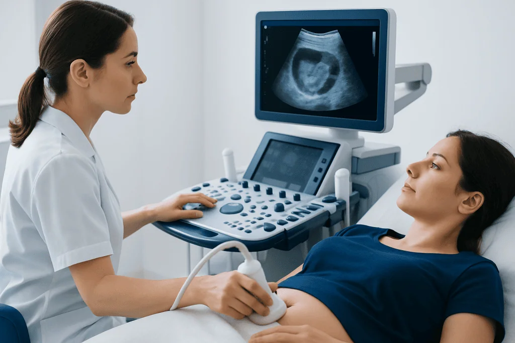
Pelvic ultrasound creates images using high-frequency sound waves. It does not use ionizing radiation. A transabdominal scan is performed across the lower abdomen, while a transvaginal scan uses a slim covered probe placed gently in the vagina to provide clearer, closer images of the uterus, endometrium, and ovaries.
A clinician may recommend ultrasound for heavy or irregular periods, bleeding after menopause, a feeling of pelvic fullness, a growing abdominal lump, pain, or a uterus that feels enlarged during a pelvic examination. It may also be used to monitor a previously identified fibroid or investigate a mass found on another imaging examination.
Ultrasound is suitable for most people and is generally well tolerated. A transvaginal ultrasound may not be appropriate or acceptable for every patient; alternatives can be discussed with the imaging team. It is important to tell the clinician about pregnancy, symptoms, prior pelvic surgery, and any known gynecologic condition.
Preparation, Step-by-Step Procedure and Recovery
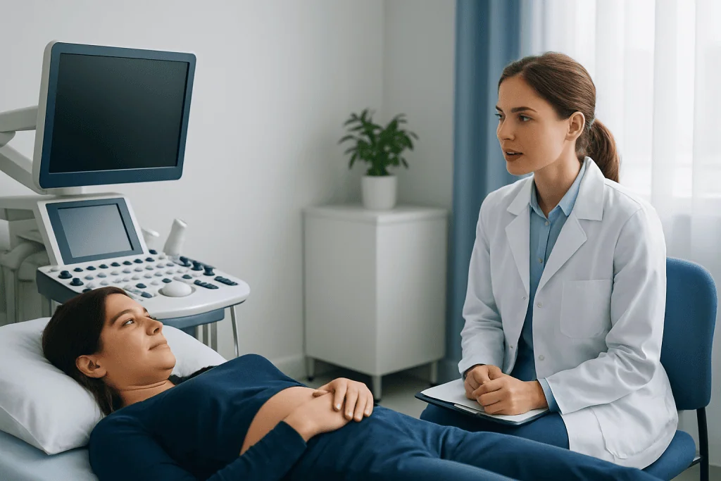
Preparation depends on the type of scan. For a transabdominal ultrasound, the patient may be asked to arrive with a comfortably full bladder because this can improve views of pelvic structures. For a transvaginal scan, an empty bladder is usually preferred. The imaging center will provide specific instructions before the appointment.
During a transabdominal examination, gel is placed on the lower abdomen and the sonographer moves a handheld probe over the skin. During a transvaginal examination, the covered and lubricated probe is inserted a short distance into the vagina. Images are obtained from several angles, and Doppler ultrasound may be used to assess blood flow within a mass. The examination commonly takes about 15 to 30 minutes, depending on the findings.
There is no recovery period after a routine pelvic ultrasound. A person can usually return to work, driving, exercise, and normal meals immediately. Mild temporary discomfort from pressure on a full bladder or probe placement can occur, but the test should not cause ongoing pain. A radiologist interprets the images, and the referring clinician explains what the findings mean in the context of symptoms and medical history.
What Are the Typical Findings of Sarcoma on Ultrasound?
There is no single ultrasound finding that proves a uterine mass is sarcoma. When sarcoma is suspected, ultrasound may show a mass that is large, irregularly shaped, or poorly defined, with a mixed pattern of solid areas, fluid-like spaces, bleeding, or tissue breakdown. A mass may also appear different from the typical well-circumscribed appearance of many fibroids.
Color Doppler imaging may show increased or irregular blood flow, particularly within solid portions of a mass. Other potentially concerning observations can include rapid apparent growth, distortion of the uterus, or a mass that extends beyond its expected boundaries. These features are not specific, and degenerating fibroids can also have uneven texture, fluid areas, and altered blood flow.
If findings are uncertain, MRI can provide more detailed information about the location and internal characteristics of a uterine mass. Imaging helps guide care, but a definitive diagnosis generally depends on examination of tissue by a pathologist, often after surgery. Decisions about further testing should be individualized by a gynecologist or gynecologic oncology team.
Can a Sarcoma Be Mistaken for a Fibroid?
Yes. A uterine sarcoma can sometimes be mistaken for a fibroid because both may cause uterine enlargement, bleeding, pelvic pressure, pain, or an abdominal mass. They can also overlap on ultrasound, particularly when a fibroid has undergone degeneration or has an unusual appearance.
Fibroids are common benign growths of uterine muscle, whereas uterine sarcomas are rare. This difference is important: the presence of a fibroid-like mass does not mean cancer is likely. At the same time, clinicians pay careful attention to atypical imaging features, new or enlarging masses after menopause, persistent unexplained symptoms, and changes that do not fit the expected pattern for a fibroid.
MRI, repeat imaging, specialist review, and surgery may be considered when the diagnosis remains unclear. Procedures that break a uterine mass into smaller pieces before removal may not be appropriate when cancer is suspected, because careful planning is needed to avoid spreading malignant tissue if it is present.
How Fast Does Uterine Sarcoma Spread?
The rate at which uterine sarcoma grows or spreads varies widely. It depends on the exact subtype, grade, size, and stage of the cancer. Some forms can behave aggressively and may spread through the bloodstream or lymphatic system to areas such as the lungs, abdomen, or other tissues, while others may follow a slower course.
Growth rate cannot be judged reliably from symptoms alone or from one ultrasound examination. A mass that seems to increase in size should be assessed in context, since benign fibroids can also grow or change, particularly before menopause. Comparing current and prior images can be helpful, but it does not replace diagnostic evaluation.
When clinicians are concerned about a possible uterine sarcoma, timely referral for specialist assessment is important. Further imaging and a carefully planned approach to surgery can help establish the diagnosis and guide treatment. Early evaluation is intended to clarify the cause of a mass rather than to assume that it is cancer.
How Serious Is Uterine Sarcoma?
Uterine sarcoma is a serious diagnosis because it is a malignant cancer and some subtypes can spread beyond the uterus. However, the prognosis is not the same for every person. Outcomes depend on whether the cancer is confined to the uterus, its subtype and grade, the success of surgery, and whether additional treatment is needed.
Treatment commonly involves surgery to remove the uterus, known as hysterectomy. Depending on the specific diagnosis and stage, care may also include radiation therapy, chemotherapy, hormone therapy, targeted treatments, or clinical trial options. A multidisciplinary team can coordinate gynecologic oncology, pathology, radiology, medical oncology, and radiation oncology input.
For people needing an individualized treatment plan, uterine cancer treatment services may include diagnostic assessment, surgery, and systemic cancer therapies according to the confirmed pathology. Acibadem International’s multidisciplinary specialists and JCI-accredited hospitals support diagnosis and treatment planning for international patients.
When to Seek Medical Care
A person should arrange a medical assessment for bleeding after menopause, unusually heavy or prolonged menstrual bleeding, bleeding between periods, persistent pelvic or abdominal pain, a new sensation of pressure, unexplained abdominal enlargement, or a newly felt pelvic mass. These symptoms have many possible causes and are more often related to noncancerous conditions, but they deserve evaluation.
More prompt review is appropriate if bleeding is heavy enough to cause dizziness, fainting, shortness of breath, or marked weakness. Urgent care may also be needed for sudden severe pelvic pain, fever with pelvic pain, or symptoms of significant blood loss. A clinician can assess immediate needs and arrange appropriate imaging or referral.
After an ultrasound, patients should ask what type of mass was seen, whether comparison with older scans is available, and whether MRI or gynecologic oncology consultation is recommended. Keeping follow-up appointments is important, especially when imaging findings are described as indeterminate, complex, atypical, or suspicious.
Frequently asked questions
Can an ultrasound diagnose uterine sarcoma?
No. Ultrasound can identify a uterine mass and describe features that may need further evaluation, but it cannot definitively distinguish sarcoma from fibroids or other benign conditions. Confirmation usually requires specialist assessment and examination of tissue by a pathologist.
Is transvaginal ultrasound painful?
A transvaginal ultrasound may cause mild pressure or temporary discomfort, but it is usually not painful. Patients can tell the sonographer if they feel uncomfortable, and the examination can be adjusted or stopped if needed.
What happens if an ultrasound shows an unusual uterine mass?
The next step may include a gynecologic examination, review of previous imaging, MRI, or referral to a gynecologist or gynecologic oncologist. The appropriate plan depends on age, symptoms, menopausal status, medical history, and the details of the imaging findings.
Do fibroids turn into uterine sarcoma?
Current evidence indicates that typical uterine fibroids do not transform into uterine sarcoma. A sarcoma may sometimes resemble a fibroid from the beginning, which is why unusual or changing masses may need closer evaluation.
Can uterine sarcoma occur before menopause?
Yes, although it is more often diagnosed in older adults and after menopause. Abnormal bleeding or a pelvic mass at any age should be assessed by a qualified clinician, particularly when symptoms are persistent or changing.
What should a patient bring to an ultrasound appointment?
It can be helpful to bring previous ultrasound, MRI, or CT reports and a list of medications, symptoms, menstrual history, and prior surgeries. The imaging center may also provide instructions about bladder preparation before the appointment.
References
- American Cancer Society
- National Cancer Institute
- American College of Obstetricians and Gynecologists
- European Society of Gynaecological Oncology
- RadiologyInfo.org
This article is for general information only and is not a substitute for professional medical advice. Please consult a qualified doctor about your individual situation.
Ultrasound in Turkey — costs, top hospitals & a free quote
JCI-accredited · board-certified surgeons · reply within 24h
Add us as a Preferred Source to see more of our trusted health content across Google Search, AI Overviews and Discover.

Check-up & Preventive Medicine
Comprehensive health screenings designed for early detection and prevention.
3 specialists in this unit