Understanding Fat Necrosis: A Complete Patient Guide

Fat necrosis is not cancer, but it can cause a lump that needs medical evaluation. It most often develops after injury, surgery, radiation, or inflammation in fatty tissue.
Key Takeaways
- Fat necrosis is not cancer, but it can cause a lump that needs medical evaluation.
- It most often develops after injury, surgery, radiation, or inflammation in fatty tissue.
- Breast fat necrosis is a common concern because it can mimic breast cancer on exam or imaging.
- Diagnosis may involve a physical exam, ultrasound, mammogram, MRI, or biopsy.
- Many cases need only monitoring, while painful or uncertain cases may need treatment or removal.
- New lumps, persistent pain, skin changes, or nipple changes should be checked promptly.
Fat necrosis is a benign change in fatty tissue that happens when fat cells are damaged, often after trauma, surgery, injections, or inflammation. It can form a firm lump, tenderness, or skin changes, and although it is not cancer, it can sometimes look similar to more serious conditions and should be assessed by a doctor.
Overview
Fat necrosis is an area of damaged fatty tissue that has broken down after an injury or another stress to the body. In many cases, the body heals by forming scar tissue or a small firm mass where the fat cells were injured. This change is benign, meaning it is not cancer.
It can happen in different parts of the body where fatty tissue is present, including the breasts, abdomen, buttocks, thighs, and under the skin after injections or surgery. The breast is one of the most commonly discussed locations because a lump caused by fat necrosis can feel worrisome and may resemble other breast conditions on an exam or scan.
Fat necrosis may go away on its own over time, stay stable, or occasionally become uncomfortable. Even though it is usually harmless, a new lump should not be self-diagnosed. A healthcare professional can confirm whether the finding is fat necrosis or another condition that needs different care.
How fat necrosis develops
Fat cells can be injured when their blood supply is disrupted or when the tissue is directly compressed, cut, stretched, or inflamed. Once damaged, the fat may leak from the cells and trigger the body’s repair process. This can lead to inflammation, calcification, oil cyst formation, or scarring.
This process explains why fat necrosis may feel different at different times. Early on, the area may be tender, bruised, or swollen. Later, it may become firmer and more defined as the tissue heals and scar tissue forms.
Doctors sometimes describe associated findings such as an oil cyst, which is a small pocket of liquefied fat, or calcifications, which are tiny calcium deposits left as part of healing. These are not necessarily dangerous, but they can influence how the area looks on imaging and whether more testing is needed.
Symptoms and what a person may notice

The most common symptom of fat necrosis is a lump. It may feel round, firm, rubbery, or irregular, depending on where it is and how long it has been present. Some people notice tenderness or mild pain, while others have no discomfort at all and find it by chance.
Changes on the skin can also occur, especially in breast fat necrosis. The skin over the area may look bruised, red, thickened, or dimpled. In some cases, the nipple may appear pulled inward if the nearby tissue has scarred. These signs can overlap with features seen in other conditions, which is why an evaluation is important.
Possible symptoms include:
- A new lump under the skin or in the breast
- Tenderness, soreness, or sensitivity
- Localized swelling after trauma or surgery
- Bruising or discoloration
- Skin dimpling or firmness
- Occasional oily or cyst-like feeling in the lump
Not every lump caused by fat necrosis is painful, and not every painful lump is fat necrosis. A doctor will consider the symptoms along with a person’s medical history, recent procedures, and imaging findings.
Causes and risk factors
Fat necrosis often follows a clear event that injures fatty tissue. Common examples include blunt trauma, a fall, a seatbelt injury, surgery, biopsy, reconstruction, liposuction, fat transfer procedures, or radiation therapy. It can also happen after inflammation in tissues that contain fat.
In the breast, fat necrosis may develop after breast surgery, reduction, reconstruction, biopsy, or radiotherapy. It can also appear after a direct blow to the chest, even if the injury seems minor at the time. Because breast lumps raise understandable concern, doctors often assess these changes carefully and may compare them with other findings seen in breast cancer.
Other risk factors include larger areas of fatty tissue, smoking, impaired blood flow, and conditions that make healing more difficult. In some cases, no clear cause is found. When fat necrosis affects other body areas, it may be part of a broader inflammatory process or related to previous injections, pressure, or surgery.
A separate but related condition is pancreatic fat necrosis, which can occur when pancreatic enzymes damage surrounding fat during inflammation of the pancreas. This is part of the disease process in pancreatitis and is different from the more localized lumps people often feel under the skin or in the breast.
Diagnosis and tests
Diagnosis starts with a medical history and physical examination. The doctor will ask when the lump appeared, whether there was any recent trauma or surgery, whether it is painful, and whether it has changed over time. A recent procedure or injury can be a useful clue, but it does not remove the need for proper assessment.
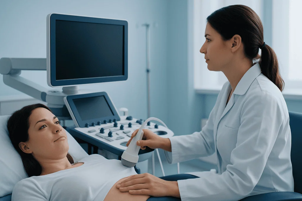
The next step often depends on where the fat necrosis is located. For breast concerns, doctors may use a clinical breast exam, mammogram, ultrasound, or sometimes MRI. These tests help show whether the finding has features typical of fat necrosis, such as an oil cyst or certain patterns of calcification. When appropriate, imaging may be part of a broader mammography assessment.
If the imaging result is not fully clear, a biopsy may be recommended. A core needle biopsy can take a small sample of tissue so a pathologist can confirm the diagnosis. This is especially helpful if the lump has an unusual appearance, is growing, or shares features with breast cancer.
For lumps outside the breast, ultrasound is often used first, and other imaging or minor procedures may be considered based on the location. The goal is to distinguish fat necrosis from cysts, infection, benign tumors, or less commonly, malignancy.
Treatment options and follow-up
Many cases of fat necrosis do not need active treatment. If the diagnosis is clear and the area is not causing symptoms, the doctor may recommend observation and follow-up. Over weeks or months, the lump may soften, shrink, or remain stable without causing harm.
When treatment is needed, it usually aims to relieve symptoms or confirm the diagnosis. Pain can often be managed with simple supportive measures such as rest, a well-fitting supportive bra for breast discomfort, or over-the-counter pain relief if a doctor says it is suitable. If there is a fluid-filled oil cyst, drainage may occasionally be considered.
Surgery is not routine, but it may be recommended if the lump is painful, persistent, enlarging, cosmetically bothersome, or uncertain after imaging and biopsy. Depending on the location, doctors may remove the area through a small procedure. Some people being evaluated for breast lumps may also need more advanced imaging, including breast MRI, if the clinical picture is not straightforward.
Care should always be individualized. Near the end of the care pathway, some patients may benefit from input from breast specialists, radiologists, plastic surgeons, or general surgeons. Acibadem International’s multidisciplinary specialists in JCI-accredited hospitals diagnose and treat conditions related to fat necrosis for international patients when specialist assessment is needed.
Self-care, monitoring, and prevention
Self-care focuses on watching for change rather than trying to treat the lump at home. It is best not to repeatedly press, squeeze, or massage the area, as this can increase irritation. If the area is sore after an injury or procedure, gentle support and following the aftercare plan given by the medical team may help comfort and healing.
People recovering from surgery should attend follow-up visits and report any new lump, redness, warmth, drainage, or worsening pain. Those who have had breast procedures may also be advised to continue routine screening and, when recommended, breast ultrasound or other imaging to document stable healing changes.
Prevention is not always possible, but reducing tissue trauma and supporting healthy healing may lower risk. General measures include not smoking, managing chronic health conditions, wearing seatbelts correctly, and carefully following surgical recovery instructions. After cosmetic or reconstructive procedures, choosing qualified medical care and keeping follow-up appointments are important.
When to seek medical care
A person should seek medical care for any new or unexplained lump, especially in the breast. Fat necrosis is usually benign, but it cannot be confirmed by touch alone. Prompt evaluation helps rule out other causes and reduces uncertainty.
Medical review is especially important if the lump is growing, painful, hard and fixed in place, or associated with skin dimpling, nipple changes, fever, redness, or discharge. These signs do not necessarily mean cancer or infection, but they do need assessment by a qualified clinician.
Urgent care may be needed if there is significant swelling, rapidly worsening pain, signs of infection, or symptoms after a recent operation such as increasing redness, warmth, or drainage from the wound. If there has been trauma, it is also sensible to seek care when a lump or bruising does not improve as expected.
Frequently asked questions
Is fat necrosis cancer?
No. Fat necrosis is a benign change that happens when fatty tissue is damaged and heals with inflammation, scarring, or cyst formation. However, because it can sometimes feel or look similar to cancer, a doctor may recommend imaging or biopsy to confirm the diagnosis.
Can fat necrosis go away on its own?
Yes, many cases improve or become less noticeable over time without specific treatment. Some lumps shrink, soften, or remain stable, while others may persist and still be harmless. Follow-up depends on symptoms, location, and how certain the diagnosis is.
What does fat necrosis feel like?
It often feels like a firm or rubbery lump under the skin or in the breast. It may be tender, but it can also be painless. Some people notice overlying bruising, skin thickening, or dimpling, especially if the area is in the breast.
How is breast fat necrosis diagnosed?
Doctors usually begin with a breast exam and imaging such as mammography or ultrasound. If the appearance is not clearly benign, MRI or a core needle biopsy may be used. The aim is to distinguish fat necrosis from cancer and other breast conditions.
Does fat necrosis always need surgery?
No. Surgery is usually reserved for cases that are painful, growing, bothersome in appearance, or still uncertain after appropriate testing. Many people only need observation and follow-up once the diagnosis is confirmed.
Can fat necrosis happen after surgery or cosmetic procedures?
Yes. It can occur after operations that affect fatty tissue, including breast surgery, reconstruction, liposuction, and fat transfer procedures. This happens because tissue handling or changes in blood supply can injure fat cells during healing.
References
- American Cancer Society
- National Health Service
- Radiological Society of North America
- Mayo Clinic
- National Cancer Institute
This article is for general information only and is not a substitute for professional medical advice. Please consult a qualified doctor about your individual situation.
Oncology care in Turkey — second opinion and treatment plan
JCI-accredited · board-certified surgeons · reply within 24h
Add us as a Preferred Source to see more of our trusted health content across Google Search, AI Overviews and Discover.