Veins Pulmonary: A Complete Medical Overview

Pulmonary veins carry oxygen-rich blood from the lungs to the left side of the heart. Most people have four pulmonary veins, but normal anatomical variations are common.
Key Takeaways
- Pulmonary veins carry oxygen-rich blood from the lungs to the left side of the heart.
- Most people have four pulmonary veins, but normal anatomical variations are common.
- Problems involving the pulmonary veins can be congenital, structural, or related to heart rhythm procedures.
- Symptoms depend on the cause and may include shortness of breath, fatigue, chest discomfort, or recurrent lung infections.
- Diagnosis often involves echocardiography, CT, MRI, and sometimes cardiac catheterization.
- Treatment ranges from monitoring to medication, catheter-based procedures, or surgery.
Veins pulmonary refers to the pulmonary veins, the blood vessels that return oxygen-rich blood from the lungs to the left atrium of the heart. Understanding how these veins work can help explain symptoms, tests, and treatments related to certain heart and lung conditions.
Overview: what “veins pulmonary” means
The term veins pulmonary refers to the pulmonary veins, which are the veins that bring oxygen-rich blood from the lungs back to the heart. In most people, there are four main pulmonary veins, two from each lung, and they empty into the left atrium, the upper chamber on the left side of the heart.
This role is different from most veins in the body. While many veins carry oxygen-poor blood back to the heart, pulmonary veins carry blood that has just picked up oxygen in the lungs. This makes them an essential part of the circulation that delivers oxygen to the brain, muscles, and organs.
Pulmonary veins can also be relevant in medicine because their anatomy matters in imaging, heart rhythm disorders, and certain congenital conditions. Some people are born with abnormal pulmonary venous connections, while others may develop narrowing of a pulmonary vein or changes after procedures for atrial fibrillation.
For patients, the most helpful way to understand pulmonary veins is to see them as the final pathway from the lungs to the heart. When they work normally, breathing and blood circulation stay well coordinated. When they are abnormal or blocked, symptoms may develop because blood flow between the lungs and heart becomes less efficient.
How pulmonary veins work in the heart and lungs
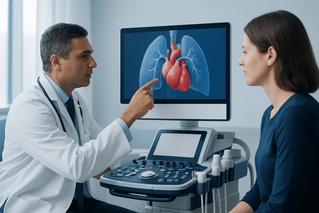
After a person breathes in, oxygen passes from the air sacs of the lungs into tiny blood vessels called capillaries. That oxygen-rich blood then collects into larger vessels that join together to form the pulmonary veins. These veins carry the blood into the left atrium.
From the left atrium, blood moves through the mitral valve into the left ventricle. The left ventricle then pumps it through the aorta to the rest of the body. In this way, pulmonary veins form the bridge between gas exchange in the lungs and circulation to all tissues.
Although the classic pattern is four pulmonary veins, the exact arrangement can vary from person to person. Some people have a common trunk on one side or an extra vein. These variations are often harmless, but they can matter during imaging studies and procedures such as atrial fibrillation treatment.
The openings of the pulmonary veins into the left atrium are also important because they can be a source of abnormal electrical signals in certain arrhythmias. This is one reason pulmonary vein anatomy is discussed not only in cardiology and radiology, but also in electrophysiology, the field that manages heart rhythm disorders.
Conditions that can affect the pulmonary veins

Several medical conditions can involve the pulmonary veins. One group includes congenital abnormalities present at birth, such as partial or total anomalous pulmonary venous return. In these disorders, some or all pulmonary veins connect to the wrong place instead of the left atrium, which can affect oxygen delivery and strain the heart.
Another condition is pulmonary vein stenosis, which means narrowing of one or more pulmonary veins. This can be congenital, but it may also occur later in life, sometimes after procedures near the veins. When a vein is narrowed, blood has more difficulty leaving the lungs, which can lead to breathlessness, cough, reduced exercise tolerance, or recurring lung problems.
Pulmonary veins are also closely linked with atrial fibrillation, a common irregular heart rhythm. Abnormal electrical activity often begins near these veins, so they may be targeted during catheter procedures. Patients learning about atrial fibrillation may hear pulmonary veins mentioned as part of the treatment plan.
In some cases, pulmonary venous congestion happens not because the veins themselves are malformed, but because the left side of the heart cannot receive blood normally. For example, certain valve disorders or heart failure can increase pressure behind the left atrium, causing blood to back up into the lungs. This is why pulmonary vein findings on imaging are interpreted together with the rest of the heart and lungs.
Symptoms and possible warning signs
Symptoms related to pulmonary vein problems vary widely depending on the underlying cause, the number of veins involved, and whether the issue developed gradually or was present from birth. Some people have no symptoms and only learn of an anatomical variation during a scan done for another reason.
When symptoms do occur, shortness of breath is one of the most common. A person may notice breathlessness with activity, poor exercise tolerance, fatigue, chest discomfort, cough, or repeated respiratory infections. In infants and children with congenital abnormalities, signs may include rapid breathing, poor feeding, slow growth, or bluish skin coloration.
If pulmonary veins are involved in a rhythm problem, palpitations may be more noticeable than lung symptoms. These may include a racing heartbeat, fluttering in the chest, dizziness, or reduced stamina. Some patients evaluated for arrhythmia undergo imaging that also shows pulmonary vein anatomy.
Symptoms that warrant prompt medical review include sudden worsening breathlessness, chest pain, coughing up blood, fainting, or signs of low oxygen. These symptoms do not always mean a pulmonary vein disorder, but they should not be ignored because they can also occur in other important heart or lung conditions.
How doctors diagnose pulmonary vein problems
Diagnosis starts with a medical history and physical examination. A doctor will ask about breathing symptoms, exercise tolerance, palpitations, prior heart procedures, childhood heart disease, and family history. The next steps depend on the age of the patient and the suspected cause.
Common tests include echocardiography, which uses ultrasound to look at heart structure and blood flow. It is often the first test because it is noninvasive and helps show how the heart chambers, valves, and nearby vessels are functioning. In some cases, transesophageal echocardiography provides more detailed images.
CT angiography and cardiac MRI can give a clearer map of pulmonary vein anatomy. These tests are especially useful for detecting abnormal connections, narrowing, or variations that may influence procedure planning. If rhythm-related treatment is being considered, detailed imaging can help guide cardiology care.
In more complex situations, doctors may use cardiac catheterization to measure pressures and assess blood flow directly. Additional tests can include electrocardiography, oxygen measurements, and lung imaging. The goal is not only to identify the pulmonary veins, but also to understand how they are affecting the lungs, heart function, and overall circulation.
Treatment options and long-term management
Treatment depends entirely on the diagnosis. Some anatomical variations are normal and do not need treatment. In those cases, the main need is accurate identification so that future doctors and imaging specialists understand the anatomy correctly.
Congenital abnormalities of pulmonary venous return may require surgery, especially when blood flow is significantly altered or the heart is under strain. Timing depends on the severity, symptoms, age, and whether the condition is causing low oxygen levels or enlargement of the right side of the heart. Care is usually coordinated by pediatric or adult congenital heart specialists.
Pulmonary vein stenosis may be managed with monitoring, medication for related symptoms, catheter-based dilation or stenting in selected cases, or surgery when appropriate. If the issue occurs after a rhythm procedure, follow-up imaging and specialist review are important. Treatment plans are individualized because the causes and recurrence risks vary.
When pulmonary veins are relevant to atrial fibrillation, treatment may include medicines, monitoring, and catheter ablation strategies designed around the vein openings. In selected patients, electrophysiology evaluation helps determine the safest and most effective approach. Near the end of the care pathway, some international patients also choose centers such as Acibadem International, where multidisciplinary specialists in JCI-accredited hospitals assess and treat complex heart and vascular conditions.
Prevention, self-care, and living with a pulmonary vein condition
There is no universal way to prevent all pulmonary vein conditions because some are congenital and others are linked to structural heart disease or necessary cardiac procedures. Still, overall heart and lung health can support better outcomes. Following medical advice, keeping follow-up appointments, and reporting changes in symptoms early are especially important.
Helpful self-care measures may include staying physically active within the limits advised by a doctor, avoiding smoking, managing blood pressure, treating sleep apnea if present, and taking prescribed medicines as directed. People with arrhythmias or heart failure should also follow guidance on fluids, exercise, and symptom monitoring.
For those who have undergone procedures involving the left atrium or pulmonary veins, follow-up imaging may be recommended even if they feel well. This is because some complications can develop gradually. Patients should ask what symptoms to watch for and when to return for routine reassessment.
Children and adults with congenital pulmonary venous disorders may benefit from long-term specialist care, even after successful treatment. As the body grows and ages, circulation needs can change. Regular review helps ensure heart function, lung health, and activity tolerance remain on track.
- Keep a record of prior heart tests and procedures.
- Report new shortness of breath, cough, palpitations, or reduced stamina.
- Attend scheduled imaging and cardiology follow-up visits.
- Seek personalized advice before starting intense exercise if a heart condition is known.
When to seek medical care
Medical care should be sought if a person has unexplained shortness of breath, repeated chest infections, palpitations, poor exercise tolerance, or persistent fatigue, especially if these symptoms are new or worsening. These symptoms can have many causes, but they deserve assessment when they interfere with daily activities or do not improve.
Urgent evaluation is important for severe breathlessness, chest pain, fainting, blue lips or fingertips, coughing up blood, or a rapid irregular heartbeat with dizziness. In infants and young children, warning signs include trouble feeding, poor weight gain, unusual sleepiness, fast breathing, or a bluish color.
Anyone with a known congenital heart condition, prior atrial fibrillation procedure, or previously identified pulmonary vein abnormality should ask their doctor about an appropriate follow-up schedule. Early review can help detect changes before they become more serious and may simplify treatment decisions.
Because pulmonary vein conditions overlap with heart and lung disease, evaluation often involves more than one specialty. A qualified doctor can decide whether symptoms are related to the pulmonary veins themselves or to another issue that needs care.
Frequently asked questions
What are veins pulmonary?
Veins pulmonary means the pulmonary veins. These are the blood vessels that carry oxygen-rich blood from the lungs to the left atrium of the heart, where it can then be pumped to the rest of the body.
How many pulmonary veins does a person usually have?
Most people have four pulmonary veins, with two coming from each lung. However, normal anatomical variations are common, and some people may have a different branching pattern without having any illness.
Are pulmonary veins the same as the pulmonary arteries?
No. Pulmonary arteries carry oxygen-poor blood from the heart to the lungs, while pulmonary veins carry oxygen-rich blood from the lungs back to the heart. They work together but move blood in opposite directions.
Can pulmonary vein problems cause shortness of breath?
Yes, they can. If the pulmonary veins are narrowed, abnormally connected, or affected by pressure changes from heart disease, blood may not flow efficiently from the lungs to the heart, which can contribute to breathlessness.
How are pulmonary vein conditions diagnosed?
Doctors often use echocardiography first, followed by CT or MRI for more detailed anatomy. In complex cases, cardiac catheterization and heart rhythm testing may also be used to understand blood flow and pressure changes.
Do all pulmonary vein abnormalities need treatment?
No. Some anatomical variations are harmless and only need to be documented. Treatment is usually considered when a condition changes blood flow, causes symptoms, affects oxygen levels, or increases strain on the heart.
This article is for general information only and is not a substitute for professional medical advice. Please consult a qualified doctor about your individual situation.
Explore treatments in Turkey — costs, top hospitals & a free quote
JCI-accredited · board-certified surgeons · reply within 24h
Add us as a Preferred Source to see more of our trusted health content across Google Search, AI Overviews and Discover.

More from the Health Library

C-pap: An Evidence-Based Guide for Patients

Diencephalon: An Evidence-Based Guide for Patients

My Chart: What Patients Need to Know

Femoral Nerve: What Patients Need to Know

Disseminated.: What Patients Need to Know