Visual Field Screening: How It Works, Results and What to Expect

Visual field screening checks central and peripheral vision one eye at a time. The test is commonly used to monitor glaucoma and assess possible optic nerve or brain-related vision changes.
Key Takeaways
- Visual field screening checks central and peripheral vision one eye at a time.
- The test is commonly used to monitor glaucoma and assess possible optic nerve or brain-related vision changes.
- An abnormal or unreliable result does not confirm a diagnosis and may need to be repeated.
- Most visual field tests take only a few minutes per eye and require no recovery time.
- Sudden loss of side vision, new blind spots or vision changes with neurological symptoms need prompt medical assessment.
Visual field screening is a simple, noninvasive test that maps how well each eye sees straight ahead and to the sides. It can identify changes in peripheral vision that may be linked to glaucoma, eye disease, optic nerve conditions or neurological problems.
Overview: What Is Visual Field Screening?
Visual field screening is an eye test that measures the full area a person can see while looking straight ahead, including central vision and side, or peripheral, vision. During the assessment, one eye is tested at a time while the person responds when small lights or targets appear in different locations. It is usually painless, does not involve radiation, and can be performed in an eye clinic.
The test helps an eye specialist identify areas where vision may be reduced or missing. These changes can be subtle and may not be noticed in everyday life, particularly when the other eye compensates. Visual field screening is often part of glaucoma assessment and follow-up, but it may also be used when symptoms or examination findings suggest a problem affecting the retina, optic nerve or visual pathways in the brain.
Screening results are interpreted alongside vision measurements, eye pressure, examination of the optic nerve and, when appropriate, imaging tests. A visual field test is an important source of information, but it is not a diagnosis by itself.
How Visual Field Screening Works and Who May Need It
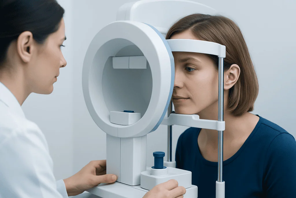
A visual field test presents light stimuli of varying brightness at different points within a curved bowl-shaped instrument or on a screen. The person keeps their gaze fixed on a central target and presses a button whenever a light is seen. The device records which lights are detected, creating a map of visual sensitivity across the field of view.
Eye specialists may recommend visual field screening for people who have raised eye pressure, suspicious optic nerve findings, a family history of glaucoma, or diagnosed glaucoma that needs monitoring. It can also be useful after certain eye injuries, for unexplained reduced vision, or when an eye or neurological examination suggests possible visual pathway involvement.
People may be assessed for conditions such as glaucoma, which can gradually affect peripheral vision without noticeable early symptoms. A clinician may also request testing when a person reports bumping into objects, difficulty seeing to the side, new blind spots, or changes in vision after a head injury or neurological event.
Visual field screening is suitable for many adults and older children who can understand and follow the instructions. Reliable testing depends on concentration and the ability to maintain fixation, so the clinician may adapt the assessment or arrange repeat testing if fatigue, learning difficulties or poor vision make the first result uncertain.
What Happens During the Procedure?
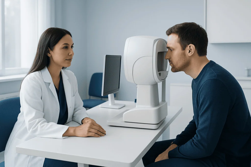
Before the test, the eye care team checks that the person has the correct glasses or trial lens for the testing distance. One eye is covered, and the other eye looks steadily at a central fixation light. The person is asked not to search for the lights with their eyes, but simply to press a handheld button when a light is noticed.
Small lights appear one at a time in different parts of the visual field. Some are bright and easy to see; others are dimmer. The machine may occasionally show a light when none is expected or repeat a light to assess response consistency. These checks help determine whether the result is reliable enough to interpret.
The test commonly takes several minutes per eye, though the exact time depends on the method used and the reason for testing. The experience can feel repetitive, and it is normal to miss some dim lights. Taking brief pauses, if offered, can help a person stay comfortable and focused.
No needles, eye drops or special recovery period are usually needed for standard visual field screening. If the appointment also includes pupil-dilating drops or other eye tests, temporary blurred near vision and light sensitivity may occur from those additional procedures rather than from the field test itself.
What Should You Not Do Before a Visual Field Test?
There is usually no extensive preparation for visual field screening. A person should bring their usual glasses and a current list of medicines, especially eye drops and treatments for glaucoma or neurological conditions. Contact lenses may often be worn, but the eye team may advise whether glasses or a trial lens will provide the most accurate correction for the test.
It is helpful to avoid arriving overly tired, rushed or distracted, because concentration affects test reliability. If possible, people should eat normally, rest adequately and allow enough time for the appointment. They should not stop prescribed eye drops or other medicines unless their clinician has specifically advised this.
Alcohol, recreational drugs and sedating medicines may affect alertness and responses. A person who feels unwell, has a severe headache, or is experiencing a major change in vision should tell the clinic before testing. The test can often be rescheduled if temporary factors are likely to make the result unreliable.
During the examination, it is important not to move the head repeatedly, look around for lights, or press the response button when uncertain. Missing an occasional light is expected; responding as naturally and consistently as possible gives the clinician the most useful information.
Understanding Visual Field Test Results
Results are usually displayed as a map showing areas where light was seen normally and areas where sensitivity was reduced. The report may compare results with an age-matched reference range and may include reliability indicators, such as missed fixation checks or responses recorded when no light was shown.
A normal result suggests that no significant field defect was detected under the conditions of the test. However, a normal screening result does not rule out every eye or neurological condition. Conversely, an abnormal result may reflect a genuine visual field defect, but it can also result from fatigue, dry eyes, incorrect glasses correction, poor fixation, drooping eyelids or difficulty understanding the test.
When changes are found, the clinician considers their pattern and whether it matches examination findings. Repeat visual field testing is common, especially after a first test, because practice improves performance and because clinicians often look for consistent changes over time. Additional assessment may include optic nerve imaging, retinal examination, eye pressure measurement or neurological evaluation.
For patients receiving glaucoma treatment, repeated visual field testing can help assess whether the condition appears stable or whether the care plan needs review. Results should always be discussed with an ophthalmologist or other qualified clinician who knows the person’s wider eye and medical history.
What Does It Mean If You Fail a Visual Field Test?
“Failing” a visual field test is not a medical diagnosis. It usually means that the screening identified an abnormal area, or that the responses were not reliable enough for the test to be interpreted with confidence. Many people need a repeat test before a clinician can decide whether there is a true visual field change.
If a defect is confirmed, its location and pattern may guide further investigation. Some patterns can occur with glaucoma or other optic nerve disorders, while others may suggest retinal disease or a problem along the visual pathways in the brain. The clinician will compare the test with eye examination findings and may arrange imaging or referral when needed.
An abnormal test should be taken seriously but calmly. It does not automatically mean vision will worsen, and timely assessment can identify treatable causes or establish an appropriate monitoring plan. People should attend follow-up appointments and report any new or rapidly changing symptoms rather than waiting for their next routine visit.
Benefits, Limitations and Possible Risks
The main benefit of visual field screening is that it can detect functional vision changes that may not be obvious to the person being tested. It is noninvasive, repeatable and useful for monitoring conditions over time. In glaucoma care, field testing complements structural tests that look at the optic nerve and retinal nerve fiber layer.
The test has few physical risks. Some people experience temporary eye strain, boredom, frustration or fatigue, particularly during longer examinations. Because attention strongly affects the result, fatigue and anxiety can contribute to false-positive or false-negative findings. This is why clinicians review reliability measures and may repeat the test.
Visual field screening also has limitations: it does not explain the cause of every abnormal area and should not be used alone to diagnose a disease. A comprehensive eye assessment remains important. Depending on symptoms, a specialist may recommend additional eye examination and testing to clarify the cause.
For international patients, Acibadem International’s multidisciplinary specialists and JCI-accredited hospitals can assess visual field changes with ophthalmology and, where indicated, neurological expertise. The appropriate pathway depends on the individual’s symptoms, examination and test findings.
Can You Still Drive If You Fail a Field Vision Test?
Whether a person can continue driving after an abnormal visual field test depends on the extent and location of any confirmed vision loss, the requirements of the country or region, and the type of driving licence held. A single unreliable or abnormal screening result does not automatically mean that a person must stop driving permanently.
Some visual field defects can affect the ability to notice pedestrians, vehicles, road signs or hazards approaching from the side. If a clinician identifies a significant field loss, they may advise temporary driving restrictions while further testing is completed. The person may also need to report the condition to the relevant licensing authority, depending on local regulations.
It is important not to make decisions based only on online advice or an initial test report. The eye specialist can explain the findings, arrange confirmatory testing and advise on safe next steps. Anyone with sudden vision loss, a new blind area, double vision or symptoms that could affect safe driving should avoid driving and seek urgent medical advice.
Is a Visual Field Test Worth It and When to Seek Medical Care
Visual field testing is worth it when recommended by an eye specialist because it provides information that cannot be obtained from a standard eye chart alone. It is particularly valuable for detecting and tracking side-vision changes in glaucoma and for investigating certain visual or neurological symptoms. The test is generally quick, painless and can help clinicians make better-informed decisions about monitoring or treatment.
Routine testing is often appropriate for people at risk of glaucoma or those already being monitored for optic nerve changes. Repeat testing may be needed even when a person feels well, because some eye conditions develop gradually and may not cause noticeable symptoms until later stages.
Medical care should be sought promptly for sudden loss of vision, a new curtain-like shadow, sudden blind spots, severe eye pain, redness with nausea, or vision changes accompanied by facial weakness, trouble speaking, severe headache, weakness or numbness. These symptoms may require urgent assessment and should not wait for a routine visual field appointment.
For non-urgent concerns, a person should book an eye examination if they notice increasing difficulty seeing to the side, frequently bump into objects, have unexplained headaches with visual changes, or have a family history of glaucoma. Early assessment supports timely investigation and appropriate care.
Frequently asked questions
How long does visual field screening take?
Many automated visual field tests take several minutes for each eye, although timing varies by the type of test and the testing strategy used. The full appointment may take longer if it includes eye pressure checks, imaging or pupil dilation.
Does a visual field test hurt?
No. The test does not touch the eye and should not be painful. Some people find it tiring because it requires steady attention, but breaks can often be requested if needed.
Why might a visual field test need to be repeated?
A repeat test may be needed if the first result has low reliability or shows an unexpected change. Practice, fatigue, dry eyes, an incorrect prescription or difficulty maintaining fixation can all influence results.
Can glaucoma be present with a normal visual field test?
Yes. Early glaucoma may cause structural changes to the optic nerve before measurable field loss is detected. Eye pressure measurements, optic nerve examination and imaging are therefore often used alongside visual field testing.
What does it mean if you fail a visual field test?
It may mean that the test detected reduced sensitivity in part of the visual field, or that the responses were too inconsistent to interpret reliably. A clinician usually reviews the pattern, checks the reliability data and may arrange repeat or additional tests before drawing conclusions.
Can you still drive if you fail a field vision test?
This depends on whether a visual field defect is confirmed, how severe it is, and local driving rules. An eye specialist can advise whether temporary restrictions, repeat testing or notification to a licensing authority may be necessary.
Is a visual field test worth it?
Yes, when an eye specialist recommends it. It is a noninvasive way to assess central and side vision, detect changes that may otherwise go unnoticed, and monitor conditions such as glaucoma over time.
References
- American Academy of Ophthalmology
- National Eye Institute
- Glaucoma Research Foundation
- National Health Service
- World Glaucoma Association
This article is for general information only and is not a substitute for professional medical advice. Please consult a qualified doctor about your individual situation.
Explore treatments in Turkey — costs, top hospitals & a free quote
JCI-accredited · board-certified surgeons · reply within 24h
Add us as a Preferred Source to see more of our trusted health content across Google Search, AI Overviews and Discover.