Visual Field Testing Explained: How Doctors Check for Brain and Optic Nerve Disease

Visual field testing maps central and peripheral vision in each eye. It can help detect glaucoma, optic nerve disorders, stroke-related vision loss, and other neurological conditions.
Key Takeaways
- Visual field testing maps central and peripheral vision in each eye.
- It can help detect glaucoma, optic nerve disorders, stroke-related vision loss, and other neurological conditions.
- The test is noninvasive, usually painless, and commonly done in an outpatient clinic.
- Results depend on concentration and technique, so repeat testing is sometimes needed.
- Visual field changes do not diagnose a condition alone; doctors interpret them together with the eye exam and imaging when needed.
Visual field testing measures how well a person sees in the center and at the edges of vision. It helps doctors find patterns of vision loss linked to eye disease, optic nerve damage, and certain brain conditions.
Overview: What Visual Field Testing Measures
Visual field testing is a way to measure the full area a person can see while looking straight ahead. This includes central vision, which is used for reading and recognizing faces, and peripheral vision, which helps with movement, orientation, and noticing objects to the side. The test creates a map that shows whether any areas of vision are reduced or missing.
Doctors use visual field testing because different eye, optic nerve, and brain disorders can cause characteristic patterns of vision loss. A problem in the retina or optic nerve may affect one eye differently from the other, while a problem in the visual pathways of the brain may create a matching defect in both eyes. These patterns can give important clues about where the problem may be located.
Visual field testing is often part of a neuro-ophthalmic evaluation. It may be requested when a person reports blurred side vision, bumping into objects, trouble reading lines of text, visual dimming, or unexplained headaches with visual symptoms. It may also be used to monitor known conditions over time and to see whether treatment is helping preserve vision.
Why Doctors Order a Visual Field Test
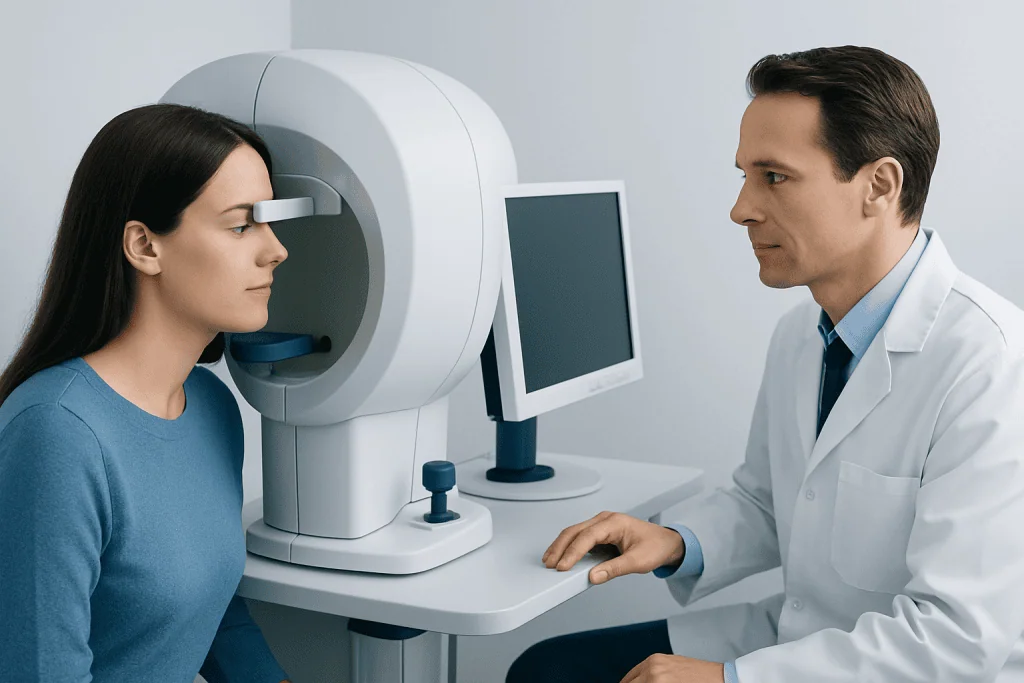
A visual field test may be ordered for many reasons. In eye care, it is widely used to help detect and monitor glaucoma, a condition that can gradually damage the optic nerve and narrow side vision. It can also help assess retinal disease, drooping eyelids that block vision, and complications from eye inflammation or injury.
In neuro-ophthalmology and neurology, visual field testing can reveal changes caused by optic neuritis, compression of the optic nerve, pituitary tumors, stroke, brain injury, and other conditions affecting the visual pathways. For example, a person with damage to one side of the brain may lose the same half of the visual field in both eyes, a pattern called homonymous hemianopia.
The test may also be used when symptoms are vague or intermittent. Some people notice only subtle difficulties, such as trouble driving, missing words while reading, or needing to turn the head more often. In these situations, a mapped visual field can turn a general complaint into clear, measurable information that guides diagnosis and follow-up.
Types of Visual Field Tests and How They Work
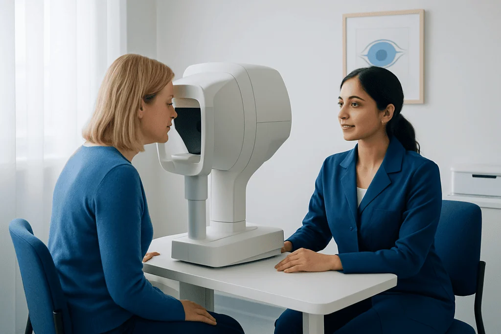
There are several ways to test the visual field. The most common method in clinics is automated perimetry. During this test, the person looks into a bowl-shaped machine and presses a button when lights appear in different parts of vision. The device measures how faint a light can be detected at many points, creating a detailed sensitivity map for each eye.
Another method is confrontation visual field testing, which is a simpler screening test performed by a doctor at the bedside or in the office. The patient looks straight ahead while reporting when fingers or a moving target are seen in the side vision. This approach is quick and useful, but it is less sensitive than automated testing for mild defects.
Doctors may also use kinetic perimetry, in which a moving target is brought in from the outer edge toward the center until it is seen. This can be especially helpful for people who have difficulty with standard automated testing, or when a large or unusual field defect is suspected. In selected cases, the eye specialist may combine field testing with other assessments such as optical coherence tomography (OCT) to compare vision loss with the structure of the optic nerve and retina.
Each test has strengths and limitations. Automated tests are detailed and reproducible, but they require attention and may feel tiring. Simpler screening tests are fast, yet they may miss subtle changes. The most useful approach depends on the person’s symptoms, age, cooperation, and the clinical question being asked.
What the Test Is Like and How to Prepare
Visual field testing is noninvasive and usually painless. The person typically sits in front of the machine with one eye covered, placing the chin on a support and looking at a fixed central target. Small lights appear one at a time, and the patient responds whenever a light is seen. The same process is then repeated for the other eye.
The test requires concentration, so it is normal for it to feel repetitive. Blinking is allowed and important for comfort, but large eye movements can affect the result because the goal is to keep looking straight ahead. Most clinics explain the instructions before starting and may pause if the person feels tired or uncertain.
Preparation is simple. Wearing regular glasses or the prescription advised by the clinic can help the machine measure vision more accurately. It is also helpful to mention dry eyes, fatigue, anxiety, or difficulty understanding the instructions, because these factors can make the test harder. If the first result seems unreliable, doctors often repeat the test rather than drawing conclusions too quickly.
What the Results Can Show
Visual field results are displayed as maps and graphs that show where vision is normal and where sensitivity is reduced. Doctors look for the size, depth, and shape of any defect. They also review reliability indicators, such as whether the person responded consistently and maintained steady fixation during the test.
Certain patterns can suggest particular causes. Patchy or arcuate defects may point toward glaucoma. Central blind spots or enlarged blind spots can occur with optic nerve swelling or inflammation. Loss of the outer half of vision on the same side in both eyes can suggest a problem in the brain’s visual pathways, sometimes after a stroke or other neurological event. In this context, visual field testing may be part of a broader evaluation for stroke or other neurological disease.
A visual field test does not usually provide a final diagnosis by itself. Instead, doctors interpret it together with the medical history, vision testing, pupil responses, eye pressure, retinal examination, and imaging when needed. If a pattern raises concern for optic nerve compression or a brain-related cause, additional studies such as MRI may be recommended.
Because results can vary from day to day, trends over time are often very important. A single abnormal test may need confirmation, while repeated testing can show whether vision is stable, improving, or gradually changing. This is especially valuable in chronic conditions such as glaucoma or in follow-up after treatment.
Conditions Commonly Linked to Visual Field Loss
Many conditions can change the visual field. Glaucoma is one of the most common examples, often affecting peripheral vision first. Some people do not notice any symptoms until significant loss has occurred, which is why regular eye care matters for those at risk. Visual field testing helps both with early detection and with long-term monitoring.
Optic nerve disorders are another important group. Inflammation, reduced blood flow, inherited optic neuropathies, pressure from nearby structures, and swelling from increased intracranial pressure can all produce characteristic defects. When the optic nerve is involved, people may also have reduced color vision, pain with eye movement, or dimming of vision.
Brain-related causes include stroke, tumors, traumatic brain injury, and lesions affecting the pathways that carry visual information from the eyes to the brain. These conditions may cause loss on the same side of the visual field in both eyes or other patterns that help localize the problem. If the visual field suggests a structural cause, doctors may coordinate care with neurology, neurosurgery, or endocrinology depending on the suspected source.
Other causes can include retinal disorders, migraine with aura, eyelid problems, medication effects, and functional or nonorganic vision symptoms. Because the range of possibilities is broad, visual field findings are most useful when considered as one part of a careful and complete assessment.
How Doctors Confirm the Cause and Plan Treatment
Treatment depends on the underlying reason for the visual field change. The test itself does not treat vision loss, but it helps doctors decide what needs attention and how urgently. In some cases, the next step is close observation and repeat testing. In others, treatment focuses on the eye, optic nerve, or brain problem causing the defect.
If glaucoma is suspected, management may include pressure-lowering eye drops, laser treatment, or surgery as advised by an eye specialist. If inflammation of the optic nerve is suspected, the doctor may consider blood tests, imaging, and tailored medical treatment. If the pattern suggests a tumor or compression near the optic pathways, imaging and referral for specialist care are important; some patients may need brain tumor treatment or endocrine evaluation depending on the cause.
For stroke-related field loss, treatment focuses on the underlying vascular event, rehabilitation, and safety counseling, including practical advice about reading and mobility. Some patients benefit from low-vision support, compensatory scanning techniques, and occupational therapy. If reduced side vision affects daily life, clinicians may discuss driving restrictions or local regulations to help protect safety.
Near the end of the diagnostic process, some patients seek care at specialized centers. Acibadem International’s multidisciplinary specialists and JCI-accredited hospitals evaluate visual field loss for international patients, particularly when eye, optic nerve, and neurological causes need coordinated assessment.
When to Seek Medical Attention
Any sudden change in vision should be assessed promptly. This includes abrupt loss of side vision, new dark or missing areas in vision, sudden double vision, or vision loss accompanied by weakness, numbness, severe headache, trouble speaking, or imbalance. These symptoms can sometimes signal a neurological emergency and should not be ignored.
A prompt appointment is also important for more gradual concerns, such as increasing difficulty reading, bumping into objects, or progressive narrowing of side vision. Even when symptoms seem mild, visual field changes can provide an early sign of eye or optic nerve disease that benefits from timely evaluation.
People already being monitored for glaucoma, optic nerve disease, or a neurological condition should attend follow-up appointments as recommended. Regular testing helps doctors detect changes early and adjust care before further vision is lost. If the instructions for the test are difficult to follow, it is useful to say so, because alternative methods may be available.
Frequently asked questions
Is visual field testing painful?
Visual field testing is usually painless and noninvasive. Most people find it more tiring than uncomfortable because it requires concentration and repeated responses to visual targets.
How long does a visual field test take?
The length depends on the type of test and the reason it is being done. In many clinics, each eye takes several minutes, but preparation, explanation, and repeat testing can make the full visit longer.
Can a visual field test detect brain problems?
Yes, certain patterns can suggest problems in the brain’s visual pathways, including changes after stroke, tumors, or other neurological conditions. The test does not confirm the cause by itself, so doctors may combine it with the examination and brain imaging when needed.
Why might a doctor repeat the test?
Results can be influenced by fatigue, inattention, dry eyes, anxiety, or difficulty keeping the gaze steady. Repeating the test helps confirm whether a finding is real and allows doctors to compare changes over time.
Do abnormal results always mean glaucoma?
No. Glaucoma is a common cause of visual field loss, but abnormal results can also occur with optic nerve inflammation, retinal disease, stroke, brain lesions, eyelid blockage, and other conditions. The pattern of loss and the rest of the evaluation help narrow the cause.
Can visual field loss be treated?
Treatment depends on what is causing the visual field change. Some causes can be treated or stabilized, while others may require rehabilitation and monitoring to help the person adapt and protect remaining vision.
References
- American Academy of Ophthalmology
- National Eye Institute
- American Academy of Neurology
- Royal College of Ophthalmologists
- National Institute of Neurological Disorders and Stroke
This article is for general information only and is not a substitute for professional medical advice. Please consult a qualified doctor about your individual situation.
Explore treatments in Turkey — costs, top hospitals & a free quote
JCI-accredited · board-certified surgeons · reply within 24h
Add us as a Preferred Source to see more of our trusted health content across Google Search, AI Overviews and Discover.

More from the Health Library



Related Specialists

Dr. Kumsal Kurucu
Acibadem Life Clinical Service
Dr. Ümit Ege Canyurt
Acibadem Life Clinical Service
Dr. Burak Sercan Erçin
Aesthetic Plastic & Reconstructive Surgery
Assoc. Prof. Dr. Mustafa Bilal Tuna
Urology