What Happens After an Abnormal Imaging Result? Next Tests, Follow-Up, and Timing
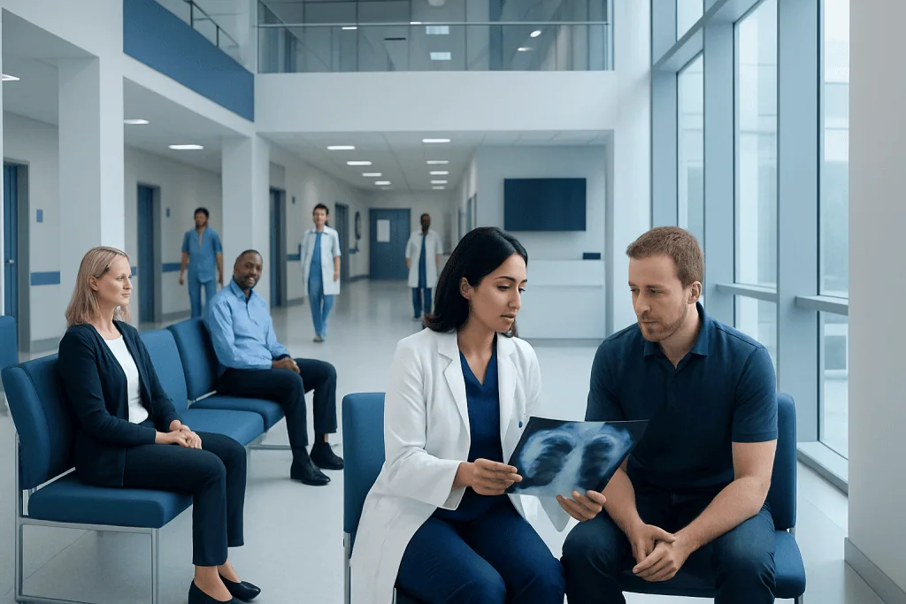
An abnormal imaging result often means an unexpected finding that needs clarification, not necessarily a dangerous diagnosis. Follow-up may include comparison with older scans, repeat imaging, blood tests, ultrasound, MRI, CT, or sometimes biopsy.
Key Takeaways
- An abnormal imaging result often means an unexpected finding that needs clarification, not necessarily a dangerous diagnosis.
- Follow-up may include comparison with older scans, repeat imaging, blood tests, ultrasound, MRI, CT, or sometimes biopsy.
- Timing depends on urgency: some findings need same-day care, while others are safely rechecked in weeks or months.
- Patients should ask what was found, how urgent it is, what test is next, and when results are expected.
- Bringing prior imaging and medical history can help avoid unnecessary repeat tests and improve accuracy.
An abnormal imaging result means a scan showed something that needs closer review, but it does not automatically mean a serious illness. The next steps depend on what was seen, which body area was scanned, and how urgent the finding appears to the radiologist and doctor.
Overview: what an abnormal imaging result means
An abnormal imaging result means that a radiology test such as an X-ray, ultrasound, mammogram, CT scan, MRI, or nuclear medicine scan showed something outside the expected appearance. This can include a shadow, nodule, cyst, mass, enlarged structure, inflammation, fracture, fluid collection, or another change that needs interpretation. In many cases, the finding is minor, benign, or unrelated to the reason the scan was ordered.
Imaging results are usually interpreted by a radiologist, a doctor trained to read scans. The radiologist sends a report to the clinician who ordered the test. That clinician then connects the report with the person’s symptoms, examination, medical history, and any previous tests. This step matters because the same imaging finding may mean different things in different people.
Many abnormal results need only follow-up rather than immediate treatment. For example, a small simple cyst may need no action, while an unclear spot may require another type of scan for a better view. The main goal is to find out whether the finding is harmless, temporary, or something that needs closer attention.
Why an abnormal result does not always mean something serious
Imaging is very sensitive, which means it can detect small changes that would otherwise go unnoticed. Some of these are called incidental findings. They are discovered by chance and may not be causing symptoms. Common examples include benign liver cysts, thyroid nodules, small lung nodules, age-related spine changes, and ovarian cysts that are often monitored rather than treated.
There can also be technical reasons why a result appears abnormal. Motion during a scan, overlapping tissues, metal artifacts, incomplete bladder filling, or differences in how the test was performed can make interpretation more difficult. In these situations, a repeat image or a more targeted study may clarify the picture.
Doctors also consider how likely a finding is to be important based on age, personal and family history, smoking history, previous cancer, infection risk, symptoms, and physical examination. This is why the same wording on a radiology report can lead to different follow-up plans in different patients.
- Some findings are clearly benign and need no further action.
- Some findings are indeterminate, meaning they are not yet fully explained.
- Some findings are suspicious and need urgent testing.
Common next steps after an abnormal scan

The next step is often a careful review of the radiology report together with the images themselves. If prior imaging is available, comparing old and new scans can be very helpful. A finding that has been unchanged for years is often less concerning than one that is new or growing.
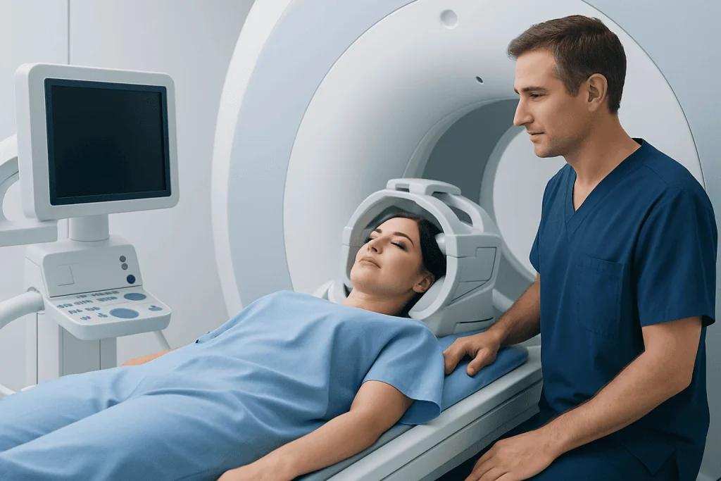
Additional imaging is a common follow-up step. The best test depends on the body area and the original scan. An ultrasound may help distinguish a fluid-filled cyst from a solid mass. A CT scan can provide more detail about the lungs, abdomen, or bones. An MRI may better evaluate soft tissues, the brain, spine, joints, liver, or pelvis. In some situations, doctors may recommend MRI or CT scan to clarify what was seen first.
Laboratory tests may be ordered if the finding could be related to infection, inflammation, hormone changes, liver or kidney function, or blood disorders. If cancer is a concern, blood tests alone usually cannot confirm or exclude it, but they may support the overall assessment.
Sometimes a procedure is needed to make a definite diagnosis. This may include aspiration of fluid, endoscopy, or a biopsy so a tissue sample can be examined under a microscope. If the result involves the breast, additional mammographic views or breast ultrasound may be recommended before any invasive testing is considered.
How follow-up timing is decided
Timing depends on how urgent the finding appears. Radiologists often classify abnormalities as routine, urgent, or critical. A critical result, such as signs of a stroke, bowel perforation, major bleeding, or a blood clot in the lung, is usually communicated quickly and may need same-day treatment. A suspicious but stable finding may need testing within days to weeks. A probably benign finding may only need repeat imaging after a few months.
The body area involved also affects timing. For example, a new brain abnormality with neurological symptoms is more urgent than a tiny simple kidney cyst found incidentally. Similarly, severe symptoms such as chest pain, difficulty breathing, weakness on one side, sudden vision changes, or high fever can make even a modest imaging abnormality more urgent.
Doctors also balance urgency against test quality and safety. Some scans require preparation, such as fasting, contrast review, or checking kidney function. In follow-up imaging, waiting a short interval can sometimes be useful because it helps show whether a finding resolves, stays stable, or changes over time. This is common with some lung nodules, ovarian cysts, inflammatory changes, and selected liver lesions.
Questions patients should ask after an abnormal imaging result
Clear communication can reduce uncertainty and help patients understand what happens next. It is reasonable to ask for a plain-language explanation of the report. Medical terms such as lesion, opacity, enhancement, nodule, or indeterminate can sound worrying, but they often simply describe an appearance rather than a final diagnosis.
Helpful questions include: What exactly was seen? How concerning is it? Is it likely benign, unclear, or suspicious? Do older scans exist for comparison? What test, if any, is recommended next? How soon should that happen? What symptoms should prompt urgent care? When and how will results be communicated?
Patients may also ask whether a specialist is needed. Depending on the finding, the next doctor may be a pulmonologist, neurologist, gastroenterologist, surgeon, gynecologist, urologist, or oncologist. If the report suggests a known condition, the doctor may explain whether it relates to issues such as lung cancer or breast cancer, or whether the appearance is more consistent with a benign process that simply needs monitoring.
How to prepare for follow-up testing and appointments
Preparation starts with collecting information. Patients should bring prior scan reports and, if possible, the images on disc or through electronic transfer. They should also share a list of medications, allergies, major illnesses, surgeries, and any personal or family history that may be relevant. This information can improve accuracy and may prevent duplicate tests.
It is helpful to write down symptoms, when they began, and whether they are changing. Even symptoms that seem unrelated may matter. For example, weight loss, fever, persistent cough, unusual bleeding, abdominal pain, headaches, or weakness can help doctors understand the significance of an imaging finding.
Patients should ask about practical details before the next test. Some scans use contrast material and may require checking kidney function or discussing allergy history. MRI may not be suitable for some people with certain implanted devices or severe claustrophobia. If a biopsy is planned, doctors may review blood thinners, fasting instructions, and recovery expectations.
If care becomes more complex, a multidisciplinary review may be useful. Near the end of the evaluation process, some patients choose centers that coordinate radiology, pathology, surgery, and specialty care. Acibadem International’s multidisciplinary specialists and JCI-accredited hospitals diagnose and treat this kind of condition for international patients when advanced follow-up is needed.
When to seek urgent medical attention
Most abnormal imaging results are not medical emergencies, but some situations should not wait. Patients should seek urgent care if they have severe chest pain, shortness of breath, sudden weakness, confusion, trouble speaking, seizures, heavy bleeding, severe abdominal pain, or a rapidly worsening condition. These symptoms may signal an urgent problem regardless of the exact wording of the scan report.
Urgent follow-up is also important when the ordering doctor or radiologist says the finding is critical or suspicious and recommends immediate evaluation. If a patient has not heard back within the promised timeframe, it is reasonable to contact the doctor’s office promptly. Delays sometimes happen because reports move through several systems, but urgent results should still be addressed without unnecessary waiting.
For non-urgent findings, follow-up should still be taken seriously. Missing a recommended repeat scan can make it harder to tell whether a finding is stable or changing. If the plan is not clear, patients should request specific dates, instructions, and a contact point for questions.
Frequently asked questions
Does an abnormal imaging result mean cancer?
No. An abnormal imaging result means the scan found something unexpected, but many findings are benign, temporary, or unrelated to serious disease. The report often needs to be interpreted with symptoms, medical history, prior scans, and sometimes additional tests.
How long does it take to find out what an abnormal scan means?
This depends on the type of finding and how urgent it appears. Some results are clarified the same day or within a few days, while others are safely followed with repeat imaging over weeks or months. The doctor should explain the expected timeline and next step.
What tests might come after an abnormal CT, MRI, or ultrasound?
Common follow-up tests include repeat imaging, a different imaging method, blood tests, or a biopsy. The goal is to better define the finding, compare it with prior studies, or obtain tissue when imaging alone cannot give a definite answer.
Should patients get a second opinion on an abnormal imaging result?
A second opinion can be helpful if the result is unclear, serious, or may lead to an invasive procedure. Another radiologist or specialist may review the images and report in the context of the full medical history. This can be especially useful when treatment decisions are complex.
What if the radiology report sounds frightening?
Radiology reports often use technical terms that describe appearance rather than diagnosis. Words such as lesion, mass, nodule, or indeterminate do not automatically mean something dangerous. Patients should ask their doctor for a plain-language explanation and the practical next step.
Can an abnormal result ever turn out to be nothing important?
Yes. Many abnormal findings are incidental or caused by harmless changes, normal variation, or technical limitations of the scan. Follow-up testing is often done to confirm that the finding is stable or clearly benign.
This article is for general information only and is not a substitute for professional medical advice. Please consult a qualified doctor about your individual situation.
Explore treatments in Turkey — costs, top hospitals & a free quote
JCI-accredited · board-certified surgeons · reply within 24h
Add us as a Preferred Source to see more of our trusted health content across Google Search, AI Overviews and Discover.

More from the Health Library

Can You Drink Water Before a Blood Test or Scan?
What Is a Diagnostic Mammogram for Men?

Blood Test or Imaging First? How Doctors Decide

Diagnostic Criteria for PTSD: How F43.1 Is Diagnosed
Brain MRI vs CT for Neurological Symptoms: How Doctors Choose Imaging