

What Happens During a Neuro-Ophthalmology Exam?
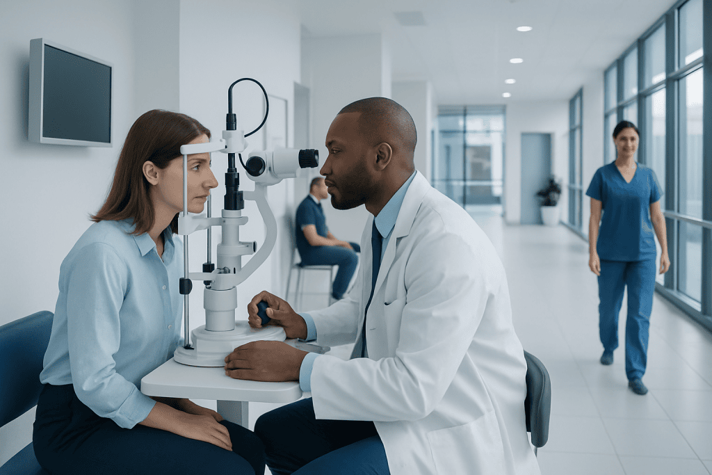
A neuro-ophthalmology exam looks at both eye health and nervous system function. It is often used to assess double vision, unexplained vision loss, abnormal pupils, eyelid drooping, and optic nerve concerns.
Key Takeaways
- A neuro-ophthalmology exam looks at both eye health and nervous system function.
- It is often used to assess double vision, unexplained vision loss, abnormal pupils, eyelid drooping, and optic nerve concerns.
- The visit may include a medical history, vision testing, pupil checks, eye movement assessment, visual field testing, and a dilated eye exam.
- Additional scans or blood tests may be recommended if the findings suggest a neurological or systemic cause.
- Most parts of the exam are noninvasive, though dilating drops can blur near vision temporarily.
A neuro-ophthalmology exam evaluates vision, eye movements, pupils, optic nerves, and related nervous system function. It helps identify whether visual symptoms are linked to the eyes themselves, the optic nerve, or the brain pathways that control sight.
Overview: What a Neuro-Ophthalmology Exam Is
A neuro-ophthalmology exam is a specialized evaluation that focuses on the connection between the eyes and the nervous system. Neuro-ophthalmologists assess how the eyes, optic nerves, eye muscles, pupils, and brain pathways involved in vision are working together. The goal is to understand why a person is having symptoms such as vision loss, double vision, blurred vision, abnormal eye movements, headaches with visual changes, or eyelid drooping.
Unlike a routine eye exam, this type of visit goes beyond checking vision and eye structures alone. It looks for signs that a problem may involve the optic nerve, the nerves controlling eye movement, or areas of the brain that process visual information. In many cases, symptoms that seem to come from the eye may actually start elsewhere in the nervous system.
The exam is tailored to the person’s symptoms. Some people are referred after a regular eye doctor notices optic nerve swelling or an abnormal visual field. Others are sent by a neurologist, emergency physician, or primary care doctor because of new double vision, unequal pupils, or unexplained visual symptoms. The process is usually careful and step-by-step, helping the specialist decide whether the cause is eye-related, neurological, or both.
Who May Need This Exam
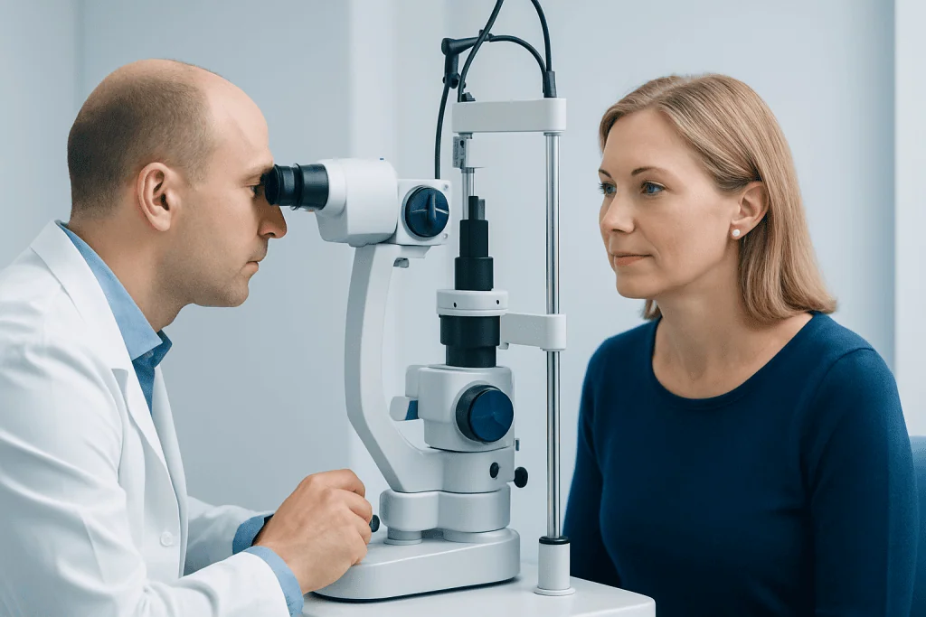
A neuro-ophthalmology exam may be recommended for people with symptoms that involve both vision and nerve function. These can include sudden or gradual vision loss, double vision, difficulty moving the eyes, eye pain with vision changes, unusual pupil size, episodes of temporary visual loss, or persistent headaches with visual symptoms.
It is also commonly used when another doctor finds signs such as optic disc swelling, pale optic nerves, unexplained visual field loss, or suspected nerve palsy. People with conditions that can affect the nervous system and vision, including autoimmune disease, diabetes, stroke, multiple sclerosis, thyroid eye disease, or raised pressure inside the skull, may also need this type of assessment.
Sometimes the exam is not prompted by a serious disease, but by symptoms that need careful sorting out. For example, migraine, dry eye, and ordinary refractive problems can sometimes mimic neurological visual complaints. A detailed evaluation helps clarify the cause and guide the next steps.
What Happens During the Visit
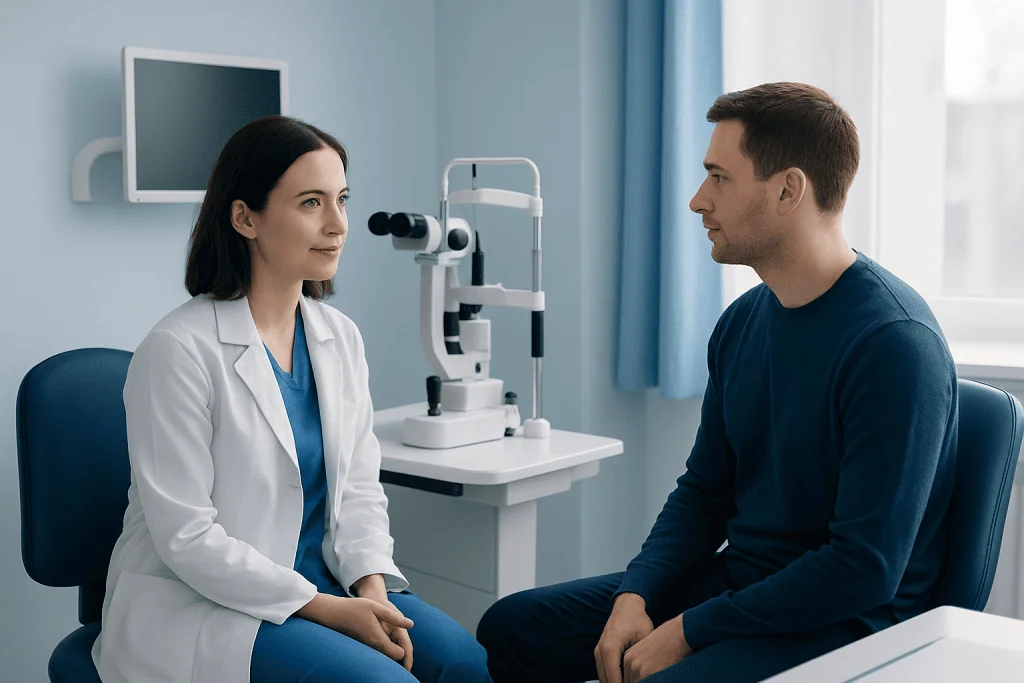
The appointment usually begins with a detailed medical history. The specialist may ask when the symptoms began, whether they came on suddenly or gradually, and whether they affect one or both eyes. They may also ask about pain, headache, weakness, numbness, balance problems, past eye disease, recent infections, medications, and family history. This part is important because the pattern of symptoms often offers strong clues.
The physical examination then focuses on vision and related nerve function. Visual acuity is checked, often with each eye separately. Color vision may be tested because some optic nerve problems affect color perception early. The doctor may also evaluate contrast sensitivity or ask the patient to read or identify targets at different distances.
Pupil testing is another key part of the exam. The specialist checks whether the pupils are equal, whether they react normally to light, and whether there is evidence that one optic nerve is transmitting signals less effectively than the other. Eye alignment and eye movements are also assessed carefully to see whether the eyes move together smoothly in all directions.
Many visits also include a general neurological screening. This may involve checking facial sensation, eyelid position, facial movement, coordination, or reflexes, depending on the symptoms. The aim is not to perform a full neurological workup in every case, but to identify signs that can help narrow the diagnosis.
Common Tests in a Neuro-Ophthalmology Exam
Several office-based tests may be used during the evaluation. Visual field testing checks side vision and can reveal patterns of loss linked to the optic nerve, retina, or brain. Eye alignment measurements help determine the cause of double vision and whether a specific eye muscle or cranial nerve may be involved. In some cases, the specialist may use colored lenses, prisms, or cover testing to measure misalignment precisely.
A slit-lamp exam may be performed to look at the front of the eye, followed by a dilated eye exam to view the optic nerve and retina at the back of the eye. Dilation allows the doctor to look for swelling, pallor, bleeding, inflammation, or other changes that may explain symptoms. Because dilation can blur near vision and increase light sensitivity for several hours, patients are often advised to plan accordingly.
Some clinics may also perform imaging or photography of the optic nerve and retina, such as optical coherence tomography, when appropriate. This can help measure the thickness of retinal nerve fiber layers and support the assessment of optic nerve disease. Depending on the findings, the doctor may recommend further testing such as an MRI scan or other imaging to examine the brain, eye sockets, or optic pathways.
Additional tests are not necessary for everyone. Blood tests, lumbar puncture, or referrals to other specialists are only used when the history and examination suggest they are needed. The overall aim is to answer the specific clinical question while avoiding unnecessary procedures.
Conditions This Exam Can Help Identify
A neuro-ophthalmology exam can help detect a wide range of conditions. These include optic neuritis, ischemic optic neuropathy, papilledema, cranial nerve palsies, myasthenia gravis, thyroid eye disease, and visual pathway disorders related to stroke or tumors. It may also help evaluate symptoms associated with migraine, concussion, and some inflammatory or autoimmune diseases.
For example, unexplained optic nerve swelling may lead the specialist to look for signs of raised pressure around the brain, while double vision may point toward a nerve palsy or an eye movement disorder. Sudden loss of part of the visual field can sometimes suggest a problem affecting the optic tract or brain rather than the eye itself. In this way, the exam acts as a bridge between ophthalmology and neurology.
In some people, the assessment helps distinguish between conditions with similar symptoms. A patient referred for suspected optic nerve disease may instead have retinal disease, while someone with blurred vision may have migraine aura rather than structural eye damage. Accurate distinction matters because treatment plans can be very different.
If symptoms suggest a broader neurological issue, a doctor may discuss related evaluations for conditions such as multiple sclerosis or other disorders affecting the nervous system. The specialist may work closely with neurologists, endocrinologists, rheumatologists, or neurosurgeons to build a complete picture.
What Happens After the Exam
At the end of the visit, the specialist usually explains the main findings and whether the results suggest a problem in the eye, optic nerve, eye movement system, or brain pathways. In some cases, the diagnosis is clear from the examination alone. In others, the visit helps narrow the possibilities and determine which additional tests, if any, are needed.
Treatment depends on the cause. Some people only need monitoring and follow-up eye exams. Others may need medication, prism correction for double vision, or treatment of an underlying neurological or systemic illness. If the problem is related to pressure on visual pathways, structural abnormalities, or certain brain disorders, the next step may involve a referral for neurosurgical care or another targeted treatment plan.
When symptoms are urgent, such as sudden visual loss, acute optic nerve swelling, or new double vision with neurological symptoms, the specialist may arrange rapid testing or hospital-based evaluation. When symptoms are stable and less urgent, follow-up may be scheduled to monitor changes over time and compare repeat exams.
For international patients or those seeking coordinated care, Acibadem International’s multidisciplinary specialists and JCI-accredited hospitals evaluate neuro-ophthalmic conditions and organize further testing and treatment when needed.
How to Prepare and When to Seek Prompt Care
Before the appointment, it can help to bring a list of symptoms, current medications, eyeglasses, and records from previous eye exams or scans. If the patient has copies of visual field tests, MRI reports, blood test results, or referral notes, these can be especially useful. Because dilating drops may be used, some people prefer not to drive themselves home, although this depends on the individual and the clinic’s advice.
Writing down a timeline of symptoms can also make the visit more efficient. Important details include whether symptoms are constant or intermittent, whether they affect one or both eyes, and whether they are associated with pain, headache, weakness, or recent illness. Photos of eyelid drooping or eye misalignment during symptomatic episodes may sometimes help if the problem fluctuates.
Prompt medical attention is important for sudden vision loss, severe eye pain with visual change, sudden double vision, new drooping of the eyelid, or visual symptoms accompanied by weakness, confusion, trouble speaking, or severe headache. These symptoms do not always mean a serious emergency, but they should be assessed quickly to protect vision and overall health.
Even when symptoms seem mild, persistent unexplained visual changes deserve professional review. Early evaluation can help identify treatable causes and offer reassurance when symptoms are not linked to a dangerous condition.
Frequently asked questions
Is a neuro-ophthalmology exam the same as a regular eye exam?
No. A regular eye exam focuses mainly on vision, glasses, and the health of the eyes, while a neuro-ophthalmology exam also evaluates the optic nerves, eye movement nerves, pupils, and visual pathways in the brain. It is used when symptoms suggest that vision problems may be related to the nervous system.
How long does a neuro-ophthalmology appointment usually take?
The length varies depending on the symptoms and the tests needed. Many visits take longer than a standard eye appointment because they involve a detailed history, multiple vision tests, and sometimes dilation. Patients should allow extra time in case additional testing is performed on the same day.
Will the exam be painful?
Most parts of the exam are not painful. Some tests may be tiring because they require concentration, and bright lights may feel uncomfortable for a short time. Dilating drops can sting briefly and may cause temporary light sensitivity and blurred near vision.
Why might I need an MRI after the exam?
An MRI may be recommended if the findings suggest a problem involving the optic nerve, eye sockets, brain, or cranial nerves. It helps the doctor look for inflammation, compression, stroke, tumors, or other structural causes. Not everyone needs imaging, and the decision depends on the individual examination results.
Can a neuro-ophthalmology exam detect neurological disease?
It can provide important clues and sometimes strongly suggest a neurological condition. However, diagnosis may also require imaging, blood tests, or evaluation by other specialists. The exam is one part of a broader process of identifying the cause of symptoms.
Should I wear contact lenses to the appointment?
It is often fine to arrive wearing contact lenses, but bringing glasses is a good idea. Some tests and dilating drops may make contact lenses less comfortable afterward. The clinic may also ask the patient to remove lenses for part of the examination.
References
- American Academy of Ophthalmology
- North American Neuro-Ophthalmology Society
- National Eye Institute
- National Institute of Neurological Disorders and Stroke
- Mayo Clinic
This article is for general information only and is not a substitute for professional medical advice. Please consult a qualified doctor about your individual situation.
Explore treatments in Turkey — costs, top hospitals & a free quote
JCI-accredited · board-certified surgeons · reply within 24h
Add us as a Preferred Source to see more of our trusted health content across Google Search, AI Overviews and Discover.

More from the Health Library



Related Specialists

Prof. Dr. Sinan Zeren
Urology
Assoc. Prof. Dr. Tunay Erden
Orthopedic Surgery & Traumatology
Dr. Ahsen Ercan
Physical Medicine & Rehabilitation
Dr. Aytekin Ünlükaplan
Anesthesiology