What Holds Bones Together in a Fibrous Joint?

Fibrous joints connect bones with strong fibrous connective tissue. Most fibrous joints allow very little movement and mainly provide stability.
Key Takeaways
- Fibrous joints connect bones with strong fibrous connective tissue.
- Most fibrous joints allow very little movement and mainly provide stability.
- The three main types are sutures, syndesmoses, and gomphoses.
- Fibrous joints are found in the skull, between certain long bones, and around the teeth.
- Pain near a fibrous joint may relate to injury, inflammation, or dental or bone conditions and should be assessed if persistent.
In a fibrous joint, bones are held together by dense fibrous connective tissue rather than a fluid-filled joint space. This design gives the joint strength and stability, with little or no movement depending on the type.
Overview
A fibrous joint is a type of joint in which two bones are connected by dense fibrous connective tissue. Unlike freely movable joints such as the knee or shoulder, a fibrous joint does not have a joint cavity filled with fluid. Instead, its structure is built for support, protection, and firmness.
The tissue that holds the bones together is rich in collagen fibers. These tough fibers act like strong bands, binding neighboring bones and helping them resist pulling, twisting, or separation. Depending on the exact type of fibrous joint, the connection may be nearly immovable or may allow a small amount of movement.
Fibrous joints are especially important where the body needs stability more than flexibility. For example, the bones of the skull must fit tightly together to protect the brain, while the connection between certain leg or forearm bones must be secure but still permit a small degree of motion. Understanding how these joints work helps explain why they are essential to everyday movement, posture, and protection.
What holds bones together in a fibrous joint?
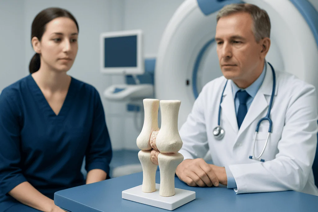
Bones in a fibrous joint are held together by dense fibrous connective tissue. This tissue is made mostly of collagen, a strong structural protein also found in tendons and ligaments. The collagen fibers create a firm connection between bone surfaces, helping the joint stay aligned and stable.
In many fibrous joints, this connective tissue is arranged in short bands or sheets that fit tightly between neighboring bones. In others, such as the connection between a tooth and its socket, specialized fibers anchor one structure to another. The exact arrangement depends on the location and the job that joint needs to perform.
Because there is no open joint cavity, fibrous joints are usually not designed for wide movement. Their main purpose is to hold structures together securely. This is why they are often found in places where protection and support matter more than flexibility.
Although fibrous joints are usually stable, they are still living tissues. They can be affected by injury, inflammation, developmental differences, or nearby bone and dental problems. When symptoms occur, a doctor or dentist may evaluate whether the issue involves the joint itself or surrounding tissues.
Types of fibrous joints
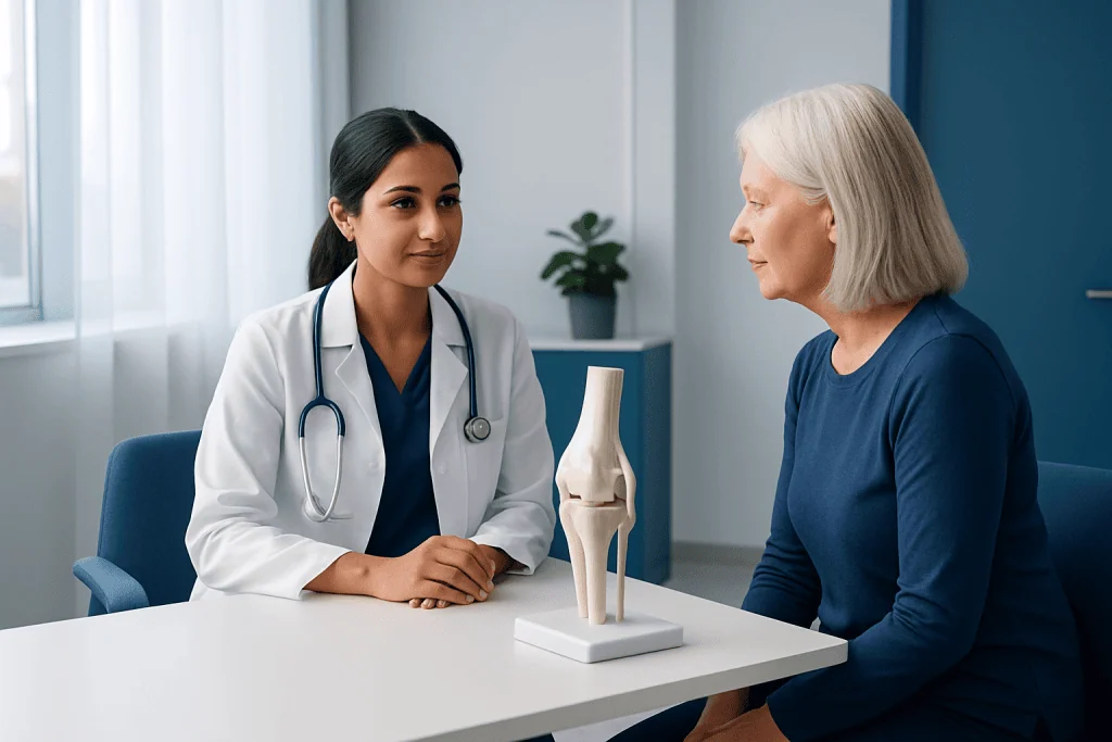
There are three main types of fibrous joints: sutures, syndesmoses, and gomphoses. Each type uses fibrous tissue to connect bones, but they differ in location and mobility.
Sutures are found between the bones of the skull. The thin layer of fibrous tissue between skull bones holds them tightly together. In infants and children, these areas also allow the skull to grow. Over time, many sutures become even less movable as the bones mature.
Syndesmoses connect bones with ligaments or an interosseous membrane. Examples include the connection between the tibia and fibula in the lower leg and between the radius and ulna in the forearm. These joints allow a small amount of movement, which helps with functions such as walking and rotating the forearm.
Gomphoses are the peg-and-socket fibrous joints that anchor teeth into the jawbone. The tooth is held in place by the periodontal ligament, a specialized fibrous tissue. This attachment is firm but not rigid, which helps the tooth absorb normal biting forces.
- Sutures: skull bones
- Syndesmoses: long bones linked by ligaments or membranes
- Gomphoses: teeth anchored in their sockets
Function and why fibrous joints matter
The main function of a fibrous joint is stability. By binding bones closely together, these joints help preserve the shape of important body structures and protect delicate organs. The skull is a clear example: sutures allow the cranium to act as a strong protective shell around the brain.
Fibrous joints also help distribute force. In the lower leg and forearm, syndesmoses allow bones to work together while sharing the stresses created by movement. Even though these joints move only slightly, that small motion is important for normal biomechanics.
Gomphoses support another essential function: chewing. The fibrous attachment around each tooth helps hold it securely while also cushioning pressure from biting. Without this supportive connection, teeth would not remain stable in the jaw.
Overall, fibrous joints show that not all joints are meant to move freely. Some are designed mainly to keep the body strong, aligned, and protected. When these joints are healthy, they usually work quietly in the background without drawing attention.
Problems that can affect fibrous joints
Fibrous joints are stable, but they can still be involved in medical or dental problems. In the skull, an abnormal early closure of sutures in infants is called craniosynostosis. This can affect skull shape and, in some cases, may require specialist assessment. In adults, pain around the skull is more often related to muscles, nerves, or nearby structures rather than the sutures themselves.
Syndesmoses may be injured by twisting or high-impact trauma. A common example is a high ankle sprain, where the ligaments between the tibia and fibula are stretched or torn. This can cause pain, swelling, bruising, and difficulty bearing weight. Imaging and an orthopedic evaluation may be needed if symptoms are significant or do not improve.
Gomphoses can be affected by gum disease, inflammation, injury, or infection around the tooth. If the supporting tissues weaken, a tooth may become loose or painful. Dental evaluation is important because early treatment can help protect the tooth and surrounding bone.
Problems near fibrous joints may also overlap with bone, ligament, and tendon conditions. Depending on the location, a doctor may consider injuries, fractures, dental disease, or inflammatory conditions. Persistent pain, deformity, swelling, or reduced function should be assessed rather than ignored.
Diagnosis
Diagnosis begins with a careful history and physical examination. A clinician asks where the pain or problem is located, how it started, whether there was an injury, and what movements make symptoms better or worse. The exam may include checking swelling, tenderness, alignment, mobility, and function of the nearby structures.
Imaging may be useful when a structural problem is suspected. Standard X-rays can help assess bones, skull sutures, and tooth support, while other imaging may be used for more detail. For some ligament-related injuries, doctors may request an MRI scan to examine soft tissues more closely. In selected cases, a CT scan can give a detailed view of bone anatomy.
Dental evaluation is often the right approach when symptoms involve a tooth socket or gums. Orthopedic, pediatric, or maxillofacial specialists may be involved depending on the site. The goal is to identify whether the issue is a normal variation, an injury, a developmental condition, or a disease affecting surrounding tissues.
Accurate diagnosis matters because treatment depends on the cause. A painful ankle syndesmosis injury is managed differently from a dental ligament problem or a childhood skull suture condition. Professional assessment helps guide safe care and reduces the chance of complications.
Treatment options
Treatment for a fibrous joint problem depends on the location and cause. Many mild syndesmosis injuries improve with rest, activity modification, support, and a guided rehabilitation plan. More severe injuries may need immobilization, and in some cases surgery is considered to restore alignment and stability.
When the problem involves the teeth, treatment may focus on the gums, periodontal ligament, or infection control. Dental cleaning, treatment of gum disease, splinting of an injured tooth, or other procedures may be recommended. If infection is suspected, prompt care is important to protect the tooth and nearby bone.
For developmental skull conditions such as craniosynostosis, children are evaluated by specialist teams. Management may include monitoring, imaging, and sometimes neurosurgery or craniofacial surgery, depending on the pattern and severity. Treatment decisions are individualized and made with careful attention to the child’s growth and overall health.
If surgery is needed for bone or ligament injury, recovery often includes follow-up imaging and rehabilitation. In complex cases, coordinated care may involve orthopedics, dentistry, radiology, pediatrics, or neurosurgery. Near the end of the care pathway, some international patients may choose centers such as Acibadem International, where multidisciplinary specialists in JCI-accredited hospitals diagnose and treat bone, dental, and related structural conditions.
Self-care, prevention, and when to see a doctor
Self-care depends on the type of fibrous joint involved. For possible ankle or forearm injuries, early rest, avoiding painful activity, and using support as advised can help while waiting for evaluation. For tooth-related concerns, regular brushing, flossing, and routine dental care help protect the supporting fibrous tissues around the teeth.
Prevention focuses on reducing injury risk and supporting overall bone and oral health. Wearing appropriate sports footwear, using protective equipment when needed, maintaining good posture and conditioning, and treating dental problems early can all be helpful. Children should also attend regular health checkups so developmental concerns can be recognized promptly.
A doctor should be consulted if there is significant pain, swelling, deformity, difficulty walking, a loose tooth, numbness, fever, or symptoms that do not improve. Urgent assessment is especially important after trauma, if a child has an unusual skull shape, or if infection is suspected. Early evaluation often makes treatment simpler and more effective.
It is reassuring to remember that many symptoms near fibrous joints have treatable causes. A qualified healthcare professional can explain what is normal, what needs monitoring, and what may benefit from treatment or specialist referral.
Frequently asked questions
What exactly holds bones together in a fibrous joint?
Bones in a fibrous joint are held together by dense fibrous connective tissue, which contains strong collagen fibers. This tissue forms a firm connection that helps keep the bones stable and properly aligned.
Do fibrous joints move at all?
Some do, but only slightly. Sutures in the skull are essentially immovable, while syndesmoses between certain long bones allow a small amount of movement that supports normal function.
What are the three types of fibrous joints?
The three types are sutures, syndesmoses, and gomphoses. Sutures are found in the skull, syndesmoses connect certain long bones, and gomphoses anchor teeth in their sockets.
Can a fibrous joint be injured?
Yes. A syndesmosis can be sprained or torn, especially around the ankle after twisting injuries. The tissues around a gomphosis can also be damaged by trauma, gum disease, or infection.
Why are skull sutures considered fibrous joints?
Skull sutures are considered fibrous joints because the bones are joined by a thin layer of fibrous connective tissue. This arrangement provides strength and protection while allowing growth during early life.
When should someone seek medical advice for a problem near a fibrous joint?
Medical or dental advice is appropriate if there is ongoing pain, swelling, visible deformity, difficulty using the area, or a loose tooth. Urgent assessment is important after significant injury, with signs of infection, or if a child has an unusual skull shape.
References
- Cleveland Clinic
- Johns Hopkins Medicine
- American Academy of Orthopaedic Surgeons
- MedlinePlus
- National Institute of Dental and Craniofacial Research
This article is for general information only and is not a substitute for professional medical advice. Please consult a qualified doctor about your individual situation.
Joint and spine care in Turkey — expert assessment & treatment
JCI-accredited · board-certified surgeons · reply within 24h
Add us as a Preferred Source to see more of our trusted health content across Google Search, AI Overviews and Discover.