What Is a CT Scan and When Is It Needed?
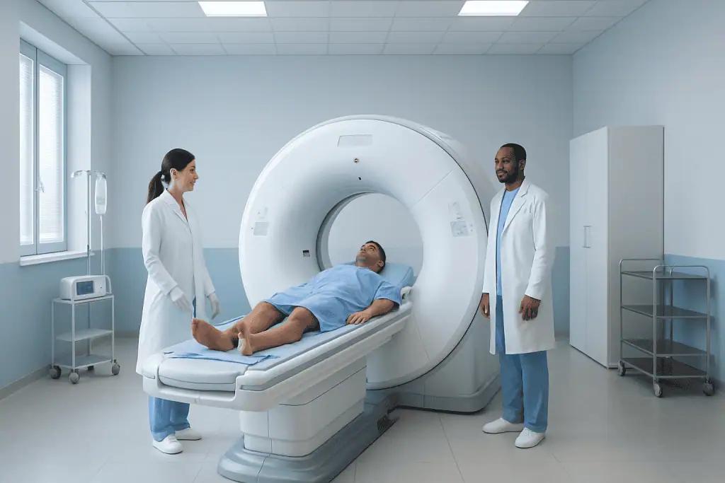
A CT scan, also called computed tomography, creates detailed cross-sectional images of bones, organs, blood vessels, and soft tissues. Doctors may recommend a CT scan after an injury, for suspected infections or tumors, to evaluate chest or abdominal symptoms, or to plan procedures.
Key Takeaways
- A CT scan, also called computed tomography, creates detailed cross-sectional images of bones, organs, blood vessels, and soft tissues.
- Doctors may recommend a CT scan after an injury, for suspected infections or tumors, to evaluate chest or abdominal symptoms, or to plan procedures.
- Some CT scans use contrast material to make blood vessels, organs, or abnormal areas easier to see.
- CT scans involve radiation, so doctors weigh the benefits and risks and choose the most suitable imaging test for each patient.
- Patients should tell the healthcare team about pregnancy, kidney disease, allergies, diabetes medicines, or previous reactions to contrast before the scan.
A CT scan is a fast, detailed imaging test that helps doctors look inside the body using X-rays and computer technology. It can support diagnosis, guide treatment, and help monitor many conditions when used appropriately.
Overview
A CT scan, or computed tomography scan, is a medical imaging test that uses a rotating X-ray machine and advanced computer processing to create detailed pictures of the inside of the body. Unlike a standard X-ray, which produces a flat image, a CT scan creates cross-sectional images, sometimes described as slices. These slices can be viewed individually or combined to form three-dimensional images.
CT imaging is used in many areas of medicine because it can show bones, organs, blood vessels, lungs, and soft tissues with a high level of detail. It is often chosen when doctors need quick information, such as after an accident, or when symptoms need further investigation. CT scans can also help guide biopsies, drainage procedures, and certain treatments.
The test is usually painless and relatively quick. Most scans take only a few minutes, although preparation and positioning may take longer. Some CT scans are performed with a contrast material, which may be given by mouth, injection, or sometimes through the rectum, depending on the area being examined.
How a CT Scan Works
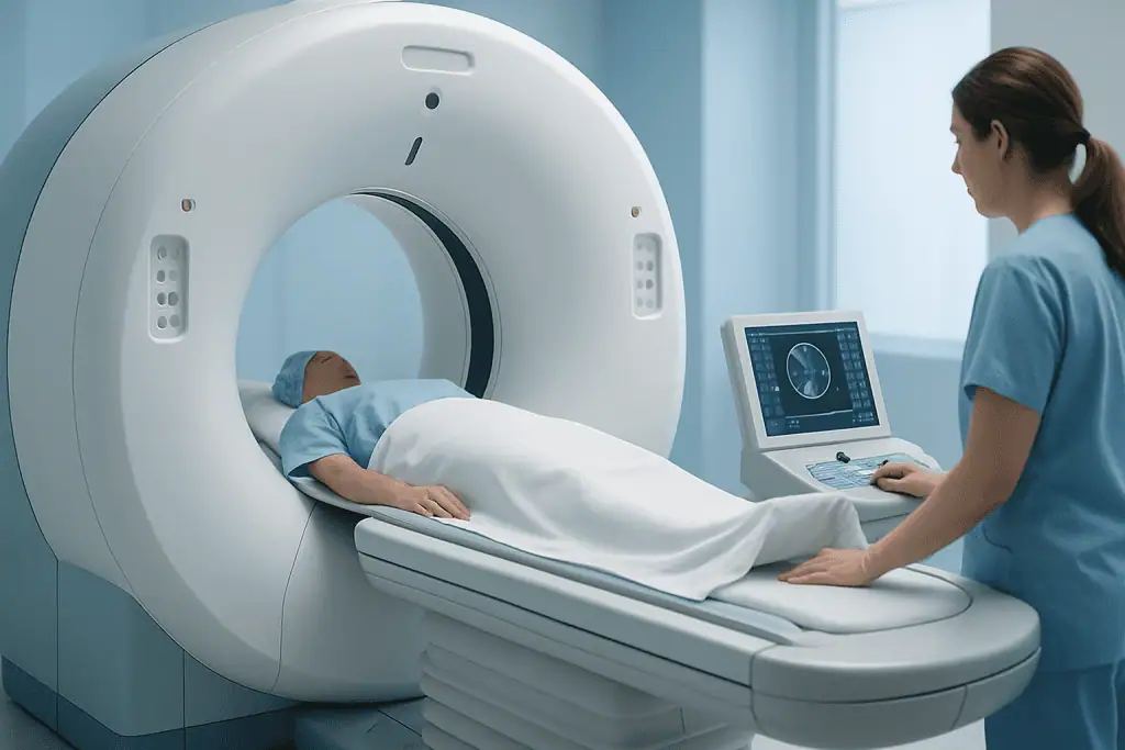
During a CT scan, the patient lies on a motorized table that moves through a doughnut-shaped scanner. Inside the scanner, an X-ray tube rotates around the body and takes multiple images from different angles. A computer then processes this information to create detailed images that radiologists and other doctors can review.
The images can show differences between tissues more clearly than many standard X-rays. For example, CT can help distinguish air, bone, fat, blood, fluid, and many types of soft tissue. This makes it especially useful for evaluating the chest, abdomen, pelvis, spine, head, and complex bone injuries.
In some cases, a contrast material is used to improve image detail. Contrast can highlight blood vessels, the urinary tract, the digestive tract, or areas of inflammation or abnormal tissue. The type of contrast used depends on the clinical question and the body part being examined.
When Is a CT Scan Needed?

A doctor may request a CT scan when more detailed imaging is needed to understand symptoms, confirm a diagnosis, or plan treatment. CT scans are commonly used in emergency care because they can quickly detect internal injuries, bleeding, fractures, stroke-related changes, and other urgent problems. They are also used in planned outpatient evaluation for many conditions.
Common reasons for a CT scan include:
- Head injury, severe headache, stroke symptoms, or suspected bleeding in the brain
- Chest symptoms such as unexplained shortness of breath, chest pain, or suspected lung disease
- Abdominal or pelvic pain, suspected appendicitis, kidney stones, bowel problems, or internal bleeding
- Evaluation of complex bone fractures, spine problems, or joint injuries
- Detection, staging, or follow-up of cancers and other masses
- Planning surgery, radiation therapy, or image-guided procedures
A CT scan is not needed for every symptom or condition. Doctors consider the patient’s medical history, examination findings, previous test results, age, pregnancy status, and whether another imaging method, such as ultrasound or MRI, may be more appropriate.
Types of CT Scans and Contrast Use
Different CT scan techniques are used depending on the clinical need. A non-contrast CT scan is performed without contrast material and may be suitable for conditions such as some head injuries, certain types of bleeding, kidney stones, or bone problems. A contrast-enhanced CT scan is used when doctors need clearer views of blood vessels, organs, tumors, infections, or inflammation.
Intravenous contrast is injected into a vein, usually in the arm. Patients may feel a brief warm sensation or a metallic taste, which typically passes quickly. Oral contrast may be used for some abdominal or pelvic scans to outline the stomach and intestines. In selected cases, rectal contrast may be used to better visualize the lower bowel.
Specialized CT examinations include CT angiography, which focuses on blood vessels; low-dose CT, which may be used in specific screening or follow-up situations; and cardiac CT, which can assess certain heart and coronary artery concerns. The imaging team selects the protocol to answer the medical question while keeping radiation exposure as low as reasonably achievable.
Preparing for a CT Scan
Preparation depends on the type of CT scan. For many scans, little or no preparation is needed. Patients may be asked to remove jewelry, glasses, hearing aids, hairpins, belts, or clothing with metal parts, as metal can interfere with image quality. A hospital gown may be provided.
If contrast is planned, the healthcare team may ask about kidney disease, diabetes, thyroid disease, asthma, allergies, previous reactions to contrast, current medicines, and pregnancy or breastfeeding. Blood tests may be needed in some patients to check kidney function before intravenous contrast. Patients taking certain diabetes medications may receive specific instructions from their doctor.
Some abdominal and pelvic CT scans require fasting for a few hours before the test, especially if intravenous contrast or sedation is planned. Patients should follow the instructions provided by the imaging center. If a patient is anxious, has difficulty lying still, or has pain that makes positioning difficult, it is helpful to inform the team in advance.
What to Expect During and After the Test
Before the scan, a radiology technologist helps position the patient on the CT table. Pillows or straps may be used to support the body and reduce movement. The patient may be asked to hold still or hold their breath for a few seconds during image capture, especially for chest or abdominal scans.
The CT scanner is open at both ends and the scan itself is usually brief. The technologist remains nearby in a control room and can see, hear, and speak with the patient throughout the exam. If intravenous contrast is used, an IV line is placed before the scan and removed afterward unless it is needed for other medical care.
After most CT scans, patients can return to normal activities unless their doctor gives different instructions. Drinking fluids may be recommended after contrast to help the body eliminate it naturally. A radiologist reviews the images and sends a report to the referring doctor, who explains the results in the context of the patient’s symptoms and overall health.
Benefits, Risks, and Safety
The main benefit of a CT scan is that it provides detailed information quickly and noninvasively. This can help doctors diagnose conditions earlier, decide whether treatment is needed, guide procedures, and monitor response to therapy. In urgent situations, CT imaging can be especially valuable because it can rapidly detect potentially serious problems.
CT scans use ionizing radiation. The amount varies depending on the body part and the scanning protocol. While radiation exposure from a medically necessary CT scan is generally considered acceptable when the benefits outweigh the risks, unnecessary repeat scans should be avoided. Radiology teams use dose-reduction techniques and tailor scans to the patient whenever possible.
Contrast material is safe for most people, but it can occasionally cause allergic-type reactions or affect kidney function in patients who already have kidney problems. This is why screening questions are important before the scan. Patients should always tell the imaging team if they are or may be pregnant, because the need for CT imaging and possible alternatives should be carefully considered.
When to Speak With a Doctor
A patient should speak with a doctor if they have symptoms that are severe, persistent, worsening, or unexplained. Imaging decisions should be made by a qualified clinician who can assess whether a CT scan is the most appropriate test. In some cases, an ultrasound, MRI, blood test, endoscopy, or careful observation may be recommended instead.
Before the scan, patients should ask why the CT scan is needed, whether contrast will be used, how to prepare, and when results will be available. It is also reasonable to ask whether previous imaging can be reviewed to avoid unnecessary repeat testing. Patients with kidney disease, prior contrast reactions, pregnancy concerns, or implanted medical devices should mention these before the appointment.
Acibadem International’s multidisciplinary specialists and JCI-accredited hospitals provide diagnostic imaging and related care for international patients, including CT examinations when clinically appropriate. As with any medical test, the decision should be individualized and discussed with the treating doctor.
Frequently asked questions
Is a CT scan painful?
A CT scan itself is not painful. Patients need to lie still on the scanning table, which may be uncomfortable for people with pain or limited mobility. If contrast is injected, there may be a brief pinch from the IV and a short warm sensation.
How long does a CT scan take?
The actual image capture often takes only a few minutes. The full appointment may take longer because of registration, changing clothes, positioning, IV placement, or drinking oral contrast if needed. The imaging center can provide more specific timing based on the type of scan.
Is CT contrast dangerous?
CT contrast is safe for most patients and helps doctors see certain structures more clearly. A small number of people may have allergic-type reactions, and patients with kidney problems may need special assessment before receiving intravenous contrast. This is why the healthcare team asks screening questions before the scan.
Can pregnant patients have a CT scan?
Pregnant patients should always tell the doctor and imaging team before any imaging test involving radiation. In some situations, CT may still be necessary if the medical benefit is important, but alternatives such as ultrasound or MRI may be considered. The decision is made carefully based on the patient’s condition and the area being scanned.
What is the difference between a CT scan and an MRI?
A CT scan uses X-rays and is often faster, making it useful for emergencies, bones, lungs, bleeding, and many abdominal conditions. MRI uses a strong magnetic field and radio waves, and it can provide excellent detail for the brain, spinal cord, joints, and many soft tissues. The best test depends on the medical question.
When will CT scan results be available?
A radiologist reviews the CT images and prepares a report for the referring doctor. In urgent situations, results may be communicated quickly, while routine results may take longer depending on the facility. Patients should ask their healthcare provider when and how they will receive the findings.
References
- World Health Organization
- American College of Radiology
- Radiological Society of North America
- National Institute of Biomedical Imaging and Bioengineering
- European Society of Radiology
This article is for general information only and is not a substitute for professional medical advice. Please consult a qualified doctor about your individual situation.
Explore treatments in Turkey — costs, top hospitals & a free quote
JCI-accredited · board-certified surgeons · reply within 24h
Add us as a Preferred Source to see more of our trusted health content across Google Search, AI Overviews and Discover.