What Is Interventional Neurology? Procedures, Conditions Treated, and How It Works

Interventional neurology treats many conditions through tiny catheters inserted into blood vessels rather than large open surgery. It is commonly used for ischemic stroke, brain aneurysms, vascular malformations, and carotid artery disease.
Key Takeaways
- Interventional neurology treats many conditions through tiny catheters inserted into blood vessels rather than large open surgery.
- It is commonly used for ischemic stroke, brain aneurysms, vascular malformations, and carotid artery disease.
- Procedures are guided by advanced imaging to reach delicate blood vessels in the brain and spine.
- Treatment may be urgent, especially for stroke, where faster care can help protect brain function.
- Recovery is often shorter than with open surgery, but every procedure still has risks and requires expert evaluation.
Interventional neurology is a subspecialty that uses minimally invasive, image-guided procedures to treat conditions affecting the brain, spine, head, and neck blood vessels. It is best known for emergency stroke treatment, but it also plays an important role in managing aneurysms, vascular malformations, and narrowed arteries.
Overview
Interventional neurology is a highly specialized field focused on diagnosing and treating disorders of the brain, spinal cord, head, and neck blood vessels using minimally invasive techniques. Instead of making a large incision, the specialist usually guides very thin tubes called catheters through blood vessels, often starting from the wrist or groin, to reach the area that needs treatment.
These procedures are performed with real-time imaging, such as X-ray-based angiography, which helps the doctor navigate through the blood vessels with precision. Because the approach is targeted, interventional neurology can often reduce tissue disruption, shorten hospital stays, and support faster recovery compared with some open surgical procedures.
Interventional neurology overlaps with stroke care, neurology, neurosurgery, and vascular medicine. Patients may be treated by a multidisciplinary team that includes neurologists, neurointerventional specialists, neurosurgeons, neuroradiologists, emergency physicians, and rehabilitation experts. The exact team depends on the condition being treated and whether care is urgent or planned.
How Interventional Neurology Works
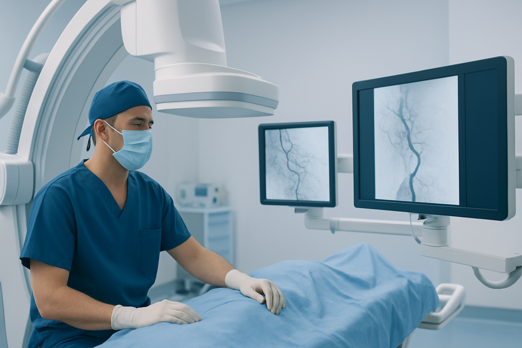
The main principle of interventional neurology is endovascular treatment. This means the specialist works from inside the blood vessels. A catheter is carefully advanced to the brain or spinal circulation while contrast dye and imaging are used to map the vessels and identify the problem.
Once the catheter reaches the target area, different tools can be used depending on the condition. These may include tiny coils to seal an aneurysm, stents to support narrowed arteries, devices to remove a blood clot during stroke, or liquid embolic agents to block abnormal blood flow. The goal is to treat the source of the problem as directly and safely as possible.
Some procedures are emergencies, especially when a blocked artery is causing a stroke. Others are planned in advance after tests show an aneurysm, vascular malformation, or severe vessel narrowing. In either setting, the treatment plan is individualized based on the patient’s symptoms, imaging results, overall health, and the anatomy of the affected blood vessels.
Conditions Treated

Interventional neurology is best known for treating acute ischemic stroke, where a blood clot blocks blood flow to part of the brain. In selected patients, specialists may perform mechanical clot removal to restore circulation quickly. This can be an important option alongside other stroke treatments and may improve the chance of preserving brain function when used promptly.
Another major area is the treatment of brain aneurysms. An aneurysm is a weak, bulging area in a blood vessel wall that can leak or rupture. Depending on its size, shape, and location, it may be treated with endovascular techniques such as coiling or flow-diverting stents. This may be part of care for brain aneurysm or other cerebrovascular conditions.
Interventional neurology also treats arteriovenous malformations and dural arteriovenous fistulas, which are abnormal connections between blood vessels. These conditions can increase the risk of bleeding, headaches, seizures, or neurological symptoms. Embolization may be used alone or as part of a broader plan that includes surgery or radiosurgery.
Additional conditions may include carotid artery stenosis, intracranial artery narrowing, certain causes of brain hemorrhage, and venous sinus problems. In some centers, neurointerventional specialists also help manage spinal vascular lesions and selected tumors by blocking blood supply before surgery.
Common Procedures
Mechanical thrombectomy is one of the most important emergency procedures in interventional neurology. It is used for some patients with a large-vessel ischemic stroke. The doctor guides a catheter to the blocked artery and uses a device to capture or suction out the clot. This treatment is often discussed as part of modern stroke treatment when imaging and timing show it is appropriate.
For aneurysms, common procedures include coil embolization, stent-assisted coiling, and flow diversion. Coils are tiny metal spirals placed inside the aneurysm to reduce blood flow into it. Flow diverters are specialized stents placed in the parent artery to redirect blood away from the aneurysm and encourage healing of the vessel wall.
Angioplasty and stenting may be used to treat narrowed arteries in the neck or brain in carefully selected patients. Embolization procedures can also block abnormal vessels feeding an arteriovenous malformation or a dural fistula. In many cases, treatment is guided by detailed neuroradiology imaging before, during, and after the procedure.
Some patients need diagnostic cerebral angiography even if no immediate treatment is planned. This test gives a very detailed view of blood vessels and can help confirm findings seen on CT, MRI, or ultrasound. It may be especially useful when doctors need more precise information to decide between monitoring, endovascular treatment, surgery, or other therapies.
Benefits, Risks, and Recovery
A major benefit of interventional neurology is that many conditions can be treated without open surgery. Smaller access points, less disruption to surrounding tissues, and targeted treatment can make recovery easier for some patients. In urgent stroke care, rapid endovascular treatment may also help reduce long-term disability in appropriately selected patients.
Even so, these are still complex medical procedures and they carry real risks. Possible complications can include bleeding, blood vessel injury, stroke, allergic reaction to contrast dye, kidney strain from contrast, infection, or device-related problems. The exact risk depends on the patient’s health, the condition being treated, and the type of procedure.
Recovery varies widely. Some people return home within a day or two after a planned procedure, while others need longer hospital care, especially after a stroke or hemorrhage. Follow-up may include medications, repeat imaging, rehabilitation, and ongoing management of blood pressure, cholesterol, diabetes, or other risk factors. When vascular disease is involved, care may be coordinated with specialists in vascular surgery and other related fields.
Diagnosis and Evaluation Before Treatment
Before recommending an interventional neurology procedure, doctors first confirm the diagnosis and assess whether the treatment is likely to help. This usually begins with a medical history, neurological examination, and imaging studies. Depending on the situation, imaging may include CT, CT angiography, MRI, MR angiography, ultrasound, or cerebral angiography.
In emergency stroke care, evaluation happens very quickly. The team needs to determine whether symptoms are caused by a blocked vessel, a hemorrhage, or another condition that can mimic stroke. They also assess how much brain tissue may still be salvageable and whether the patient is within a suitable treatment window.
For planned procedures, the evaluation is often more detailed. Doctors consider the size and location of an aneurysm, the pattern of abnormal blood flow in a vascular malformation, or the degree of vessel narrowing. They also review medications, kidney function, bleeding risk, and any history of contrast allergy so the procedure can be planned as safely as possible.
Prevention, Follow-Up, and When to Seek Medical Care
Not every condition treated by interventional neurology can be prevented, but many vascular risks can be reduced. Managing high blood pressure, diabetes, high cholesterol, and smoking is especially important. Regular exercise, a balanced diet, and adherence to prescribed medications may help protect blood vessel health and lower the risk of stroke and other complications.
Follow-up care matters after any neurointerventional procedure. Some patients need repeat scans to check an aneurysm repair, monitor a stent, or confirm that an arteriovenous malformation remains closed. Others may need blood-thinning or antiplatelet medications for a period of time, along with routine visits to review recovery and symptoms.
Urgent medical attention is needed for sudden signs of stroke, such as facial drooping, arm weakness, speech difficulty, confusion, severe dizziness, or sudden vision loss. A sudden severe headache, especially one described as the worst headache of a person’s life, also needs immediate evaluation. For non-emergency concerns, new neurological symptoms, recurrent headaches, or questions after a procedure should be discussed promptly with a qualified doctor.
For international patients who need advanced evaluation, Acibadem International’s multidisciplinary specialists and JCI-accredited hospitals diagnose and treat complex neurovascular conditions using modern imaging and interventional techniques.
Frequently asked questions
What is the difference between interventional neurology and neurosurgery?
Interventional neurology usually treats conditions from inside the blood vessels using catheters and imaging guidance. Neurosurgery more often involves open or minimally invasive operations on the brain, spine, or nerves. In many cases, the two specialties work together to choose the safest and most effective approach.
Is interventional neurology only used for stroke?
No. Stroke treatment is one of its most important uses, but interventional neurology also treats aneurysms, arteriovenous malformations, fistulas, and certain narrowed arteries. Some procedures are urgent, while others are planned after detailed imaging and specialist review.
Are interventional neurology procedures considered surgery?
They are generally considered minimally invasive procedures rather than traditional open surgery. They still require specialized expertise, hospital-based care, and careful monitoring before and after treatment. Because they are performed inside blood vessels, they can often avoid large incisions.
How long does recovery take after a neurointerventional procedure?
Recovery depends on the condition being treated and the patient’s overall health. Some people recover quickly after a planned procedure, while others need longer hospitalization and rehabilitation, especially after a stroke or brain hemorrhage. The treating team usually gives specific advice about activity, medications, and follow-up imaging.
Are these procedures safe?
Interventional neurology procedures are widely used and can be very effective in appropriate patients, but they are not risk-free. Possible complications include bleeding, stroke, vessel injury, and reactions to contrast dye. The doctor balances these risks against the potential benefits before recommending treatment.
When should someone go to the emergency department instead of waiting for an appointment?
Emergency care is needed for sudden stroke symptoms such as weakness on one side, trouble speaking, facial drooping, severe confusion, or sudden loss of vision. A sudden severe headache or collapse also needs immediate evaluation. Fast treatment can be especially important in brain and blood vessel conditions.
References
- World Stroke Organization
- American Stroke Association
- National Institute of Neurological Disorders and Stroke
- Society of NeuroInterventional Surgery
- Radiological Society of North America
This article is for general information only and is not a substitute for professional medical advice. Please consult a qualified doctor about your individual situation.
Interventional Neurology in Turkey — costs, top hospitals & a free quote
JCI-accredited · board-certified surgeons · reply within 24h
Add us as a Preferred Source to see more of our trusted health content across Google Search, AI Overviews and Discover.

More from the Health Library



Related Specialists

Prof. Dr. Işık Aslay
Radiation Oncology
Prof. Dr. Faysal Dane
Medical Oncology
Prof. Dr. Aziz Kaya Alturfan
Orthopedic Surgery & Traumatology
Prof. Dr. V. Melih Kara
General Surgery