X-Ray Imaging: Common Uses, Radiation Safety, and Next Steps

X-ray imaging uses a small amount of ionizing radiation to create pictures of bones, the chest, abdomen, teeth, and some soft tissues. Doctors request X-rays when the expected diagnostic benefit outweighs the small radiation exposure.
Key Takeaways
- X-ray imaging uses a small amount of ionizing radiation to create pictures of bones, the chest, abdomen, teeth, and some soft tissues.
- Doctors request X-rays when the expected diagnostic benefit outweighs the small radiation exposure.
- Preparation is usually simple, but patients should tell the imaging team if they may be pregnant or have had recent imaging tests.
- Most X-ray exams are quick, painless, and performed by a trained radiology technologist.
- Results are interpreted by a radiologist and shared with the referring doctor, who explains the findings and next steps.
X-ray imaging is a widely used diagnostic test that helps doctors look inside the body quickly and safely. Understanding why it is ordered, how radiation exposure is controlled, and what happens next can help patients feel prepared and informed.
Overview
X-ray imaging is one of the most common and familiar forms of diagnostic imaging. It uses a controlled beam of ionizing radiation to create images of structures inside the body. Dense tissues, such as bone, absorb more X-rays and appear lighter on the image, while air-filled areas, such as the lungs, appear darker. This contrast helps doctors identify fractures, infections, certain lung conditions, bowel problems, dental concerns, and many other medical issues.
An X-ray may be performed as a standard radiograph, such as a chest X-ray or hand X-ray, or as part of a more specialized test. Fluoroscopy, for example, uses continuous or pulsed X-rays to show movement in real time, often with a contrast material. Mammography is a specialized X-ray technique for breast imaging. Although these tests share the same basic principle, each has specific equipment, technique, and safety considerations.
For many patients, an X-ray is a first-line imaging test because it is quick, widely available, and helpful for answering focused clinical questions. It does not provide the same level of soft-tissue detail as MRI or the cross-sectional detail of CT, but it can guide diagnosis and help doctors decide whether further testing is needed. The most appropriate imaging test depends on the symptoms, physical examination, medical history, and the question the doctor is trying to answer.
Common Uses of X-Ray Imaging
X-ray imaging is frequently used in emergency, outpatient, dental, orthopedic, respiratory, and surgical settings. One of its best-known uses is evaluating bones and joints after an injury. X-rays can show many fractures, dislocations, arthritis-related changes, bone alignment, and the position of orthopedic implants. They may also be used to monitor healing after treatment.
Chest X-rays are commonly requested when a person has symptoms such as cough, fever, shortness of breath, chest discomfort, or after certain injuries. They can help doctors assess the lungs, heart size, major airways, and the chest wall. A chest X-ray may show signs that suggest pneumonia, fluid around the lungs, collapsed lung, chronic lung disease, or other findings that require further evaluation.
Other common uses include abdominal X-rays for selected bowel or urinary concerns, dental X-rays to evaluate teeth and jaw structures, and X-rays before or after medical procedures. In some cases, X-rays are performed with contrast materials to outline the digestive tract, urinary system, blood vessels, or joints. Common reasons for X-ray imaging include:
- Checking for a bone fracture or joint dislocation after a fall or injury
- Evaluating chest symptoms or monitoring certain lung and heart-related findings
- Locating swallowed objects or certain foreign bodies
- Assessing dental disease, tooth position, or jaw problems
- Guiding or confirming placement of tubes, lines, casts, or implants
How to Prepare for an X-Ray

Preparation for a standard X-ray is usually minimal. Patients are often asked to remove jewelry, eyeglasses, belts, or clothing with metal parts because metal can block the image. Depending on the body area being examined, a hospital gown may be provided. It is helpful to bring previous imaging reports or images if available, especially when the X-ray is being done to compare changes over time.
Patients should tell the radiology team about pregnancy or possible pregnancy before any X-ray, particularly when the abdomen, pelvis, lower back, or hips may be exposed. The team can then consider whether the exam is necessary at that time, whether another test may be more appropriate, or whether shielding and technique adjustments are needed. Breastfeeding usually does not need to be interrupted after a standard X-ray, but patients should ask their doctor if contrast material will be used.
For most routine X-rays, patients can eat, drink, and take regular medications as usual. However, some contrast studies or fluoroscopy exams may require fasting, bowel preparation, or medication instructions. If the appointment notice includes preparation steps, patients should follow them carefully or contact the imaging center for clarification before the exam.
Radiation Safety: Benefits, Risks, and Protection
X-ray imaging uses ionizing radiation, which means radiation exposure is an important safety consideration. In medical imaging, the guiding principle is that an exam should be justified by a clear clinical benefit and performed using the lowest radiation dose that can produce images of sufficient quality. This approach is often described as keeping exposure as low as reasonably achievable while still obtaining useful diagnostic information.
For most routine diagnostic X-rays, the radiation dose is low, and the expected benefit of an appropriate exam generally outweighs the small potential risk. Risk can vary depending on the body part examined, the number of images needed, age, pregnancy status, and whether fluoroscopy or CT is involved. Children and pregnant patients require special attention because developing tissues can be more sensitive to radiation.
Radiology departments use several methods to reduce exposure. These include properly maintained equipment, trained technologists, careful positioning, limiting the beam to the area of interest, using appropriate exposure settings, and avoiding repeat images when possible. Protective shields may be used when they do not interfere with the diagnostic image. Patients can support radiation safety by informing their doctor about recent imaging tests and asking whether the X-ray is needed for their current medical question.
What Happens During and After the Exam
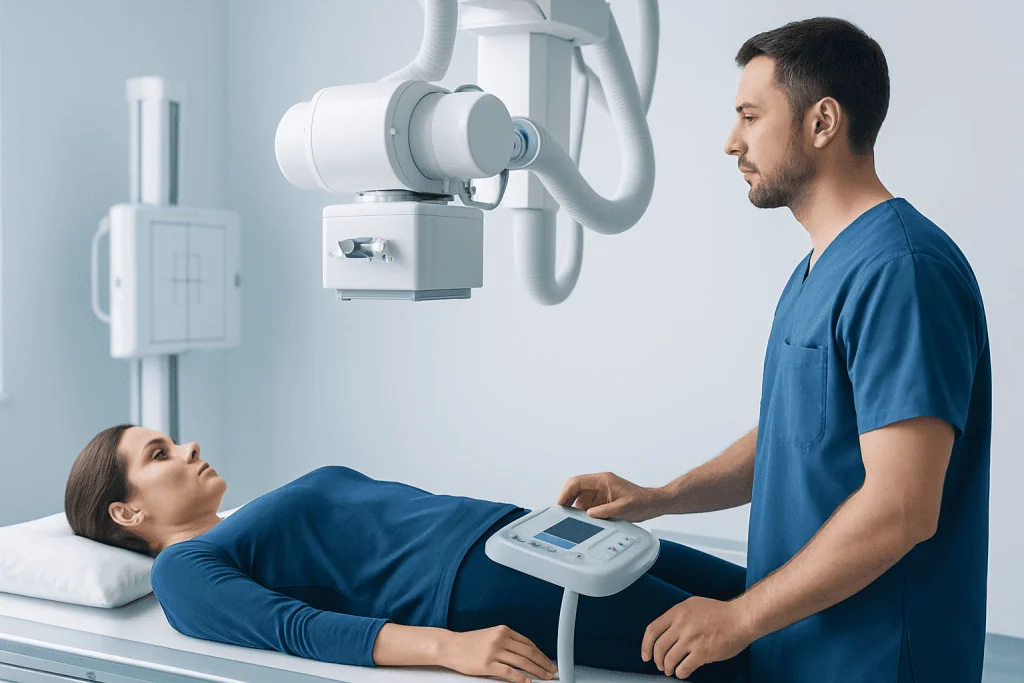
A standard X-ray exam is usually brief and painless. A radiology technologist positions the patient standing, sitting, or lying on an imaging table, depending on the body part being examined. The patient may be asked to hold still or briefly hold their breath so the image is sharp. The technologist steps behind a protective barrier or moves to a control area while taking the image.
Some exams require more than one view. For example, a suspected wrist fracture may need images from different angles, and a chest X-ray may include front and side views. If an injury is painful, the technologist will help position the patient as gently as possible while still aiming for a useful image. Parents may be asked to help comfort a child, with protective measures used when appropriate.
After a routine X-ray, most patients can return to normal activities immediately. There is no radiation left in the body after the image is taken. If contrast material was used, the care team may provide instructions about drinking fluids, monitoring for delayed reactions, or resuming medicines. Any unexpected symptoms after a contrast study should be reported to a healthcare professional.
Understanding X-Ray Results
X-ray images are usually reviewed by a radiologist, a doctor trained to interpret medical images. The radiologist prepares a report for the referring doctor, who combines the imaging findings with the patient’s symptoms, examination, medical history, and laboratory tests if available. Sometimes a result is clear and directly answers the clinical question; at other times, it may suggest several possibilities or recommend another test.
Common report terms can be confusing. A finding may be described as acute, meaning recent or active, or chronic, meaning long-standing. The phrase no acute abnormality may mean that no urgent or new X-ray finding was seen, but it does not always rule out every possible cause of symptoms. Some conditions, such as early stress fractures, certain soft-tissue injuries, or very early infections, may not be visible on a standard X-ray.
If symptoms persist despite a normal X-ray, the referring doctor may recommend follow-up, repeat imaging, ultrasound, CT, MRI, laboratory tests, or specialist evaluation. Patients should avoid interpreting images on their own without medical guidance. The most useful discussion is usually with the doctor who ordered the test, because that doctor knows the reason for the exam and the next steps in care.
When to See a Doctor and Next Steps
Patients should seek medical advice when symptoms are new, persistent, worsening, or linked to an injury. Examples include pain after a fall, difficulty bearing weight, chest symptoms, shortness of breath, ongoing cough with fever, abdominal swelling, suspected swallowed object, or unexplained swelling near a bone or joint. A doctor can decide whether an X-ray is appropriate or whether another type of imaging would be more helpful.
It is also important to speak with a healthcare professional if symptoms continue after a normal X-ray, if pain or function does not improve as expected, or if a follow-up appointment was recommended. In some cases, repeat imaging after a short interval can reveal changes that were not visible at first. In other cases, a different test may be better suited to evaluate soft tissues, organs, blood vessels, or nerves.
For international patients, Acibadem International’s multidisciplinary specialists and JCI-accredited hospitals provide diagnostic imaging and coordinated care for a wide range of conditions, including those evaluated with X-ray imaging. Patients should bring prior reports, medication lists, and relevant medical records to support safe and efficient decision-making.
Frequently asked questions
Is an X-ray painful?
The X-ray itself is painless. Some patients may feel discomfort from holding a position, especially after an injury, but the technologist will try to make positioning as gentle as possible. The image is usually taken within seconds once the patient is in place.
How much radiation is used in an X-ray?
The amount depends on the body area and type of exam. Routine diagnostic X-rays generally use a low amount of radiation, and modern equipment is designed to keep exposure as low as reasonably achievable. The doctor orders the exam when the expected benefit is greater than the small potential risk.
Can a pregnant patient have an X-ray?
Pregnancy should always be discussed with the doctor and radiology team before an X-ray. Some exams may be postponed, replaced with another imaging test, or performed with special precautions if the information is medically important. The decision depends on the area being imaged and the urgency of the medical question.
Why might an X-ray be normal even if symptoms continue?
Some conditions are not visible on a standard X-ray, especially early or soft-tissue problems. Ligament injuries, muscle injuries, some early fractures, and certain infections may require follow-up or another imaging test. Patients should return to their doctor if symptoms persist or worsen.
Who reads the X-ray?
A radiologist usually interprets the images and prepares a written report. The referring doctor then explains the results in the context of the patient’s symptoms and examination. This team approach helps connect the imaging findings with the most appropriate care plan.
Does radiation stay in the body after an X-ray?
No. After a standard X-ray image is taken, there is no radiation left in the body. Patients can usually return to normal activities immediately unless they received contrast material or were given different instructions for a specific procedure.
This article is for general information only and is not a substitute for professional medical advice. Please consult a qualified doctor about your individual situation.
Explore treatments in Turkey — costs, top hospitals & a free quote
JCI-accredited · board-certified surgeons · reply within 24h
Add us as a Preferred Source to see more of our trusted health content across Google Search, AI Overviews and Discover.

More from the Health Library

Transcranial Doppler Ultrasound: How It Evaluates Brain Blood Flow

Male Breast Imaging: What to Expect From Mammogram and Ultrasound

Diagnostic Medical Ultrasound: Who May Need It and What It Can Detect

Quest Diagnostics Appointment: How to Book, Prepare, and Check In

Urine Test Results Explained: Infection, Protein, Blood, and Other Common Findings