Male Breast Imaging: What to Expect From Mammogram and Ultrasound
Men may need breast imaging if they have a lump, swelling, pain, nipple discharge, or skin changes. A mammogram uses low-dose X-rays, while ultrasound uses sound waves; many people have one or both tests.
Key Takeaways
- Men may need breast imaging if they have a lump, swelling, pain, nipple discharge, or skin changes.
- A mammogram uses low-dose X-rays, while ultrasound uses sound waves; many people have one or both tests.
- Most male breast symptoms are caused by benign conditions, especially gynecomastia.
- Imaging helps guide next steps, which may include reassurance, follow-up, or a biopsy.
- The tests are usually quick, outpatient procedures and do not require major preparation.
Male breast imaging helps doctors assess symptoms such as a lump, pain, nipple changes, or breast enlargement. Mammogram and ultrasound are commonly used to identify benign conditions like gynecomastia and to check for less common but important causes, including breast cancer.
Overview of Male Breast Imaging
Male breast imaging refers to tests used to examine breast tissue in men when there is a symptom or a finding on physical examination. Although breast imaging is more commonly discussed in women, men can also develop breast lumps, tenderness, nipple discharge, or enlargement of breast tissue. Imaging helps doctors understand the cause and decide whether any further testing is needed.
The two main imaging tests are mammography and ultrasound. A mammogram uses low-dose X-rays to create detailed images of the breast. Ultrasound uses sound waves to show the tissue beneath the skin and can help distinguish a fluid-filled area from a solid one. Depending on the person’s age, symptoms, and examination findings, the radiologist may recommend one test first and then add the other if needed.
In many cases, male breast symptoms are due to benign conditions, especially gynecomastia, which is a noncancerous enlargement of breast tissue. Imaging can often identify patterns that strongly suggest benign change. At the same time, it is an important way to evaluate for less common causes, including infection, cysts, lipomas, and male breast cancer.
When Mammogram or Ultrasound May Be Recommended
Doctors may recommend male breast imaging when a man notices a new lump, one-sided breast enlargement, persistent pain in one area, nipple discharge, nipple inversion, or skin changes such as dimpling or redness. Imaging may also be advised if a doctor feels an abnormality during an exam, even if the person has no pain.
Not every symptom means cancer. In fact, most male breast concerns are not cancerous. However, imaging is useful because it provides more information than a physical examination alone. It can help show whether a change is centered behind the nipple, as often happens with gynecomastia, or whether it looks like a more distinct mass that needs closer attention.
Breast imaging may be especially important in men who have risk factors for breast cancer, such as a strong family history of breast cancer, a known BRCA mutation, prior chest radiation, Klinefelter syndrome, liver disease, or hormone-related medication use. A doctor will consider the full clinical picture before deciding which test is most appropriate.
What to Expect During a Male Mammogram
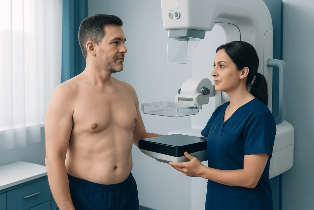
A male mammogram is performed similarly to a mammogram in women, but the amount of breast tissue is usually smaller. During the test, a technologist positions the breast on the imaging plate and gently compresses it for a few seconds while the X-ray image is taken. Compression can feel uncomfortable, but it is brief and helps produce clearer images with the lowest necessary radiation dose.
Most mammograms take only a short time to complete. Patients are usually asked not to wear deodorant, powders, or creams on the chest or underarm area on the day of the exam, because these products can sometimes affect the images. Wearing a two-piece outfit may make changing easier, as the upper body will need to be uncovered for the study.
A mammogram is often the first imaging test in adult men with a suspicious breast symptom because it can be very effective at showing the pattern of tissue change. In many cases, the images can strongly suggest benign gynecomastia. If more detail is needed, the radiologist may add an ultrasound on the same day.
If anxiety is a concern, it may help to know that the examination is routine and typically completed as an outpatient procedure. The technologist will explain each step and try to keep the person as comfortable as possible throughout the test.
What to Expect During a Breast Ultrasound in Men
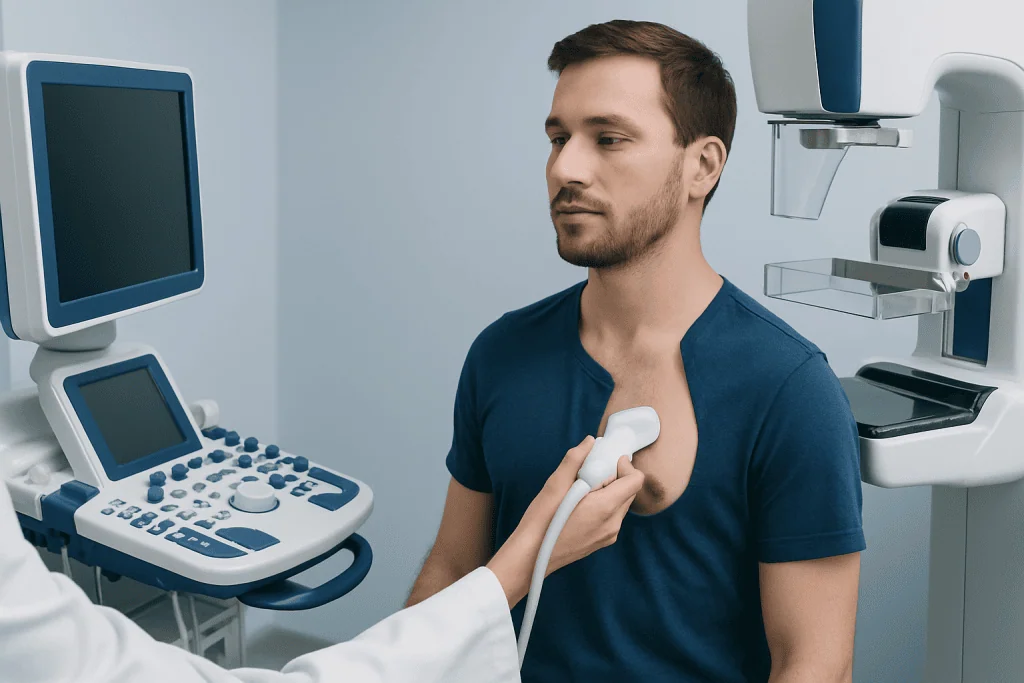
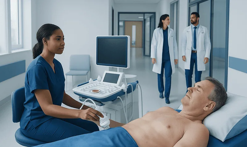
Breast ultrasound in men is a painless imaging test that uses a handheld probe placed over the skin with a small amount of gel. The person usually lies on an exam table while the sonographer or radiologist moves the probe over the area of concern. Sound waves create real-time images on a monitor, allowing the doctor to assess the structure of the tissue.
Ultrasound is especially helpful for looking more closely at a specific lump or area seen on examination or mammography. It can help determine whether an abnormality is solid or fluid-filled, assess its shape and margins, and examine nearby tissues such as the underarm region if needed. Unlike mammography, ultrasound does not use radiation.
The test is usually quick and does not require recovery time. Patients can return to normal activities immediately afterward. Because it is comfortable and widely available, ultrasound is often used as a complementary test rather than a replacement for mammography when the diagnosis is uncertain.
What the Images Can Show
One of the most common findings on male breast imaging is gynecomastia. This appears as an increase in glandular tissue, usually centered behind the nipple, and may affect one or both sides. Gynecomastia can occur during puberty, aging, medication use, hormonal imbalance, obesity, liver disease, thyroid disease, or other medical conditions. Imaging can help confirm this typical benign pattern and reduce uncertainty.
Imaging may also show other noncancerous causes of symptoms, such as a lipoma, oil cyst, abscess, or skin-related lesion. In some cases, the problem is not in the breast tissue itself but in the surrounding area, such as a swollen lymph node or chest wall change. Ultrasound is particularly useful for clarifying these possibilities.
When a finding looks suspicious, imaging helps the radiologist describe its size, shape, location, and other features. Suspicious signs can include an irregular mass, tissue distortion, or associated skin or nipple changes. Imaging alone cannot always confirm whether a lesion is cancerous, so a biopsy may be recommended to obtain a tissue sample for diagnosis.
If a biopsy is needed, the radiologist may use ultrasound guidance to target the area accurately. This is often part of a broader diagnostic pathway for breast cancer, although many biopsied lesions ultimately prove to be benign.
How Results Are Reported and What Happens Next
After the images are reviewed, the radiologist prepares a report that describes the findings and whether more evaluation is needed. Sometimes the result is clearly benign, and no further breast testing is necessary. In other situations, the radiologist may recommend additional views, ultrasound, short-term follow-up imaging, or biopsy.
If the imaging suggests gynecomastia and the symptoms fit that diagnosis, the next step may be a clinical discussion about possible causes. Doctors may review medications, alcohol use, hormone-related issues, weight changes, and other health conditions. In selected cases, blood tests or referral to another specialist may be appropriate to look for an underlying cause.
When a suspicious mass is found, tissue sampling is often the next step. This may involve a breast biopsy performed with imaging guidance. If a confirmed cancer diagnosis is made, treatment planning may include medical oncology care and, when appropriate, breast cancer surgery.
Results are best interpreted together with the physical examination and medical history. Patients should feel comfortable asking what was seen, whether the finding appears benign, and what follow-up is recommended.
Preparation, Safety, and Emotional Concerns
Male breast imaging usually requires very little preparation. Patients may be advised to avoid deodorant, lotions, or powders on the chest and underarm area before mammography. Bringing previous breast imaging, if available, can help the radiologist compare findings over time. It is also useful to tell the team about current medications, hormone use, or any personal or family history of breast cancer.
Mammography uses a low dose of radiation, and for most people the benefit of evaluating a concerning symptom outweighs the minimal exposure. Ultrasound does not involve radiation. Both tests are generally considered safe, and complications are very uncommon. The main temporary discomfort is usually brief compression during the mammogram.
Some men feel embarrassed or anxious about having a breast exam or imaging study because these tests are often associated with women’s health. This is understandable, but breast symptoms in men are medically important and deserve evaluation without stigma. Clear communication with the imaging team can make the experience more comfortable.
For international patients who need coordinated assessment, Acibadem International’s multidisciplinary specialists and JCI-accredited hospitals diagnose and treat breast conditions using modern imaging and follow-up care.
When to See a Doctor
A man should see a doctor if he notices a new breast lump, breast enlargement on one side, nipple discharge, nipple inversion, persistent localized pain, or skin changes over the breast. These symptoms do not necessarily mean cancer, but they should be evaluated promptly so the cause can be identified.
Medical attention is especially important if a lump feels firm, seems fixed in place, is getting larger, or is associated with underarm swelling. Redness, warmth, fever, or significant tenderness may suggest infection and also deserve timely assessment. If there is a strong family history of breast or ovarian cancer, that history should be shared with the doctor.
Early evaluation often leads to reassurance when the cause is benign, and it allows faster treatment if a more serious condition is found. Men should not ignore persistent symptoms or assume they are too minor to discuss.
Frequently asked questions
Do men really get mammograms?
Yes. Men may have a mammogram when they develop symptoms such as a lump, nipple discharge, or one-sided breast enlargement. The test helps doctors evaluate the cause and decide whether any further testing is needed.
Is male breast imaging usually done for cancer?
Not usually. Most male breast symptoms are caused by benign conditions, especially gynecomastia. Imaging is still important because it helps distinguish common noncancerous changes from less common but more serious problems.
Is a male mammogram painful?
A mammogram can cause brief discomfort because the breast tissue is compressed to get clear images. For most people, the discomfort lasts only a few seconds at a time. The technologist tries to keep the exam as comfortable as possible.
Why might a doctor order both mammogram and ultrasound?
The two tests provide different kinds of information. Mammography is often good at showing the overall tissue pattern, while ultrasound can look more closely at a specific area or lump. Using both can improve diagnostic clarity.
What does gynecomastia look like on imaging?
Gynecomastia usually appears as glandular tissue enlargement centered behind the nipple. Radiologists often recognize this pattern on mammography or ultrasound. When the appearance is typical, it can strongly support a benign diagnosis.
Will I need a biopsy if imaging finds a lump?
Not always. If the imaging features look clearly benign, a biopsy may not be necessary. A biopsy is more likely if the finding is suspicious or if the images do not fully explain the symptoms.
How should a man prepare for breast imaging?
Preparation is simple. It is often recommended to avoid deodorant, powder, or lotion on the chest and underarm area before mammography, and to bring any prior imaging if available. Patients should also tell the doctor about medications, hormone use, and family history.
This article is for general information only and is not a substitute for professional medical advice. Please consult a qualified doctor about your individual situation.
Ultrasound in Turkey — costs, top hospitals & a free quote
JCI-accredited · board-certified surgeons · reply within 24h
Add us as a Preferred Source to see more of our trusted health content across Google Search, AI Overviews and Discover.

Check-up & Preventive Medicine
Comprehensive health screenings designed for early detection and prevention.
1 specialists in this unitMore from the Health Library

How a Diagnosis Is Made: From Symptoms to Tests and Results

EMG and Nerve Conduction Studies: Diagnosing Neuromuscular Disease

When Should You Repeat a Lab Test or Imaging Exam?
Echocardiogram: Heart Ultrasound for Valves, Pump Function, and Symptoms
Cerebral Angiography vs MRI: Which Test Shows Brain Blood Vessels Better?