Cerebral Angiography vs MRI: Which Test Shows Brain Blood Vessels Better?
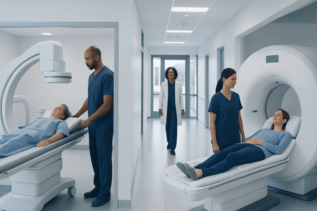
MRI and MR angiography can show many brain blood vessel problems without using a catheter. Cerebral angiography usually gives the most precise, detailed view of brain arteries and veins.
Key Takeaways
- MRI and MR angiography can show many brain blood vessel problems without using a catheter.
- Cerebral angiography usually gives the most precise, detailed view of brain arteries and veins.
- The best test depends on the clinical question, urgency, and whether treatment may be needed at the same time.
- Cerebral angiography is more invasive than MRI and carries higher procedural risks.
- Doctors may use these tests together rather than choosing only one.
Both cerebral angiography and MRI can evaluate brain blood vessels, but they do not provide exactly the same information. MRI and MR angiography are often the first choice because they are noninvasive, while cerebral angiography is usually the most detailed test when doctors need a closer look or plan treatment.
Overview: How These Tests Differ
When comparing cerebral angiography vs MRI, the main difference is how the images are created and how much detail they show. MRI uses a strong magnet and radio waves to create images of the brain. A related technique called magnetic resonance angiography, or MRA, focuses on blood vessels and blood flow. These tests are noninvasive because they do not require a catheter to be threaded into an artery.
Cerebral angiography, often called catheter angiography or digital subtraction angiography, is an invasive imaging test. During the procedure, a doctor inserts a thin catheter into an artery, usually in the wrist or groin, and guides it toward the blood vessels supplying the brain. Contrast dye is then injected while X-ray images are taken in rapid sequence.
In general, MRI and MRA are excellent for screening and evaluating many brain and blood vessel conditions. However, cerebral angiography often shows finer details of vessel shape, size, narrowing, branching, and blood flow patterns. For that reason, it is often considered the reference standard when the diagnosis is uncertain or when a procedure may be needed.
Rather than competing tests, they are often complementary. A person may first have an MRI or MRA and later undergo cerebral angiography if the findings need confirmation, if treatment planning is required, or if symptoms suggest a serious vascular problem such as stroke.
What MRI and Cerebral Angiography Can Show
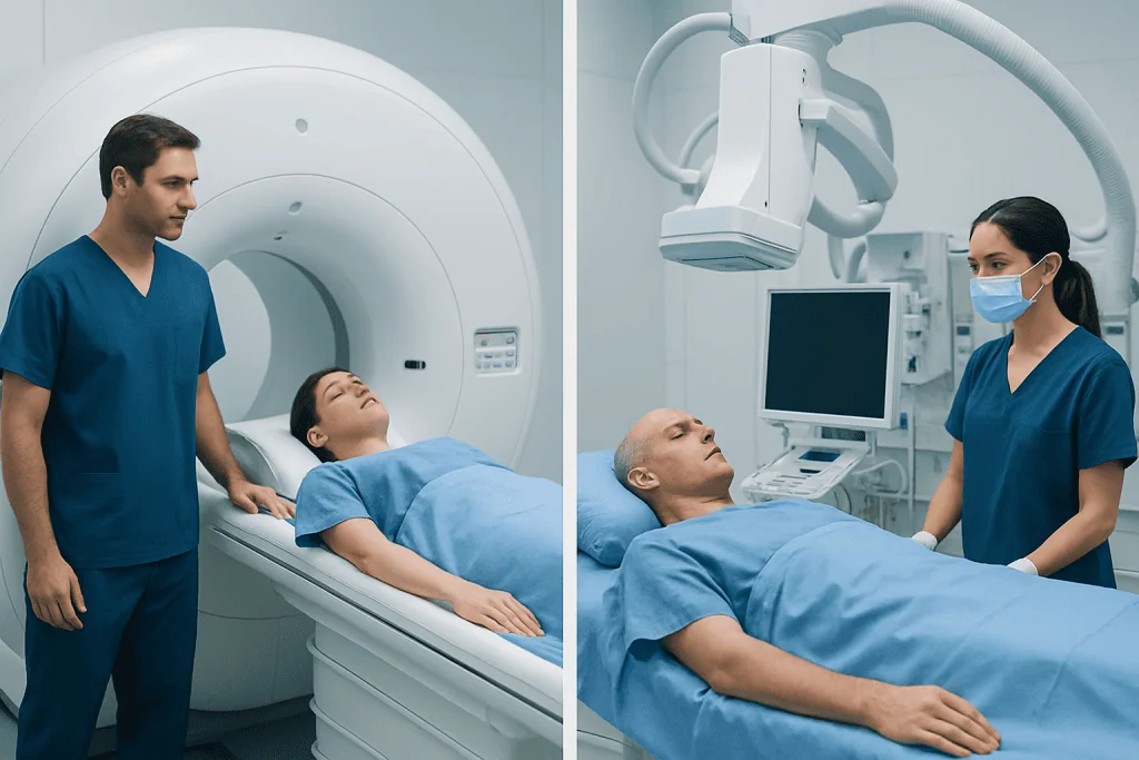
MRI provides detailed pictures of the brain tissue itself. It can reveal areas of stroke, bleeding, swelling, tumors, infection, inflammation, and damage from reduced blood flow. When combined with MRA, it can also show larger and many medium-sized blood vessels, helping doctors look for aneurysms, narrowing, blockage, or vascular malformations.
Cerebral angiography is designed specifically to map blood vessels with very high spatial and temporal detail. It can show subtle abnormalities that may not be fully visible on MRI or MRA, especially in small vessels or complex vessel networks. It is particularly helpful for evaluating aneurysms, arteriovenous malformations, fistulas, vasculitis patterns, and intricate areas of stenosis.
Another important difference is that cerebral angiography shows blood flowing through vessels in real time. This can help doctors understand how blood moves through abnormal connections, whether a vessel is severely narrowed, and which areas of the brain may be at risk. MRI can provide valuable flow information too, but catheter angiography often gives a more precise dynamic picture.
In some situations, doctors also compare MRI with other vessel studies such as CT angiography. Depending on the clinical question, a person may need several types of imaging. For example, if an MRI suggests an aneurysm, cerebral angiography may be used to define it more clearly before aneurysm treatment.
Which Test Shows Brain Blood Vessels Better?

If the question is strictly which test shows brain blood vessels in the greatest detail, cerebral angiography usually shows them better. It can detect very small vascular changes and provides a highly detailed map of arteries and veins. This is why it remains the preferred test in selected complex cases.
However, “better” does not always mean “best first.” MRI and MRA are often the better initial choice for many patients because they are safer, noninvasive, and still provide a great deal of useful information. If these tests answer the clinical question clearly, catheter angiography may not be necessary.
The answer also depends on what doctors need to know. If they want to evaluate brain tissue, signs of recent stroke, inflammation, or surrounding structures in addition to blood vessels, MRI has a major advantage. If they need exact vessel anatomy before an intervention or need to understand a complicated vascular abnormality, cerebral angiography may be more informative.
In practical terms, MRI is often the first-line test, while cerebral angiography is the most definitive test for certain blood vessel disorders. Doctors choose based on symptoms, urgency, previous imaging, medical history, and whether treatment might be performed during the same session, such as endovascular treatment.
When Doctors Choose MRI First
MRI or MRA is commonly chosen first when a person has neurological symptoms that need evaluation but is stable enough for noninvasive imaging. This may include headaches, dizziness, visual symptoms, suspected small strokes, or concerns about an incidental aneurysm seen on another test. MRI is also useful when doctors want to evaluate both the blood vessels and the brain tissue at the same time.
Another reason MRI is often preferred initially is that it avoids arterial catheterization. That means no catheter is inserted into a blood vessel, which lowers the risk of bleeding, arterial injury, or procedure-related stroke. For many patients, this makes MRI a more comfortable and practical option.
Doctors may also choose MRI for follow-up after treatment or to monitor a known condition over time, depending on the condition and the quality of images needed. It can be especially useful in patients who need repeated imaging because it does not use ionizing radiation.
That said, MRI is not right for everyone. Some people cannot undergo MRI because of certain implanted devices, severe claustrophobia, or inability to lie still. In emergency settings or if vessel detail is critical, another test may be more appropriate.
When Cerebral Angiography Is Preferred
Cerebral angiography is usually preferred when doctors need the clearest possible view of the brain’s blood vessels. This is common when noninvasive imaging is inconclusive, when the abnormality appears complex, or when a procedure is being planned. It can be especially important in evaluating aneurysms, arteriovenous malformations, dural arteriovenous fistulas, and certain types of vessel narrowing or spasm.
It is also often chosen when treatment and diagnosis may happen together. During the same session, specialists may sometimes move from imaging to an endovascular procedure if appropriate. This can be helpful in urgent or carefully planned situations involving bleeding, high-risk aneurysms, or vascular malformations.
For example, if imaging suggests an abnormal artery-to-vein connection, catheter angiography may be necessary to fully map it before treatment. It may also guide planning for AVM treatment or help clarify whether symptoms relate to a vascular lesion that was only partly seen on MRI.
Because this test is invasive, doctors recommend it only when the expected benefit outweighs the risks. The decision is individualized and usually made by a neurologist, neurosurgeon, or interventional neuroradiology team after reviewing prior imaging and symptoms.
Risks, Limitations, and Safety Considerations
MRI is generally very safe, but it has limitations. Some patients cannot have MRI because of metallic implants that are not MRI-compatible, and some may feel anxious in the scanner. In certain cases, contrast material may be used, and doctors consider kidney function and allergy history before giving it.
MRA may not always show very tiny vessels or subtle abnormalities as clearly as catheter angiography. Motion during the scan can also reduce image quality. In some situations, blood flow patterns may make a vessel look narrower or more abnormal than it truly is, which is one reason follow-up imaging may be needed.
Cerebral angiography has greater procedural risks because it involves entering an artery and injecting contrast dye. Possible complications include bruising or bleeding at the catheter site, allergic reaction to contrast, kidney strain in susceptible patients, blood vessel injury, and, rarely, stroke. Although serious complications are uncommon in experienced centers, they are an important part of informed decision-making.
Doctors reduce these risks by reviewing medical history, medications, kidney function, and contrast allergies in advance. They also monitor the patient during and after the procedure. If the test is being considered for a condition such as brain aneurysm, the care team will explain why its added detail may justify the invasive approach.
How to Prepare and What to Expect
For MRI, preparation is usually simple. Patients are asked about pacemakers, implants, metal fragments, or other device safety concerns. They may need to remove jewelry and metallic objects, and some people receive contrast through a vein. The scan itself is painless, though it can be noisy and may take some time.
For cerebral angiography, preparation is more involved. Patients may be asked not to eat or drink for a certain period before the procedure. The medical team reviews medications, especially blood thinners, as well as kidney health and allergy history. Sedation or local anesthesia may be used depending on the situation and the hospital protocol.
After cerebral angiography, patients are observed while the catheter site is monitored. They may need to lie flat or limit movement for a period, depending on how the artery was accessed. Mild soreness or bruising can occur, but patients are given instructions about what symptoms require urgent attention after going home.
At centers with multidisciplinary neurovascular care, specialists use these test results together to plan next steps. Acibadem International’s multidisciplinary specialists and JCI-accredited hospitals diagnose and treat complex brain vessel conditions for international patients, with imaging and procedural care tailored to the individual case.
How Doctors Decide Which Test Is Right
The choice between cerebral angiography and MRI is based on the clinical question rather than a simple ranking. If the goal is broad evaluation of the brain and blood vessels without an invasive procedure, MRI and MRA are often the logical first step. If the goal is the most exact vessel map or treatment planning, cerebral angiography may be the more useful test.
Doctors also consider urgency. In emergency situations involving suspected bleeding, blocked blood flow, or rapidly changing symptoms, several imaging tools may be used quickly and in sequence. The patient’s age, kidney function, medical history, implanted devices, and ability to tolerate the test also matter.
Many patients ultimately benefit from both tests at different points in care. An MRI may detect the problem, while cerebral angiography confirms it and helps guide the next decision. This combined approach is common in neurovascular medicine and helps balance safety with diagnostic accuracy.
Anyone who is unsure which test is appropriate should discuss the reasons with a qualified doctor. Asking what information the team needs, what alternatives exist, and whether the result could change treatment can help patients make an informed, confident choice.
Frequently asked questions
Is cerebral angiography more accurate than MRI for brain blood vessels?
For fine detail of brain blood vessels, cerebral angiography is usually more accurate than MRI or MRA. It can show very small vessel changes and real-time blood flow patterns. However, MRI is often accurate enough for many situations and is commonly used first because it is noninvasive.
Why would a doctor order cerebral angiography after an MRI?
A doctor may order cerebral angiography if MRI or MRA findings are unclear, incomplete, or need confirmation. It is also used when a vascular procedure is being planned and the team needs the most detailed possible map of the blood vessels. In some cases, treatment can be performed during the same session.
Can MRI detect aneurysms and vascular malformations?
Yes, MRI and MRA can detect many aneurysms and vascular malformations. They are often very helpful as initial tests and may be enough for diagnosis or follow-up in some patients. Still, catheter angiography may be needed if the abnormality is small, complex, or requires treatment planning.
Is cerebral angiography painful?
Most people do not describe cerebral angiography as painful, though it can be uncomfortable. Patients may feel a pinch at the access site and sometimes a warm sensation when contrast is injected. The care team uses local anesthesia and monitoring to make the procedure as comfortable as possible.
Which test is safer, MRI or cerebral angiography?
MRI is generally safer because it is noninvasive and does not require a catheter in an artery. Cerebral angiography has more risks, including bleeding at the access site, contrast-related issues, and a small risk of stroke. Even so, it is considered safe when performed for the right reasons by experienced specialists.
Can these tests replace each other completely?
Not always. MRI and MRA are excellent for many patients, but they do not fully replace cerebral angiography when the highest level of vessel detail is needed. In practice, doctors often use them as complementary tests rather than direct substitutes.
References
- American College of Radiology
- Radiological Society of North America
- National Institute of Neurological Disorders and Stroke
- Society of NeuroInterventional Surgery
- World Stroke Organization
This article is for general information only and is not a substitute for professional medical advice. Please consult a qualified doctor about your individual situation.
Explore treatments in Turkey — costs, top hospitals & a free quote
JCI-accredited · board-certified surgeons · reply within 24h
Add us as a Preferred Source to see more of our trusted health content across Google Search, AI Overviews and Discover.