Pet Scan: What Patients Need to Know
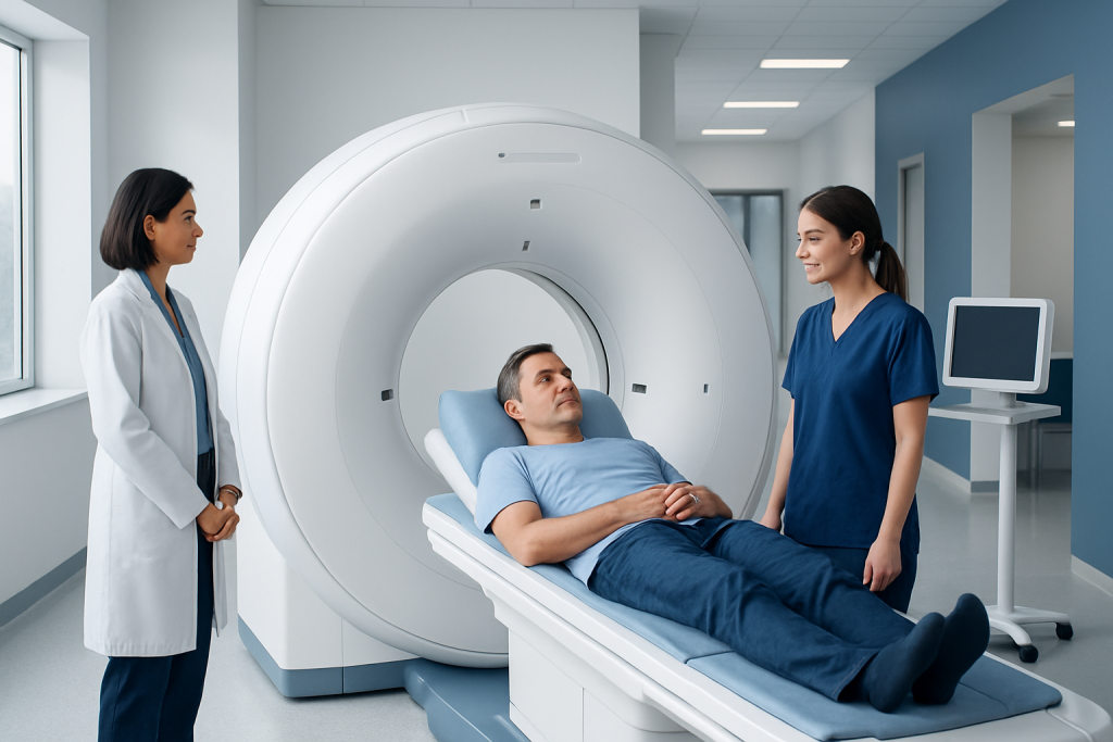
A PET scan shows metabolic activity and can reveal disease before structural changes appear on other scans. It is commonly used for cancer assessment, heart evaluation, and certain neurological conditions.
Key Takeaways
- A PET scan shows metabolic activity and can reveal disease before structural changes appear on other scans.
- It is commonly used for cancer assessment, heart evaluation, and certain neurological conditions.
- Preparation may include fasting, avoiding strenuous exercise, and following instructions about medicines and blood sugar.
- The test usually involves a small injection of a radioactive tracer, a waiting period, and then imaging while lying still.
- PET scans are generally safe, but patients should tell their doctor about pregnancy, breastfeeding, diabetes, or difficulty staying still.
A PET scan is an imaging test that helps doctors see how tissues and organs are functioning, not just how they look. It is often used to evaluate cancer, heart conditions, brain disorders, and some unexplained symptoms when more detail is needed.
Overview: What a PET Scan Is
A pet scan is a type of imaging test that shows how organs and tissues are working inside the body. Unlike scans that mainly show anatomy or structure, a PET scan highlights metabolic activity, which can help doctors identify areas that are more or less active than expected. This can provide important clues about disease.
PET stands for positron emission tomography. The test uses a small amount of radioactive tracer, usually given by injection into a vein. The tracer travels through the body and collects in tissues in different ways depending on how active they are. A special camera then detects this activity and creates images for the doctor to review.
Many PET scans are combined with CT, and sometimes MRI, so doctors can compare function with detailed anatomy in the same session. This combination can improve accuracy when evaluating a suspicious area, planning treatment, or checking how well treatment is working.
Why Doctors Order a PET Scan
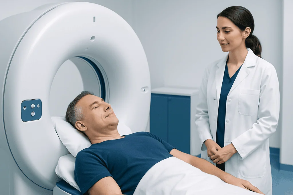
Doctors use a pet scan when they need more information about how the body is functioning at a cellular level. One of the most common uses is cancer care. A PET scan may help detect cancer, show whether it has spread, guide biopsy planning, or assess response after treatment. In this setting, it is often part of a broader evaluation that may also include cancer care and other imaging tests.
A PET scan can also help assess heart health. It may show whether parts of the heart muscle are getting enough blood flow or whether tissue has been damaged after a heart problem. In some cases, this information helps doctors decide whether procedures such as coronary angiography or other heart treatments should be considered.
In neurology, PET imaging may support the evaluation of memory problems, seizures, or certain movement disorders. It can sometimes help distinguish between different causes of symptoms when MRI or CT does not provide the full picture. Doctors may also use it in inflammatory or infectious conditions when they are searching for hidden active disease.
How a PET Scan Works and What It Can Show
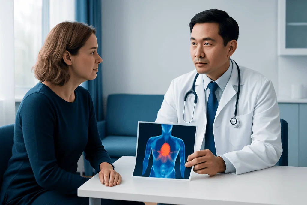
The tracer used in a PET scan is designed to behave like a substance the body normally uses, such as glucose. Cells that are more active often absorb more of the tracer. The scanner detects signals from the tracer and turns them into images that show patterns of activity throughout the body.
This is why PET can sometimes detect problems before they are clearly visible on structural scans. For example, cancer cells often use energy differently than normal cells, and inflamed tissues may also appear more active. At the same time, not every bright area on a PET scan means cancer, because infection, inflammation, healing tissue, and some normal organs can also take up tracer.
Results are always interpreted in context. Radiologists and treating physicians compare the PET findings with symptoms, blood tests, physical examination, and other imaging studies. If needed, additional tests such as biopsy or MRI may help confirm what the scan suggests.
How to Prepare and What Happens During the Test
Preparation for a pet scan depends on the reason for the test and the tracer being used, but many patients are asked not to eat for several hours beforehand. Drinking water is usually encouraged unless the care team gives different instructions. Strenuous exercise is often avoided for a day before the scan because it can change how muscles absorb the tracer.
Patients should tell the medical team about pregnancy, breastfeeding, diabetes, claustrophobia, recent illness, and all medicines they take. Blood sugar is especially important for some PET scans because high glucose levels can affect image quality. People with diabetes may receive tailored instructions about meals and medication timing.
During the appointment, a small IV is placed and the tracer is injected. Then there is usually a waiting period, often around 30 to 60 minutes, while the tracer circulates. The scan itself involves lying still on a table that moves through the scanner. The test is painless, though staying still can feel uncomfortable for some people. The full visit may take a few hours, even though the actual imaging time is shorter.
- Wear comfortable clothing if advised.
- Remove metal objects if requested.
- Follow instructions carefully about food, exercise, and medications.
- Arrange support if anxiety or mobility issues may make the visit harder.
Understanding PET Scan Results
PET scan results are not usually given immediately at the scanner. A radiologist or nuclear medicine specialist reviews the images and sends a report to the doctor who ordered the test. That doctor then explains what the findings mean in the context of the patient’s health history and symptoms.
Results may be described as normal, abnormal, or indeterminate. A normal result generally means tracer distribution appears as expected for the type of scan performed. An abnormal result means there are areas of activity that need explanation, but this does not automatically mean a serious illness. Benign conditions can also change tracer uptake.
Sometimes a PET scan answers the question clearly, but in other situations it leads to follow-up testing. A doctor may recommend blood tests, repeat imaging, biopsy, or referral to a specialist. In cancer care, PET findings can help guide whether treatments such as chemotherapy are appropriate, but decisions are not based on PET images alone.
Benefits, Limits, and Safety
One of the main benefits of a pet scan is that it can reveal functional changes early in the course of disease. This can support earlier diagnosis, more accurate staging, and better treatment planning. It can also help show whether a treatment is working, which may prevent unnecessary changes or delays in care.
However, PET scans have limits. They do not diagnose every condition on their own, and some small lesions may not be detected. False-positive findings can happen when inflammation or infection looks similar to cancer on the scan. False-negative results are also possible in some slow-growing or low-activity diseases.
The amount of radiation from the tracer is generally low and medically controlled, but it is still an important consideration. Doctors recommend PET only when the expected benefit outweighs the risk. Patients are often advised to drink fluids after the scan and may be told to limit close contact with pregnant people or very young children for a short time, depending on the tracer used and the local protocol.
For international patients who need coordinated imaging and specialist review, Acibadem International’s multidisciplinary teams and JCI-accredited hospitals diagnose and treat conditions that may require PET imaging as part of broader care planning.
When to Seek Medical Care
A pet scan itself is usually a planned test rather than an emergency procedure. A person should seek medical care if a doctor recommends further evaluation for symptoms such as unexplained weight loss, a new lump, persistent pain, ongoing fevers, chest discomfort, shortness of breath, seizures, or concerning neurological changes. These symptoms do not always mean serious disease, but they deserve proper assessment.
Patients should also contact their doctor if they are unsure how to prepare for the scan, especially if they have diabetes, are pregnant or breastfeeding, or have trouble lying still. It is better to ask questions in advance than to risk a delayed or less accurate study.
After the scan, urgent medical attention is rarely needed because complications are uncommon. However, if there is significant swelling, rash, trouble breathing, or another unexpected reaction after the tracer injection, prompt medical advice is important. Most patients complete the test without major problems and can return to normal activities soon afterward.
Frequently asked questions
What is a PET scan used for?
A PET scan is used to show how tissues and organs are functioning. Doctors commonly use it in cancer care, heart evaluation, and certain brain and neurological conditions, as well as in some cases of infection or inflammation.
Is a PET scan the same as a CT scan or MRI?
No. A PET scan mainly shows metabolic or functional activity, while CT and MRI mainly show structure and anatomy. Many patients have PET combined with CT so doctors can see both function and detailed body structures together.
How should a patient prepare for a PET scan?
Preparation varies, but fasting for several hours is common. Patients may also be asked to avoid strenuous exercise and to discuss medications, diabetes management, pregnancy, or breastfeeding with their doctor before the test.
How long does a PET scan take?
The full appointment often takes a few hours because there is usually a waiting period after the tracer injection. The actual scan time is shorter, but patients should expect extra time for preparation and image acquisition.
Is a PET scan painful or unsafe?
The scan is usually not painful. Patients may feel a brief pinch from the IV injection, and some may find it uncomfortable to lie still, but the test is generally well tolerated. It uses a small amount of radioactive tracer, and doctors order it only when the expected benefit outweighs the risk.
Can a PET scan confirm cancer?
A PET scan can strongly suggest whether an area is suspicious, but it does not confirm cancer by itself. In many cases, doctors combine PET findings with other imaging, examination, and sometimes biopsy results before making a diagnosis.
This article is for general information only and is not a substitute for professional medical advice. Please consult a qualified doctor about your individual situation.
Oncology care in Turkey — second opinion and treatment plan
JCI-accredited · board-certified surgeons · reply within 24h
Add us as a Preferred Source to see more of our trusted health content across Google Search, AI Overviews and Discover.

More from the Health Library

Trampoline Workout: What Patients Need to Know

Mrsa on Face: A Complete Medical Overview

Cross Contamination: What Patients Need to Know

Poop Chart: What Patients Need to Know

Beet Root Powder: An Evidence-Based Guide for Patients