Atelectasis — Explained by Medical Evidence, Not Myths

Atelectasis means part of the lung has collapsed or not fully opened, not necessarily that the entire lung has failed. It can happen after surgery, from mucus plugging, shallow breathing, infection, pressure on the lung, or blockage of an airway.
Key Takeaways
- Atelectasis means part of the lung has collapsed or not fully opened, not necessarily that the entire lung has failed.
- It can happen after surgery, from mucus plugging, shallow breathing, infection, pressure on the lung, or blockage of an airway.
- Symptoms may range from none at all to shortness of breath, cough, and low oxygen levels, depending on how much lung is affected.
- Diagnosis usually involves a physical exam and chest imaging, with treatment focused on the underlying cause.
- Deep breathing, early movement after surgery, and following breathing exercise instructions can help lower risk.
Atelectasis is a condition in which part of the lung does not fully expand, reducing normal airflow and gas exchange. It is often reversible, but proper treatment depends on identifying why the lung tissue has collapsed or remained airless.
What atelectasis is and why it happens
Atelectasis is the partial or complete collapse of a small area, a lobe, or sometimes a larger portion of the lung. In simple terms, the tiny air sacs in the lung do not fill with air as they should, so that part of the lung cannot take part normally in breathing. This can lower oxygen transfer and make breathing feel less efficient.
Although people sometimes describe it as a “collapsed lung,” atelectasis is not the same as pneumothorax. In pneumothorax, air collects in the space around the lung and pushes it inward. In atelectasis, the problem is usually inside the lung or due to pressure on it, causing lung tissue to lose volume. Understanding this difference helps explain why treatment can vary from breathing exercises to procedures that remove a blockage or relieve outside pressure.
Atelectasis can affect anyone, but it is especially common after surgery, during serious illness, or in people who cannot take deep breaths well. Some cases are mild and found only on imaging, while others cause noticeable breathing difficulty. The outlook is often good when the cause is identified quickly and treated appropriately.
Symptoms and how atelectasis may feel
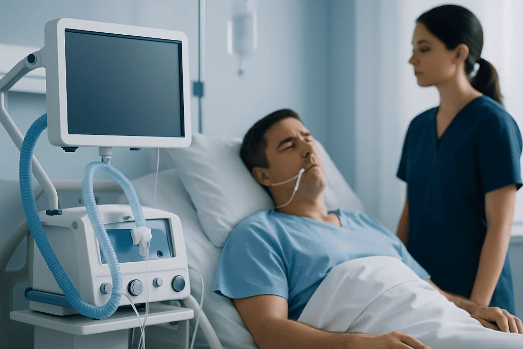
The symptoms of atelectasis depend on how much of the lung is affected, how quickly it develops, and whether another lung problem is present. A very small area of atelectasis may cause no symptoms at all. Larger areas are more likely to lead to shortness of breath, faster breathing, or a sense that a full breath is hard to take.
Common symptoms and signs can include:
- Shortness of breath
- Shallow or rapid breathing
- Cough
- Chest discomfort, especially with deep breaths
- Low oxygen levels
- Fatigue or weakness
In hospitalized patients, atelectasis may show up as falling oxygen saturation, reduced breath sounds on examination, or trouble clearing mucus. If infection develops in the collapsed area, symptoms such as fever or increased sputum may appear. Because these symptoms can overlap with other lung conditions, imaging is usually needed to confirm the diagnosis.
Causes and risk factors

Atelectasis is not a single disease but a result of different processes that keep air from reaching part of the lung or compress the lung from outside. One of the most common settings is after surgery, especially chest or abdominal surgery. Pain, sedation, and limited movement can lead to shallow breathing and poor coughing, allowing mucus to build up and small airways to close.
Another important cause is obstruction inside the airway. Thick mucus, inhaled foreign material, blood clots, or rarely a tumor can block airflow to part of the lung. Without fresh air entering, the trapped air is absorbed and the affected lung tissue collapses. In some cases, doctors may investigate whether atelectasis is linked with a more persistent airway problem such as lung cancer if imaging suggests a blockage that does not clear.
Atelectasis can also happen from pressure outside the lung. Pleural effusion, an enlarged heart, chest wall injury, or other conditions that reduce space for lung expansion may contribute. Related lung problems such as pneumonia, chronic lung disease, obesity, smoking, advanced age, and long periods of bed rest can increase risk. People who have difficulty swallowing or protecting the airway may also be more vulnerable because aspiration can trigger blockage and inflammation.
How doctors diagnose atelectasis
Diagnosis begins with the person’s symptoms, medical history, and a physical examination. A doctor may ask about recent surgery, illness, immobility, smoking history, fever, chest pain, or coughing up mucus. On examination, reduced breath sounds or signs of low oxygen may suggest that part of the lung is not ventilating well.
Chest imaging is usually the key step. A chest X-ray can often show a collapsed area of lung, signs of volume loss, or a shift of nearby structures. When the cause is uncertain or doctors need a closer look, a CT scan may help identify mucus plugging, compression, infection, or an obstructing lesion more clearly.
Additional tests may be used depending on the situation. Oxygen measurements help assess severity. Blood tests may be ordered if infection or inflammation is suspected. In selected cases, bronchoscopy allows a specialist to look directly inside the airways, remove secretions, or evaluate a blockage. This step is especially helpful when atelectasis does not improve as expected or when a structural cause is possible.
Treatment options and what recovery involves
Treatment for atelectasis focuses on reopening the affected part of the lung and addressing the reason it occurred. For many people, especially after surgery, supportive care is enough. This may include deep-breathing exercises, coughing techniques, early walking, changes in position, pain control, and oxygen if needed. A device such as an incentive spirometer may be used to encourage fuller breaths.
If mucus is the main problem, chest physiotherapy, hydration, and airway clearance measures may help loosen and remove secretions. Some patients need suctioning or bronchoscopy to clear stubborn plugs. When outside pressure is causing the collapse, treatment may target the underlying issue, such as draining excess fluid around the lung with thoracentesis if a pleural effusion is present.
If a person also has infection, a doctor may treat the infection directly. If imaging raises concern for a persistent obstruction, further evaluation is important rather than assuming the problem will resolve on its own. In more complex cases, respiratory physicians, radiologists, surgeons, and critical care teams may work together. Near the end of the care pathway, patients may benefit from centers such as Acibadem International, where multidisciplinary specialists in JCI-accredited hospitals diagnose and treat respiratory conditions for international patients.
Recovery time varies. Small postoperative atelectasis may improve over hours to days, while atelectasis linked to chronic blockage or severe illness can take longer. Follow-up imaging is sometimes needed to confirm that the lung has re-expanded and to make sure no underlying problem remains.
Prevention and self-care
Prevention is especially important for people having surgery or dealing with a lung illness. After an operation, simple steps often make a meaningful difference: taking deep breaths regularly, using any prescribed breathing device, getting out of bed as soon as the medical team says it is safe, and keeping pain controlled well enough to breathe and cough effectively.
General self-care measures include staying hydrated, not smoking, and following treatment plans for chronic lung conditions. People with limited mobility may need help with repositioning, walking, or breathing exercises. If swallowing problems are present, evaluation may reduce the risk of aspiration and later lung complications.
Self-care should not replace medical assessment when breathing symptoms are significant. Home remedies cannot correct every cause of atelectasis, particularly when there is a blockage, infection, or fluid around the lung. The safest approach is to use preventive habits consistently and seek professional guidance when symptoms persist or worsen.
When to seek medical care
Medical care is important if shortness of breath begins suddenly, breathing becomes rapidly worse, the lips or fingertips look bluish, or there is severe chest pain. These symptoms need urgent assessment because they can happen with atelectasis but also with other serious chest conditions that require prompt treatment.
A person should also contact a doctor if cough, fever, low oxygen readings, or fatigue develop after surgery or during recovery from a respiratory illness. Atelectasis can be mild, but it may also signal mucus obstruction, infection, fluid around the lung, or another underlying problem that should not be overlooked.
If symptoms continue despite initial treatment, further testing may be needed to rule out conditions such as chronic obstructive pulmonary disease complications or a persistent airway blockage. Early review by a qualified clinician often leads to simpler treatment and helps prevent longer-lasting lung problems.
Frequently asked questions
Is atelectasis the same as a collapsed lung?
Not exactly. Atelectasis means part of the lung has lost air and volume, while a pneumothorax is caused by air collecting around the lung and compressing it. People sometimes use the phrase “collapsed lung” for both, but doctors treat them differently.
Can atelectasis go away on its own?
Small areas sometimes improve with deep breathing, movement, and treatment of the underlying cause. However, it should not be assumed to resolve on its own, especially if symptoms are present. Persistent or worsening atelectasis needs medical evaluation.
Why is atelectasis common after surgery?
After surgery, pain, anesthesia, sedation, and lying still can make breathing shallow and coughing less effective. This allows small airways to close or mucus to collect. Breathing exercises and early mobilization are commonly used to reduce this risk.
How serious is atelectasis?
Severity depends on how much of the lung is involved and what caused it. A very small area may cause little trouble, while larger areas can reduce oxygen levels and lead to complications. It is usually manageable, but the cause should be identified carefully.
What tests are used to confirm atelectasis?
Doctors often begin with a chest X-ray and a clinical examination. If more detail is needed, a CT scan may help show blockage, compression, or infection. Some people also need bronchoscopy to inspect the airways directly.
Can children get atelectasis?
Yes. Children can develop atelectasis from mucus plugging, infections, inhaled foreign objects, or after procedures and surgery. Because symptoms can be less specific in children, parents should seek medical advice if there is persistent breathing difficulty or unusual fatigue.
References
- American Thoracic Society
- National Heart, Lung, and Blood Institute
- Mayo Clinic
- Merck Manual
- Cleveland Clinic
This article is for general information only and is not a substitute for professional medical advice. Please consult a qualified doctor about your individual situation.
Persistent digestive symptoms? Get evaluated in Turkey
JCI-accredited · board-certified surgeons · reply within 24h
Add us as a Preferred Source to see more of our trusted health content across Google Search, AI Overviews and Discover.