Posterior Vitreous Detachment — Explained by Medical Evidence, Not Myths

Posterior vitreous detachment happens when the vitreous gel pulls away from the retina, usually with aging. Common symptoms include new floaters and brief flashes of light, especially in one eye.
Key Takeaways
- Posterior vitreous detachment happens when the vitreous gel pulls away from the retina, usually with aging.
- Common symptoms include new floaters and brief flashes of light, especially in one eye.
- Most cases do not threaten vision, but a retinal tear can occur at the same time and needs urgent treatment.
- A dilated eye examination is the main way doctors confirm the diagnosis and rule out retinal damage.
- Sudden worsening of floaters, repeated flashes, or a curtain-like shadow in vision requires immediate medical care.
Posterior vitreous detachment is a common age-related change in which the gel inside the eye separates from the retina. It is often harmless, but new floaters, flashes, or a shadow in vision should be checked promptly because similar symptoms can occur with a retinal tear or detachment.
Overview: what posterior vitreous detachment means
Posterior vitreous detachment, often called PVD, is a change that happens when the vitreous gel inside the eye separates from the retina at the back of the eye. In many people, this is part of normal aging rather than a disease in itself. It can feel unsettling because the first signs are often new floaters or flashes, but the condition is commonly manageable once the retina has been carefully checked.
The key medical point is not only whether a person has posterior vitreous detachment, but whether the vitreous has pulled strongly enough to create a retinal tear. A simple PVD may cause annoying visual symptoms without damaging sight. However, because a tear can lead to retinal detachment, any sudden new symptoms deserve a prompt eye examination.
The vitreous is a clear, gel-like substance that helps the eye keep its shape. Over time, it becomes more liquid and less firmly attached to the retina. As it shrinks and shifts, it can peel away from the retinal surface. This process is common in older adults, but it can also occur earlier in people who are very nearsighted, have had eye surgery, or have experienced eye trauma.
How it feels: common symptoms and what people notice
The most common symptom of posterior vitreous detachment is seeing floaters. These may look like dots, threads, cobwebs, rings, or drifting shadows that move when the eye moves. Floaters can be more noticeable against a bright background such as a white wall or the sky.
Another common symptom is flashes of light, often described as brief streaks or flickers in the peripheral vision. These flashes happen when the vitreous tugs on the retina. They may be more noticeable in dim lighting or when moving the eyes quickly.
Vision may also seem slightly blurry or as if something is floating across the line of sight. Some people notice a large ring-shaped floater, sometimes called a Weiss ring, which can appear when the vitreous separates near the optic nerve. Symptoms often affect one eye first, although the other eye may develop similar changes later.
Not every visual disturbance is caused by posterior vitreous detachment. Migraine aura, inflammation inside the eye, bleeding into the vitreous, and retinal problems can also cause flashing lights or moving spots. That is why an eye specialist does not rely on symptoms alone and usually recommends a dilated examination.
Why it happens: causes and risk factors

The main cause of posterior vitreous detachment is age-related change in the vitreous gel. As the gel naturally becomes more watery and less stable, it can collapse inward and separate from the retina. This is why PVD becomes more common with increasing age.
Several factors can make posterior vitreous detachment more likely or cause it to happen earlier. These include nearsightedness, previous cataract surgery, eye injury, and sometimes inflammation inside the eye. Diabetes and other retinal conditions do not directly cause every PVD, but they can complicate the picture and make a thorough examination more important.
Doctors are especially careful when symptoms appear suddenly in people with risk factors for retinal tears. These may include a family history of retinal detachment, prior retinal tears, or surgery involving the eye. People who already have conditions affecting the retina may need closer follow-up, including evaluation for retinal diseases when appropriate.
- Older age
- Nearsightedness (myopia)
- Previous cataract or other eye surgery
- Eye trauma
- Inflammation inside the eye
- Past retinal tear or detachment in either eye
How doctors diagnose it and rule out urgent problems
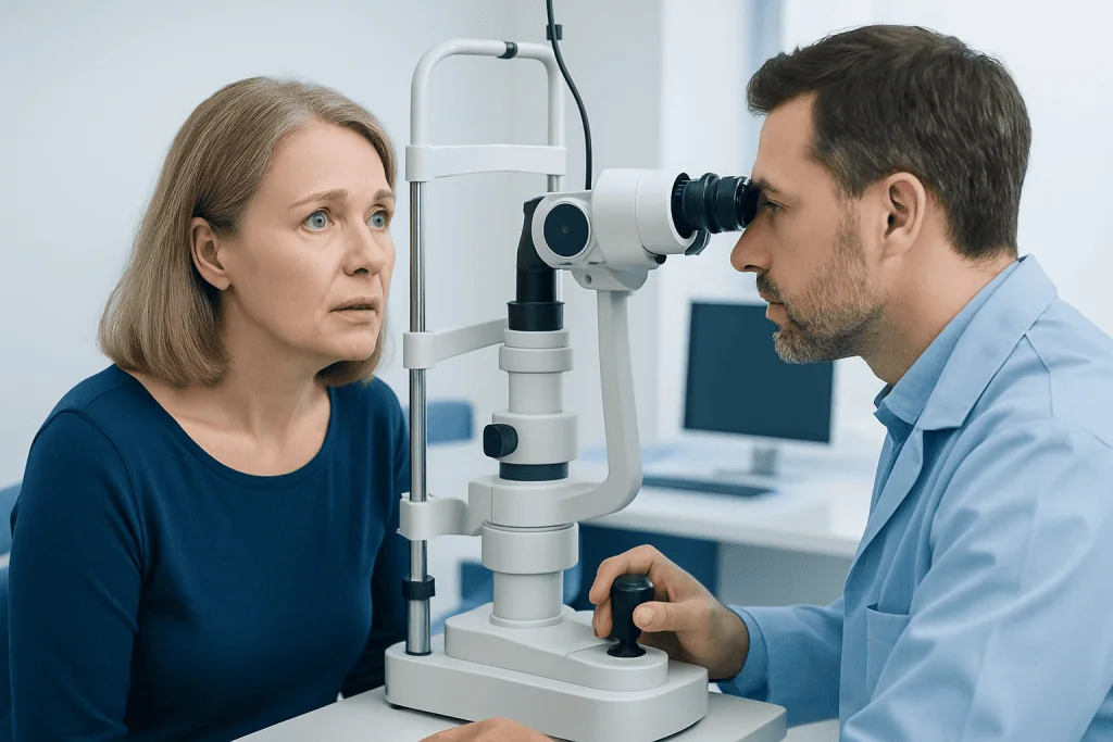
Posterior vitreous detachment is diagnosed with an eye examination, usually by an ophthalmologist. The most important part is a dilated retinal exam, which allows the doctor to look through the pupil and check whether the retina is intact. This helps distinguish a simple PVD from a retinal tear, bleeding, or detachment.
During the visit, the doctor asks when the symptoms started, whether they are getting worse, and whether there is any curtain-like shadow, vision loss, or eye pain. Visual acuity is checked, and the front and back of the eye are examined. In many cases, slit-lamp examination and indirect ophthalmoscopy are enough to make the diagnosis.
If the view of the retina is blocked by blood or dense floaters, the doctor may use ocular ultrasound to assess the back of the eye. Imaging is chosen based on the clinical situation rather than used routinely for every patient. The goal is to make sure symptoms are not due to a sight-threatening problem that needs timely treatment.
Because retinal tears may appear after the first symptoms begin, some patients are advised to return for follow-up even if the initial exam is reassuring. The timing depends on symptoms, findings, and individual risk factors. Patients are usually told what warning signs to watch for between visits.
Treatment options and what recovery is usually like
Most cases of posterior vitreous detachment do not need a procedure. Once a doctor confirms that there is no retinal tear or detachment, treatment usually focuses on observation and reassurance. Floaters often become less noticeable over time as the brain adapts and the debris settles lower in the eye.
If a retinal tear is found, treatment is aimed at sealing the tear before it progresses. This may be done with retinal laser treatment or other methods chosen by the ophthalmologist. If a true retinal detachment has already developed, surgery such as vitrectomy may be needed to protect vision.
Persistent floaters without retinal damage are usually monitored rather than removed, because invasive procedures can carry risks. In selected situations, specialists may discuss intervention if symptoms are severe and significantly affect daily life, but this decision is individualized. For many people, the safest and most effective plan is careful observation.
If symptoms are linked to another eye problem, treatment is directed at that cause. For example, patients with a cataract affecting vision may be evaluated for cataract surgery, while those with more complex findings may need assessment by a retina specialist. Near the end of the care pathway, some international patients choose centers such as Acibadem International, where multidisciplinary eye specialists in JCI-accredited hospitals diagnose and treat vitreoretinal conditions.
Self-care, daily precautions, and practical expectations
There is no proven home remedy that can reattach the vitreous or instantly remove floaters caused by posterior vitreous detachment. The most helpful step is following the eye specialist’s advice and attending any recommended follow-up visits. People can usually continue normal daily activities unless their doctor gives specific restrictions.
It may help to understand that floaters often become less distracting with time. Good lighting, regular eye care, and managing any existing eye conditions can support overall visual comfort. Protective eyewear during sports or hazardous work may reduce the risk of eye injury, which is important for long-term eye health in general.
People should avoid dismissing new symptoms just because they have been told PVD is common. A person who already has posterior vitreous detachment can still develop a retinal tear later, especially in the early period after symptoms begin. Keeping track of changes in one eye compared with the other can help when reporting symptoms to a doctor.
Routine eye examinations remain important, particularly for older adults and those with myopia, diabetes, or prior eye surgery. These visits do not prevent every vitreous change, but they can help identify retinal problems early. Prompt assessment is especially useful when symptoms are sudden rather than gradual.
When to seek medical care
New floaters or flashes should be assessed promptly, even if they seem mild. The reason is not that posterior vitreous detachment is always dangerous, but that its symptoms can overlap with retinal tears and detachments. Early evaluation gives the best chance to detect problems before they affect vision more seriously.
Urgent same-day or immediate care is especially important if there is a sudden shower of floaters, repeated flashes, blurred vision that is getting worse, or a dark curtain, veil, or shadow across any part of the visual field. These symptoms can suggest bleeding, a retinal tear, or a detachment rather than an uncomplicated PVD.
Eye pain is not a classic feature of simple posterior vitreous detachment. If pain, marked redness, severe headache, or nausea accompanies visual symptoms, another eye condition may be present and should also be assessed promptly. Patients should not drive themselves if vision is significantly impaired.
- Seek prompt eye evaluation for new floaters or new flashes
- Seek urgent care for a curtain-like shadow or sudden loss of part of the visual field
- Seek urgent care if floaters rapidly increase or vision becomes noticeably worse
- Follow the timing of recheck visits recommended after the first exam
Frequently asked questions
Is posterior vitreous detachment serious?
Posterior vitreous detachment is often a common age-related change and by itself may not threaten vision. It becomes more concerning because the same process can sometimes cause a retinal tear or detachment, which is why new symptoms should be checked promptly.
What does posterior vitreous detachment look like?
Many people notice floaters that look like spots, threads, cobwebs, or ring-shaped shadows drifting in vision. Some also notice brief flashes of light at the edge of vision, especially in dim settings.
Do floaters from posterior vitreous detachment go away?
Floaters may not disappear completely, but they often become much less noticeable over time. The brain adapts to them, and the vitreous debris may settle lower in the eye.
Can posterior vitreous detachment cause blindness?
A simple posterior vitreous detachment usually does not cause blindness. The main risk is if it leads to a retinal tear or detachment that is not treated promptly.
How is posterior vitreous detachment different from retinal detachment?
Posterior vitreous detachment means the gel inside the eye has separated from the retina. Retinal detachment means the retina itself has lifted away from the back of the eye, which is more urgent and can threaten vision.
Who is more likely to get posterior vitreous detachment?
It is more common with aging and in people who are nearsighted. It can also happen earlier after cataract surgery, after eye injury, or in people with a history of retinal problems.
References
- American Academy of Ophthalmology
- National Eye Institute
- American Society of Retina Specialists
- Mayo Clinic
- Royal College of Ophthalmologists
This article is for general information only and is not a substitute for professional medical advice. Please consult a qualified doctor about your individual situation.
Explore treatments in Turkey — costs, top hospitals & a free quote
JCI-accredited · board-certified surgeons · reply within 24h
Add us as a Preferred Source to see more of our trusted health content across Google Search, AI Overviews and Discover.