

Papilledema: What Patients Need to Know

Papilledema means optic nerve swelling due to increased intracranial pressure. It can cause headaches, brief visual changes, double vision, nausea, or sometimes no early symptoms.
Key Takeaways
- Papilledema means optic nerve swelling due to increased intracranial pressure.
- It can cause headaches, brief visual changes, double vision, nausea, or sometimes no early symptoms.
- Doctors confirm papilledema with an eye exam and usually investigate the cause with brain imaging and other tests.
- Treatment focuses on lowering pressure and treating the underlying condition.
- Prompt evaluation helps reduce the risk of permanent vision loss or complications from the cause of raised pressure.
Papilledema is swelling of the optic disc caused by increased pressure inside the skull. It is not a disease by itself, but an important sign that the brain and eyes need prompt medical assessment to identify the cause and protect vision.
Overview: what papilledema means
Papilledema is swelling at the point where the optic nerve enters the back of the eye, called the optic disc. This swelling happens because pressure inside the skull is higher than normal and that pressure is transmitted along the optic nerve. In simple terms, papilledema is usually a warning sign of increased pressure around the brain rather than a primary eye disease.
Some people learn about papilledema after they notice headaches or visual changes. Others are told about it during a routine eye examination, even before symptoms become obvious. Because the optic nerve carries visual information from the eye to the brain, ongoing swelling can affect vision if the pressure remains elevated.
Papilledema can be linked to several conditions, including brain tumors, bleeding, infections, blockages of normal cerebrospinal fluid flow, certain medicines, and idiopathic intracranial hypertension. Not every patient with a swollen optic disc has papilledema, so careful examination is important to distinguish it from other optic nerve disorders.
How papilledema can affect vision and daily life
The effects of papilledema vary from person to person. In the early stages, vision may still seem normal, which is one reason the condition can be overlooked. As pressure persists, patients may experience short episodes of blurred or dimmed vision, especially when standing up, coughing, or bending over.
Some people describe a feeling that their vision briefly “grays out” for a few seconds. Others notice difficulty focusing, reduced side vision, or double vision. These changes may come and go at first, but repeated episodes should not be ignored because they can signal strain on the optic nerve.
Headache and visual symptoms can interfere with reading, driving, work, and exercise. When papilledema is caused by a neurologic problem, other symptoms may also affect daily activities. Identifying the source of the pressure is the key step in protecting vision and overall health.
Symptoms and signs to watch for
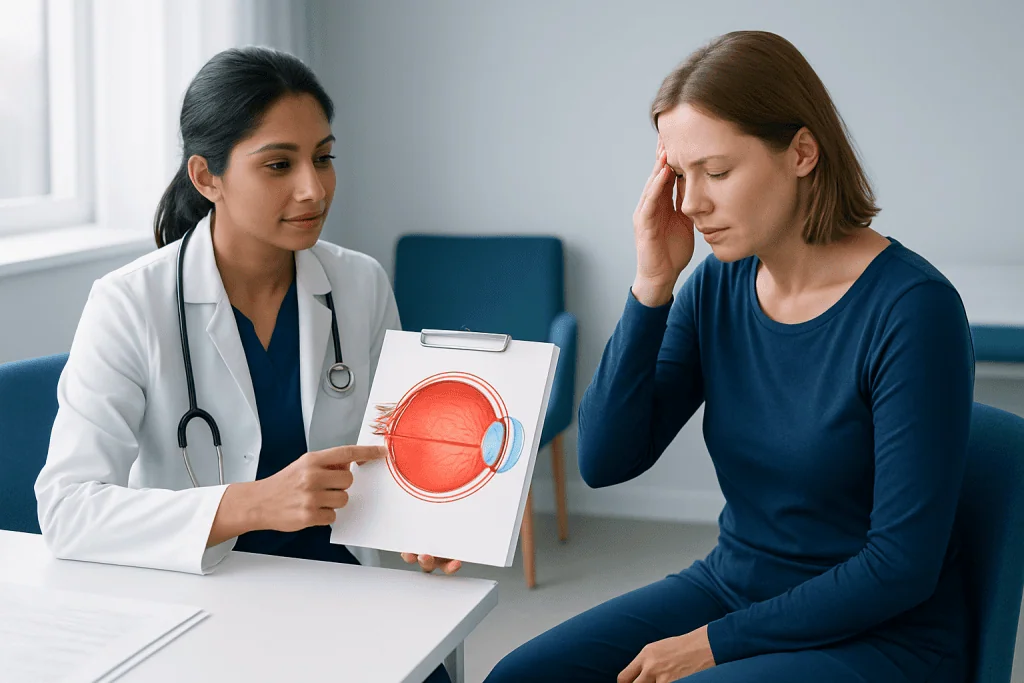
Papilledema itself may not always cause immediate symptoms, particularly early on. When symptoms do occur, they often reflect raised intracranial pressure or the condition causing it. A patient may have one symptom, several symptoms, or occasionally none at the beginning.
Common symptoms and signs include:
- Headache, often worse in the morning or with straining
- Brief episodes of blurred, dim, or blacked-out vision
- Double vision
- Nausea or vomiting
- Pulsing or “whooshing” sound in the ears
- Transient loss of peripheral vision
- Eye exam findings of swollen optic discs
In some cases, the symptoms relate more to the underlying cause than to the eye swelling itself. For example, fever and neck stiffness may suggest infection, while weakness, confusion, or seizures may point to a neurologic emergency. This is why papilledema should always be evaluated in a broader medical context.
Persistent or worsening visual symptoms deserve prompt attention. Vision loss from long-standing papilledema may become permanent if the pressure is not relieved in time.
Causes and risk factors
The direct cause of papilledema is increased pressure inside the skull, also called increased intracranial pressure. That pressure can build up for different reasons. The brain, blood vessels, and cerebrospinal fluid share a fixed space inside the skull, so any problem that adds volume or blocks normal fluid circulation may raise pressure.
Possible causes include brain masses, swelling after injury, bleeding in or around the brain, brain infections, inflammation, hydrocephalus, and blood clots in the veins that drain the brain. Papilledema may also occur with brain tumors or with idiopathic intracranial hypertension, a condition in which pressure is elevated without a mass being found on imaging.
Certain medicines have been associated with raised intracranial pressure in some patients. Examples may include excess vitamin A, some antibiotics, growth hormone, and steroid withdrawal. Risk factors for idiopathic intracranial hypertension may include obesity, recent weight gain, and being of childbearing age, although the condition can occur in other groups as well.
Because the list of causes ranges from relatively manageable to urgent, self-diagnosis is not reliable. A clinician needs to determine whether papilledema is present and what is driving the pressure increase.
How doctors diagnose papilledema
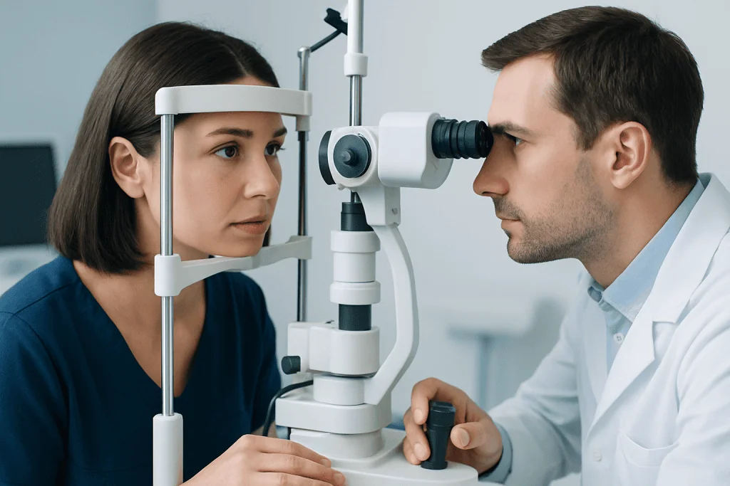
Diagnosis begins with a medical history and a detailed eye examination. An eye specialist looks at the optic disc through dilated pupils to assess whether true swelling is present. In some patients, special photographs or scans such as optical coherence tomography can help document the optic nerve appearance and monitor changes over time.
When papilledema is suspected, doctors usually arrange urgent evaluation to find the cause of raised pressure. This often includes brain imaging, such as MRI or a CT scan, to look for masses, bleeding, swelling, hydrocephalus, or venous problems. Depending on the clinical picture, additional imaging of the brain’s veins may also be needed.
After imaging rules out conditions that could make the procedure unsafe, a lumbar puncture may be performed to measure cerebrospinal fluid pressure and analyze the fluid. This test can help confirm increased pressure and may provide clues about infection or inflammation. Visual field testing is also commonly used to check whether side vision has been affected.
Diagnosis is often shared between ophthalmology, neurology, neuroradiology, and sometimes neurosurgery. In complex cases, a coordinated team approach helps ensure that both vision and the underlying neurologic problem are addressed.
Treatment options and follow-up care
Treatment for papilledema is aimed at the cause of raised intracranial pressure. There is no single treatment that fits every patient. Doctors first focus on whether the pressure increase is due to an emergency, such as a brain bleed, infection, hydrocephalus, or a mass that needs urgent management.
When papilledema is related to idiopathic intracranial hypertension, treatment may include weight management when appropriate, medicines that reduce cerebrospinal fluid production, and regular eye monitoring. If vision is worsening or pressure remains high despite medical treatment, procedures may be considered to protect vision or divert fluid. In selected cases, this may involve neurosurgery or other specialist interventions.
If imaging finds a treatable brain lesion, care is directed at that problem. A patient with hydrocephalus or a structural blockage may need procedures to restore fluid drainage, while someone with a clot in the brain’s venous system may need treatment tailored to that diagnosis. If papilledema is linked to a mass, options can include brain tumor surgery when appropriate.
Follow-up is a crucial part of care. Even after symptoms improve, repeat eye examinations and visual field tests help confirm that optic nerve swelling is resolving and that vision is stable. Near the end of the care pathway, some patients may be evaluated in multidisciplinary centers such as Acibadem International, where JCI-accredited hospitals and specialist teams assess and treat neurologic and eye conditions in international patients.
Prevention and self-care
Papilledema itself cannot always be prevented because it is a sign of another condition. Still, some general steps may lower risk in certain situations or support recovery after diagnosis. The most important self-care principle is to take symptoms seriously and follow the treatment plan closely.
Helpful measures may include keeping follow-up appointments, reporting any new visual symptoms quickly, and taking medicines exactly as prescribed. A patient should not stop or start medications linked to intracranial pressure without medical advice. If idiopathic intracranial hypertension is the cause, a doctor may recommend gradual, supervised weight reduction as part of treatment.
It can also help to keep a symptom diary that notes headaches, episodes of blurred vision, ringing in the ears, nausea, and triggers such as posture changes. This information may help the care team judge whether pressure is improving. During recovery, patients should ask their doctor about driving, strenuous exercise, and work duties if vision is affected.
When to seek medical care
Papilledema is a sign that warrants prompt medical evaluation, especially if it is newly diagnosed or suspected. The condition may be linked to serious causes of raised pressure, so waiting to see if it resolves on its own is not recommended.
A person should seek urgent medical care for severe or sudden headache, rapidly worsening vision, double vision, vomiting, confusion, weakness, seizure, fainting, fever with headache, or symptoms after head injury. These features can suggest a neurological emergency that needs immediate assessment.
Even without emergency symptoms, anyone told they may have papilledema on an eye exam should follow through with further testing as soon as advised. Early diagnosis and treatment offer the best chance of preserving vision and addressing the underlying cause safely.
Frequently asked questions
Is papilledema an eye disease?
Papilledema is not usually a primary eye disease. It is swelling of the optic disc caused by increased pressure inside the skull, so doctors look beyond the eye to find the reason for that pressure.
Can papilledema go away on its own?
Papilledema improves when the cause of increased intracranial pressure is treated effectively. It should not be assumed to resolve on its own, because ongoing pressure can damage the optic nerve and threaten vision.
Does papilledema always cause vision loss?
Not always. Some patients have normal vision at first or only brief episodes of blurred vision, but untreated papilledema can lead to lasting visual field loss or reduced vision over time.
How is papilledema different from optic neuritis?
Papilledema is optic disc swelling caused by raised pressure around the brain. Optic neuritis is inflammation of the optic nerve itself and often causes eye pain and more direct vision loss; the two conditions need different evaluation and treatment.
What tests are usually needed for papilledema?
Evaluation often includes a dilated eye exam, visual field testing, and brain imaging such as MRI or CT. After imaging, some patients also need a lumbar puncture to measure cerebrospinal fluid pressure and look for infection or inflammation.
Is papilledema an emergency?
Papilledema should be treated as an urgent medical finding because it may signal a serious problem causing raised intracranial pressure. Emergency care is especially important if there is severe headache, rapidly worsening vision, vomiting, confusion, weakness, or seizures.
References
- American Academy of Ophthalmology
- National Eye Institute
- National Institute of Neurological Disorders and Stroke
- Mayo Clinic
- Merck Manual
This article is for general information only and is not a substitute for professional medical advice. Please consult a qualified doctor about your individual situation.
Explore treatments in Turkey — costs, top hospitals & a free quote
JCI-accredited · board-certified surgeons · reply within 24h
Add us as a Preferred Source to see more of our trusted health content across Google Search, AI Overviews and Discover.

More from the Health Library



Related Specialists

Prof. Dr. Ufuk Topuz
Anesthesiology
Dr. Abdülkadir Oran
Otorhinolaryngology
Asst. Prof. Dr. Zeynep Tuğçe Sarıkaya
Intensive Care
Assoc. Prof. Dr. Halim Ulugöl
Anesthesiology