Keratoacanthoma: Symptoms, Causes, and Treatment Options

Keratoacanthoma usually appears as a fast-growing, round skin nodule with a central crust or keratin-filled core. It most often develops on sun-exposed skin, especially in older adults and people with significant ultraviolet exposure.
Key Takeaways
- Keratoacanthoma usually appears as a fast-growing, round skin nodule with a central crust or keratin-filled core.
- It most often develops on sun-exposed skin, especially in older adults and people with significant ultraviolet exposure.
- Because it can look very similar to squamous cell carcinoma, doctors often recommend biopsy or complete removal.
- Treatment commonly involves surgical removal, with other options considered in selected cases.
- Any new, rapidly enlarging skin lesion should be checked by a qualified dermatologist or surgeon.
Keratoacanthoma is a rapidly growing skin tumor that often appears as a dome-shaped bump with a central keratin plug. Although it may sometimes shrink on its own, it can closely resemble squamous cell carcinoma, so prompt medical assessment and treatment are usually recommended.
Overview
Keratoacanthoma is a skin tumor that usually grows quickly over a period of weeks and often appears as a firm, rounded bump with a central plug of keratin, the protein found in skin, hair, and nails. It is generally considered a low-grade tumor of the hair follicle unit, but in day-to-day practice it is treated carefully because it can be very difficult to distinguish from squamous cell carcinoma, a common form of skin cancer.
One reason keratoacanthoma causes uncertainty is its behavior. It may enlarge rapidly, reach a dome-shaped appearance, and in some cases begin to regress. Even so, doctors do not usually rely on spontaneous shrinkage because the lesion can be uncomfortable, leave a scar, and most importantly may actually represent or hide a cancerous process. For that reason, evaluation by a skin specialist is important.
Keratoacanthoma most often occurs on sun-exposed areas such as the face, forearms, hands, and lower legs. It is more common in middle-aged and older adults, especially those with long-term sun exposure. People may first notice it as what seems like a pimple, boil, or wart that grows unusually fast.
In many cases, diagnosis and treatment happen together, because removing the lesion allows a pathologist to examine it under the microscope. This helps confirm whether the growth is a true keratoacanthoma, a related lesion, or squamous cell carcinoma.
What keratoacanthoma looks and feels like
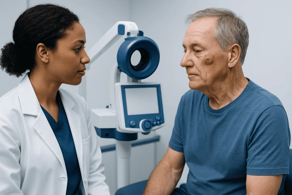
The typical keratoacanthoma begins as a small papule or bump that enlarges quickly over a few weeks. It often becomes a symmetrical, dome-shaped nodule with a central crater or plug filled with keratin. The surface may look shiny, flesh-colored, pink, red, or slightly brown, depending on skin tone and the degree of inflammation.
Many lesions are painless, but some can feel tender, irritated, or inflamed. If the surface cracks or rubs against clothing, it may bleed or develop a crust. The lesion is usually firm to the touch and may look quite dramatic compared with how recently it appeared.
Keratoacanthomas can occur as a single lesion or, less commonly, as multiple lesions. They are usually found on skin that receives regular sun exposure, but they can develop elsewhere as well. Because the appearance overlaps with several other skin conditions, a visual exam alone may not be enough for a definite diagnosis.
- Rapid growth over days to weeks
- Round or dome-shaped raised nodule
- Central keratin-filled plug or crater
- Possible redness, crusting, or minor bleeding
- Most often on the face, ears, scalp, hands, forearms, or legs
Causes and risk factors

The exact cause of keratoacanthoma is not fully understood, but several risk factors are well recognized. Chronic ultraviolet exposure is one of the strongest associations, which helps explain why these lesions commonly appear on sun-exposed areas. Damage from sunlight over many years may alter skin cells in ways that contribute to the development of this type of tumor.
Age is another important factor. Keratoacanthoma is seen more often in older adults, likely because the skin has accumulated more environmental damage over time. Fair skin, a history of frequent sunburns, and outdoor work or hobbies may increase risk. Some lesions have also been linked to prior skin injury, irritation, or exposure to certain chemical carcinogens.
In some people, immune system changes may play a role. Keratoacanthoma can be more likely in individuals who are immunosuppressed, such as transplant recipients or those taking long-term immune-modifying medicines. Rare inherited syndromes can lead to multiple keratoacanthomas, though this is uncommon.
Doctors also consider whether the lesion could instead be another condition with a similar appearance. A rapidly growing nodule may overlap clinically with skin cancer or benign lesions such as warts, inflamed cysts, or certain follicular tumors. This is why a specialist assessment is so important.
How doctors diagnose keratoacanthoma
Diagnosis starts with a careful history and skin examination. A doctor will ask when the lesion first appeared, how quickly it grew, whether it has bled or become painful, and whether there is a history of excessive sun exposure, previous skin cancers, or immunosuppression. The speed of growth is often a major clue.
Even when a lesion strongly suggests keratoacanthoma, the diagnosis usually needs confirmation with a biopsy or excision. This is because keratoacanthoma and squamous cell carcinoma can look very similar both clinically and, at times, microscopically. Pathology helps determine the nature of the lesion and whether the margins are clear if it has been completely removed.
Depending on the lesion’s size and location, the doctor may perform a shave biopsy, punch biopsy, or complete excision. In many cases, complete removal is preferred because it treats the lesion while also providing enough tissue for accurate diagnosis. Dermoscopy, a close-up examination of the skin using a special handheld device, may support assessment but does not replace pathology.
If there are many lesions, unusual features, or concern about deeper tissue involvement, the care team may investigate further. Specialists in dermatology and skin surgery can help guide the most appropriate diagnostic approach for each patient.
Treatment options
Treatment for keratoacanthoma is usually active rather than watchful waiting. Although some lesions may regress over time, doctors generally recommend removal because of the difficulty of excluding squamous cell carcinoma and the possibility of local tissue damage or scarring if the lesion is left untreated.
Surgical excision is a common treatment and often the preferred option. It allows the entire lesion to be removed and examined under the microscope. In some situations, especially when tissue preservation is important on the face or other sensitive areas, a specialist may recommend Mohs surgery to remove the growth with careful margin control.
Other treatment approaches may be considered in selected cases, such as curettage with electrosurgery, cryotherapy, or intralesional medications. These options depend on the lesion’s size, location, number, and the patient’s overall health. They may be helpful when surgery is less suitable, but pathology confirmation remains important.
After treatment, the site is monitored for healing, recurrence, and the appearance of new lesions. People who develop keratoacanthoma may also benefit from regular skin checks, since sun-damaged skin can produce other concerning lesions over time. If reconstruction is needed after removal, skin cancer surgery teams may help restore both function and appearance.
Prevention and self-care
There is no guaranteed way to prevent keratoacanthoma, but protecting the skin from ultraviolet radiation can lower the risk of many sun-related skin lesions. Daily sun protection is especially important for people with fair skin, a history of outdoor exposure, or previous skin tumors.
Practical steps include using broad-spectrum sunscreen, wearing wide-brimmed hats and protective clothing, and seeking shade during the strongest midday sun. Avoiding tanning beds is also important, as artificial ultraviolet exposure can damage the skin in similar ways. Consistent habits matter more than occasional intense efforts.
Regular self-examination can help people notice suspicious changes earlier. A new bump that grows quickly, bleeds, forms a crust, or develops a central plug should not be ignored. Taking a clear photo with the date can help track changes between the time it is first noticed and the medical visit.
It is also wise not to pick, squeeze, or try to remove a lesion at home. Home treatment can delay diagnosis, increase irritation, and make it harder for the doctor to assess the lesion accurately. If the area is bothersome, covering it lightly and arranging a skin evaluation is the safest step.
When to seek medical care
A person should seek medical care for any skin lesion that appears suddenly and grows quickly, especially on sun-exposed skin. Keratoacanthoma can resemble skin cancer, so prompt assessment is the safest approach even if the bump is not painful. Early evaluation often makes treatment simpler and helps clarify the diagnosis without delay.
Medical attention is particularly important if the lesion bleeds, crusts repeatedly, becomes painful, changes shape, or does not heal. People with a history of skin cancer, organ transplantation, significant sun damage, or a weakened immune system should be especially cautious about new or changing skin growths.
In practice, patients may be assessed by dermatologists, plastic surgeons, or other specialists experienced in skin tumors. Near the end of the care pathway, a multidisciplinary team may help coordinate diagnosis, treatment, and follow-up. Acibadem International’s multidisciplinary specialists and JCI-accredited hospitals diagnose and treat skin lesions, including suspicious fast-growing tumors, for international patients.
If a lesion is large, located in a cosmetically sensitive area, or likely to need specialized removal and closure, referral for plastic and reconstructive surgery may be considered alongside dermatologic care.
Frequently asked questions
Is keratoacanthoma cancer?
Keratoacanthoma is often described as a low-grade skin tumor, but it is closely related to and can strongly resemble squamous cell carcinoma. Because it can be hard to tell the difference based on appearance alone, doctors usually recommend biopsy or removal for accurate diagnosis.
Can keratoacanthoma go away on its own?
Some keratoacanthomas may shrink or regress over time. However, waiting is usually not advised because the lesion can mimic skin cancer and may continue to grow, ulcerate, or leave a scar.
What does keratoacanthoma look like?
It often looks like a round, dome-shaped bump that grows quickly and develops a central crater or keratin plug. The lesion may be pink, red, flesh-colored, or crusted, and it commonly appears on sun-exposed skin.
Who is more likely to develop keratoacanthoma?
It is more common in older adults, especially those with long-term sun exposure or fair skin. Risk may also be higher in people with weakened immune systems, previous skin cancers, or significant sun-damaged skin.
How is keratoacanthoma treated?
Treatment usually involves complete removal of the lesion so it can be examined under the microscope. Depending on the case, options may include surgical excision, Mohs surgery, curettage, cryotherapy, or other specialist-directed treatments.
Is keratoacanthoma painful?
Many lesions are painless, especially early on. Some can become tender, inflamed, or bleed if they are irritated, cracked, or rubbed by clothing.
When should someone worry about a fast-growing skin bump?
Any new skin bump that enlarges quickly over days to weeks should be checked by a doctor. Prompt evaluation is especially important if it bleeds, crusts, hurts, changes shape, or appears on sun-exposed skin.
References
- American Academy of Dermatology
- National Cancer Institute
- British Association of Dermatologists
- DermNet
- National Comprehensive Cancer Network
This article is for general information only and is not a substitute for professional medical advice. Please consult a qualified doctor about your individual situation.
Hair transplant in Turkey — costs, graft counts & a free assessment
JCI-accredited · board-certified surgeons · reply within 24h
Add us as a Preferred Source to see more of our trusted health content across Google Search, AI Overviews and Discover.