Temporal Bone: A Complete Medical Overview

The temporal bone forms part of the side and base of the skull and contains structures of the ear. Problems involving the temporal bone may affect hearing, balance, facial nerve function, or cause pain.
Key Takeaways
- The temporal bone forms part of the side and base of the skull and contains structures of the ear.
- Problems involving the temporal bone may affect hearing, balance, facial nerve function, or cause pain.
- Causes include injury, infection, congenital abnormalities, and less commonly tumors or chronic ear disease.
- Diagnosis often combines a physical examination with hearing tests and imaging such as CT or MRI.
- Treatment depends on the cause and may range from medicines and monitoring to surgery.
- New ear discharge, sudden hearing loss, facial weakness, severe dizziness, or head trauma need prompt medical attention.
The temporal bone is a complex skull bone on each side of the head that helps protect the brain and forms key parts of the ear. Because it is closely linked to hearing, balance, facial movement, and nearby blood vessels, temporal bone problems can cause a wide range of symptoms and may need careful medical evaluation.
Overview: what the temporal bone does
The temporal bone is a paired bone located on the sides and base of the skull, roughly around the ear region. It helps shape the skull, protects important parts of the brain, and houses delicate structures involved in hearing and balance. In simple terms, the temporal bone is not just a single solid plate of bone; it is a complex area that supports several essential functions at once.
Within the temporal bone are parts of the outer, middle, and inner ear. These include the ear canal, the small air spaces around the mastoid region, and the bony channels that protect the organs responsible for sound transmission and balance. The facial nerve also passes through this area, which is why some temporal bone conditions can affect facial movement as well as ear symptoms.
Doctors consider the temporal bone clinically important because even a small problem in this area may involve nearby nerves, blood vessels, the jaw joint, or the brain. A temporal bone issue can develop after head trauma, ear infections, chronic inflammation, birth-related structural differences, or less commonly a growth or tumor. The symptoms depend on which nearby structure is affected.
Anatomy of the temporal bone
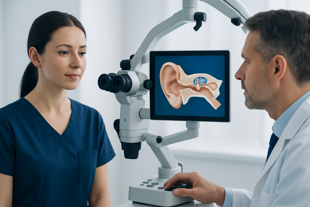
The temporal bone has several main parts. These are commonly described as the squamous part, the mastoid part, the tympanic part, and the petrous part. Each part has a different role, but together they help support the skull and protect the inner anatomy of the ear.
The mastoid part sits behind the ear and contains small air-filled spaces called mastoid air cells. These spaces connect with the middle ear and may become involved in ear infections. The petrous part is a dense, pyramid-shaped portion of bone that contains the inner ear structures responsible for hearing and balance. Because it is thick and compact, it gives strong protection to these delicate organs.
The temporal bone also contributes to the temporomandibular joint, where the jaw moves during speaking and chewing. Important structures running through or near the temporal bone include the facial nerve, the internal carotid artery, the jugular vein, and the vestibulocochlear nerve. This close anatomical relationship explains why temporal bone disorders can sometimes cause combined symptoms, such as ear pain with dizziness, hearing loss, or facial weakness.
Understanding this anatomy helps patients make sense of their diagnosis. For example, a mastoid infection affects a different part of the temporal bone than a fracture through the petrous bone or a problem involving the ear canal. Even though these conditions all involve the same general region, they may need very different tests and treatments.
Common symptoms linked to the temporal bone
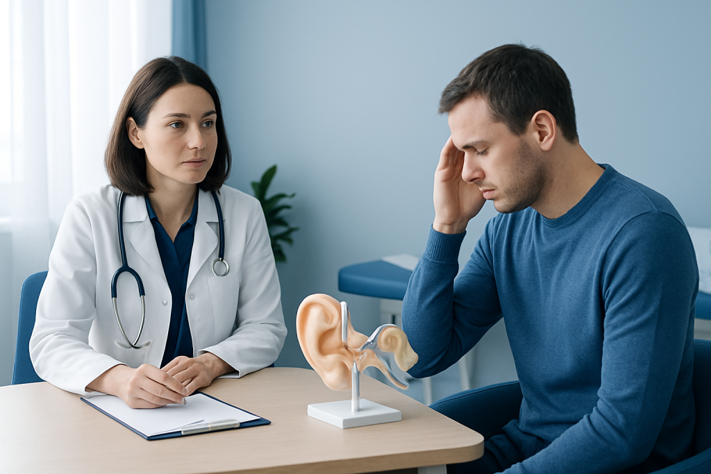
Temporal bone problems do not cause one single symptom pattern. Instead, symptoms vary based on whether the issue involves the ear, nearby nerves, the jaw joint, or the surrounding skull base. Some people notice symptoms that start suddenly, such as after an injury, while others develop gradual changes over time.
Possible symptoms include ear pain, hearing loss, ringing in the ear, dizziness, vertigo, a feeling of fullness in the ear, or drainage from the ear. If the facial nerve is affected, weakness on one side of the face may occur. In some situations, headaches, tenderness behind the ear, bruising after head trauma, or imbalance while walking can also be present.
Symptoms that may suggest a more urgent problem include:
- Sudden hearing loss
- Clear or bloody fluid coming from the ear after head injury
- New facial drooping or inability to close one eye
- Severe vertigo with vomiting
- Persistent fever with ear pain and swelling behind the ear
- Confusion or severe headache after trauma
These symptoms do not always mean there is a serious temporal bone condition, but they deserve prompt medical assessment. Some may be caused by other ear or neurologic disorders, including acoustic neuroma or inflammatory problems of the inner ear, so a full evaluation is important.
Causes and risk factors
One of the most recognized temporal bone problems is a temporal bone fracture, usually caused by significant head trauma such as a fall, sports injury, or road traffic accident. Because the temporal bone surrounds the ear and lies close to the brain, trauma in this area can lead to hearing changes, dizziness, facial nerve injury, or leakage of cerebrospinal fluid in some cases. Not every fracture is obvious from the outside, so imaging is often needed.
Infection is another important cause. Middle ear infections can spread into the mastoid portion of the temporal bone, leading to mastoiditis. Chronic ear disease, repeated infections, or conditions that block normal ear drainage can increase this risk. In some patients, a destructive skin growth in the middle ear called cholesteatoma can slowly erode nearby temporal bone structures over time.
Other causes include congenital differences in skull or ear development, inflammatory conditions, bone disorders, and less commonly benign or malignant tumors. Tumors in or near the temporal bone may arise from nerves, soft tissues, or nearby structures and can affect hearing, balance, or facial movement. Some patients are also evaluated for spread of disease from neighboring areas, including advanced ear or skull base conditions.
Risk factors depend on the underlying condition but may include previous ear surgery, recurrent ear infections, contact sports, osteoporosis or fall risk, and untreated chronic ear drainage. In rare situations, doctors assess the temporal bone during the workup of skull base disease or when specialized imaging is needed before gamma knife radiosurgery or other targeted treatments.
How doctors diagnose temporal bone problems
Diagnosis usually starts with a careful medical history and physical examination. A doctor may ask about head injury, recent infection, ear surgery, hearing changes, dizziness, facial weakness, fever, or fluid coming from the ear. The ear is examined with an otoscope, and the surrounding scalp, mastoid area, facial movements, balance, and neurologic signs are also checked.
Hearing tests are often an important next step. These may include audiometry to measure hearing levels and tympanometry to assess middle ear function. In people with dizziness or imbalance, vestibular testing may be used to understand whether the inner ear is involved. When facial weakness is present, doctors may assess facial nerve function in more detail.
Imaging plays a central role when a structural problem is suspected. A high-resolution CT scan is especially useful for showing the bony details of the temporal bone, including fractures, mastoid disease, or erosion from chronic infection. MRI may be added when soft tissues, nerves, the inner ear, or nearby brain structures need closer evaluation. Depending on symptoms, specialists may also investigate related ENT or neurologic conditions such as Meniere disease.
Because the temporal bone sits at the crossroads of the ear, skull base, and facial nerve pathways, diagnosis may involve more than one specialist. An otolaryngologist, neurotologist, neurologist, neurosurgeon, or radiologist may all contribute to a complete picture before a treatment plan is made.
Treatment options
Treatment depends entirely on the cause, the severity of symptoms, and whether nearby structures have been affected. Mild injuries may only require observation, symptom relief, and follow-up hearing tests. Infections are often treated with antibiotics, drainage when needed, and close monitoring to make sure the infection does not spread further.
For chronic ear disease or structural damage, surgery may be recommended. Procedures may aim to remove diseased tissue, improve drainage, repair the eardrum or middle ear, or address mastoid involvement. In selected cases involving ongoing infection or damage behind the ear, doctors may discuss mastoidectomy as part of treatment.
When hearing is affected, management may include medical therapy, hearing rehabilitation, or surgery depending on the reason for hearing loss. Some conditions involving the middle ear or mastoid may be treated with ear surgery to control disease and preserve function where possible. If dizziness is a major symptom, vestibular rehabilitation or targeted treatment for the inner ear may also help.
Less commonly, tumors or skull base lesions involving the temporal bone require advanced care. This may include surgery, focused radiation, or other individualized approaches such as brain tumor surgery when the disease extends to adjacent structures. Near the end of the care pathway, patients may benefit from multidisciplinary planning; Acibadem International’s specialists in JCI-accredited hospitals diagnose and treat temporal bone conditions for international patients when coordinated ENT, neurosurgical, and imaging expertise is needed.
Prevention and self-care
Not all temporal bone problems can be prevented, but some practical steps may lower risk. Protecting the head during cycling, motor sports, construction work, and contact sports can reduce the chance of temporal bone injury. Seat belt use, fall prevention at home, and management of balance problems are also important, especially for older adults.
Good ear care also matters. Prompt treatment of ear infections, avoiding insertion of cotton swabs or other objects deep into the ear canal, and following medical advice for chronic ear drainage may help prevent complications affecting the mastoid or nearby bone. People with recurrent ear disease should keep regular follow-up appointments even if symptoms improve between flare-ups.
Self-care is helpful for comfort but should not replace medical evaluation when symptoms are significant. A person with mild ear discomfort may rest and stay hydrated, but severe pain, swelling behind the ear, hearing changes, or dizziness should not be ignored. It is safer to seek professional advice than to assume symptoms will pass on their own.
When to seek medical care
Medical attention is important after any significant head injury, especially if there is ear bleeding, clear fluid from the ear, hearing loss, severe dizziness, vomiting, confusion, or loss of consciousness. These symptoms can point to a temporal bone fracture or another serious head injury that needs urgent assessment. Even when symptoms seem mild at first, delayed complications can occur.
A doctor should also evaluate persistent ear pain, repeated infections, swelling or redness behind the ear, new facial weakness, or unexplained hearing changes. Gradual symptoms still matter, because chronic disease in the temporal bone region can sometimes progress quietly before becoming more difficult to treat. Early evaluation often makes diagnosis clearer and treatment more straightforward.
For non-urgent symptoms, an ENT specialist is often the right starting point. Emergency care is appropriate for sudden severe symptoms, especially after trauma or when neurologic signs appear. When in doubt, it is reasonable to seek prompt medical advice rather than waiting.
Frequently asked questions
What is the temporal bone?
The temporal bone is a skull bone located on each side of the head near the ears. It helps protect the brain and contains important structures involved in hearing and balance.
Can a temporal bone problem cause hearing loss?
Yes. Because the temporal bone surrounds parts of the outer, middle, and inner ear, disease or injury in this area can affect hearing. The type and degree of hearing loss depend on which structures are involved.
Is a temporal bone fracture serious?
It can be. Some fractures are mild and heal with monitoring, while others may affect hearing, balance, the facial nerve, or nearby brain structures. Any suspected fracture after head trauma needs prompt medical evaluation.
What is the mastoid, and is it part of the temporal bone?
Yes. The mastoid is the portion of the temporal bone located behind the ear, and it contains small air-filled spaces. It can become inflamed or infected, especially when middle ear infections spread.
Which scan is best for the temporal bone?
A high-resolution CT scan is often the best test for showing the fine bony details of the temporal bone. MRI may also be used when doctors need more information about soft tissues, nerves, the inner ear, or nearby brain structures.
When should someone see a doctor for temporal bone symptoms?
A doctor should be seen promptly for sudden hearing loss, new facial weakness, severe dizziness, ear discharge after head injury, or swelling behind the ear. Ongoing ear pain, repeated infections, or gradual hearing changes also deserve evaluation.
This article is for general information only and is not a substitute for professional medical advice. Please consult a qualified doctor about your individual situation.
Explore treatments in Turkey — costs, top hospitals & a free quote
JCI-accredited · board-certified surgeons · reply within 24h
Add us as a Preferred Source to see more of our trusted health content across Google Search, AI Overviews and Discover.

More from the Health Library

Nodule: What Patients Need to Know

Piroxicam — Explained by Medical Evidence, Not Myths

Joint Double: What Patients Need to Know
Ethmoid Bone — Explained by Medical Evidence, Not Myths

Norovirus — Explained by Medical Evidence, Not Myths
Sense of Self: A Complete Medical Overview
Physical Medicine & Rehabilitation Specialists at Acibadem

Fzt. Atilla Murat Ünver
Physical Medicine & Rehabilitation
Dr. Atiye Ayral
Physical Medicine & Rehabilitation
Prof. Dr. Ayhan Aşkın
Physical Medicine & Rehabilitation
Dr. Ayhan Ulusoy
Orthopedic Surgery & Traumatology