Real Pictures of Breast Cancer Lumps Explained: Causes, Management, and When to See a Doctor
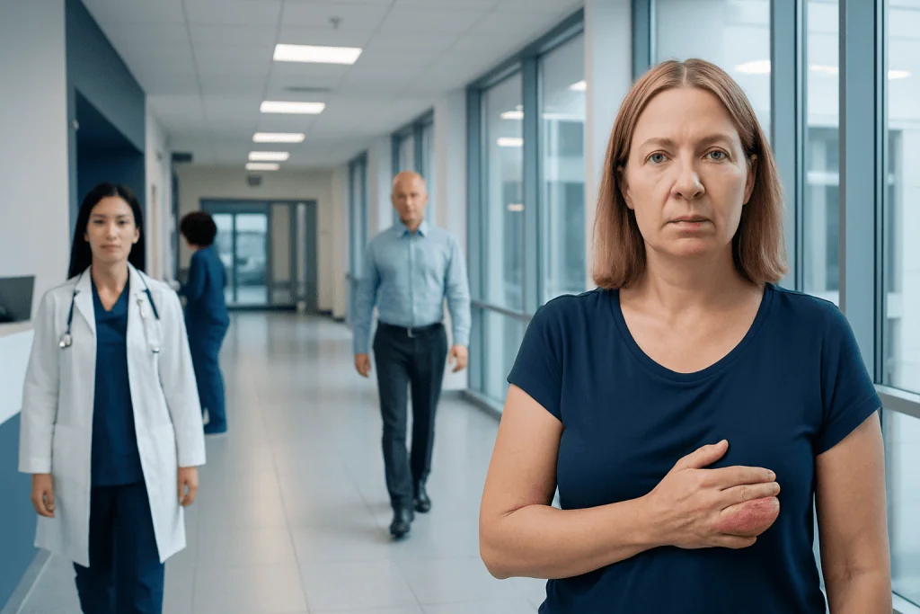
Real pictures of breast cancer lumps are only examples; breast changes vary widely from person to person. A breast cancer lump may feel hard, fixed, irregular, or painless, but some cancers do not form a classic lump at all.
Key Takeaways
- Real pictures of breast cancer lumps are only examples; breast changes vary widely from person to person.
- A breast cancer lump may feel hard, fixed, irregular, or painless, but some cancers do not form a classic lump at all.
- Many breast lumps are not cancer and may be caused by cysts, fibroadenomas, infections, or normal hormonal changes.
- Clinical breast examination, mammography, ultrasound, and sometimes biopsy are used to confirm the cause of a lump.
- Prompt medical assessment is important if a lump is new, growing, persistent, or associated with skin or nipple changes.
Real pictures of breast cancer lumps may help people recognize changes that deserve attention, but appearance alone cannot diagnose cancer. A new breast lump, skin change, or nipple change should be assessed by a qualified clinician, because many breast lumps are benign and imaging is usually needed to tell the difference.
Overview: What real pictures of breast cancer lumps can and cannot show
People often search for real pictures of breast cancer lumps because they want a clear visual answer. Real images can be useful for understanding that breast cancer does not always look the same, but they have important limits. Some cancers cause a visible lump or skin change, while others are only found on screening mammograms and cannot be recognized from an external photo.
Breast cancer lumps may appear as a localized bulge, skin dimpling, nipple inversion, redness, thickened skin, or a change in breast shape. However, these changes are not specific to cancer. Benign conditions such as cysts, inflammation, scar tissue, or fibrocystic changes can sometimes look or feel concerning as well.
The safest way to interpret a breast change is to think of pictures as examples, not proof. A photograph cannot show whether a lump is soft or hard, mobile or fixed, tender or painless, or whether there are changes on mammogram or ultrasound. That is why doctors rely on a combination of symptoms, physical examination, imaging, and sometimes biopsy.
For patients trying to make sense of a new finding, the key message is reassuring but practical: a visible or palpable change deserves attention, yet many breast lumps are not cancer. Early evaluation helps clarify what is happening and, if needed, supports timely treatment of breast cancer.
How a breast cancer lump may look and feel
There is no single “typical” appearance of a breast cancer lump. In many cases, a cancerous lump is described as hard, irregular in shape, and less movable than the surrounding tissue. It is often painless, but pain can still occur, so discomfort does not rule cancer in or out.
On the outside of the breast, some people notice a new contour change rather than a distinct round lump. The skin may look pulled inward, puckered, or dimpled, sometimes compared to the texture of an orange peel. The nipple may flatten, turn inward, or develop crusting or unusual discharge, especially if the change is new.
Some breast cancers do not present as a clearly felt lump. Instead, they may cause thickening in one area, persistent swelling, or a feeling that one part of the breast is different from the rest. Inflammatory breast cancer, for example, may cause redness, warmth, heaviness, and skin thickening rather than a discrete mass.
It also helps to know what a photo cannot capture. A clinician will want to know whether the change appeared suddenly or gradually, whether it changes with the menstrual cycle, and whether there are associated symptoms in the armpit such as swollen lymph nodes. These details often guide the next steps in assessment.
Other causes of breast lumps and breast changes

A new lump can be worrying, but many breast changes have non-cancerous explanations. Common benign causes include simple cysts, fibroadenomas, areas of dense breast tissue, fat necrosis after injury, and fibrocystic changes related to hormones. Infections, especially during breastfeeding, may also cause tender lumps, warmth, redness, or swelling.
Age, menstrual status, and personal history matter. Younger women are more likely to have benign causes such as fibroadenomas or cysts, although cancer can still occur at any adult age. After menopause, a new persistent lump is more likely to need prompt investigation because normal hormonal fluctuation is less often the explanation.
Not every abnormality is a lump. Some people notice nipple discharge, scaling of the nipple, a new asymmetry in breast size, or enlarged lymph nodes in the underarm. These findings can be linked to benign conditions, but they also sometimes accompany cancer and should not be ignored.
Because overlap is common, guessing based on feel or appearance alone is unreliable. A smooth lump may still need imaging, and a painful change may still deserve evaluation. This is one reason breast specialists often combine examination with breast ultrasound or other imaging tests rather than depending on symptoms alone.
Risk factors and warning signs that deserve attention
Anyone can develop breast cancer, including people without a known family history, but certain factors can raise risk. These include increasing age, inherited gene mutations such as BRCA1 or BRCA2, a strong family history of breast or ovarian cancer, prior chest radiation, certain previous breast biopsy findings, and some reproductive or hormonal factors. Lifestyle factors such as alcohol use, obesity after menopause, and physical inactivity may also contribute to risk over time.
Even so, risk factors do not diagnose cancer, and their absence does not exclude it. A person with no known risk factors can still develop a suspicious lump. For this reason, breast symptoms should be judged on their own merit rather than dismissed because someone feels otherwise healthy.
Warning signs that usually justify prompt assessment include a new lump that lasts beyond one menstrual cycle, a lump that is growing, skin dimpling, nipple inversion that is new, bloody or spontaneous nipple discharge, persistent redness, swelling of part or all of the breast, or enlarged underarm nodes. Changes that affect only one breast are often more concerning than generalized tenderness affecting both sides.
Doctors also pay attention to symptoms that seem subtle but persistent, such as one area that repeatedly feels thicker or firmer than before. The goal is not to create alarm, but to recognize patterns that should not be watched indefinitely without medical input.
How doctors diagnose a suspicious breast lump
Assessment usually begins with a medical history and clinical breast examination. The doctor may ask when the lump was first noticed, whether it changes during the menstrual cycle, whether there is pain or discharge, and whether there is a personal or family history of breast disease. This helps identify the most likely causes and the most appropriate tests.
Imaging is often the next step. Depending on age and the clinical situation, doctors may use mammography and breast ultrasound to learn whether the area is solid or fluid-filled and whether it has features that appear benign or suspicious. In selected cases, breast MRI may be recommended for additional detail.
If imaging shows an area that cannot be confidently classified as benign, a biopsy may be advised. A needle biopsy removes a small sample of tissue so it can be examined under a microscope. This is the only way to confirm whether cells are cancerous and, if so, what type of breast cancer is present.
Diagnosis should be individualized. Some clearly benign findings can be safely monitored, while others need tissue sampling without delay. If a person has already been diagnosed and is learning about treatment planning, related information on breast cancer treatment can help explain the broader care pathway.
Treatment and management if a lump is benign or cancerous
Management depends entirely on the cause. Benign cysts may simply be monitored if they are not causing symptoms, while a painful or large cyst may be drained. Fibroadenomas can sometimes be observed with follow-up imaging, especially if they have classic benign features and remain stable.
If infection is the cause, treatment may include antibiotics and supportive care, and any abscess may need drainage. Hormonal or fibrocystic changes may improve with time and do not always need intervention beyond symptom management and routine follow-up. The main aim is to match treatment to the confirmed diagnosis rather than to the fear associated with the word “lump.”
If breast cancer is diagnosed, treatment may involve surgery, radiation therapy, systemic treatments such as hormone therapy, chemotherapy, targeted therapy, or a combination of these. The plan depends on the cancer type, stage, receptor status, overall health, and patient preferences. Many people benefit from coordinated care involving breast surgeons, radiologists, pathologists, and medical and radiation oncologists.
Near the end of the diagnostic process, patients may also seek care at specialized centers. Acibadem International’s multidisciplinary specialists and JCI-accredited hospitals diagnose and treat breast conditions for international patients, with care plans tailored to the individual clinical picture.
Breast self-awareness, screening, and practical self-care
Rather than relying only on formal self-exams, many experts encourage breast self-awareness. This means becoming familiar with what is usual for the breasts in everyday life so that a new lump, thickening, visible change, or nipple symptom is easier to notice. It is not necessary to examine the breasts in an anxious or overly frequent way, but consistent awareness can be helpful.
Routine screening is also important because some breast cancers do not cause symptoms. Screening recommendations vary by age and risk level, so patients should discuss mammography timing with their clinician. People at higher risk may need earlier or additional imaging based on family history, genetic findings, or previous breast conditions.
General self-care supports breast health but does not replace medical evaluation. Practical steps include maintaining a healthy weight, staying physically active, limiting alcohol, and attending recommended screening appointments. Keeping a simple note of when a change was first noticed can also help during a medical consultation.
If a lump seems to come and go with the menstrual cycle, it may still be reasonable to seek advice if it persists, enlarges, or is associated with other changes. Waiting for a short interval may be appropriate in some situations, but long delays are best avoided when a breast finding is new or clearly different from baseline.
When to seek medical care
Medical care should be sought if a person notices a new breast lump, an area of persistent thickening, skin dimpling, nipple inversion, nipple discharge, unexplained swelling, or enlarged lymph nodes in the armpit. These changes do not automatically mean cancer, but they are important enough to justify professional assessment.
Urgent evaluation is especially sensible when a lump feels hard, is growing, remains present after a menstrual cycle, or is associated with redness, warmth, or significant breast shape change. People who are pregnant, breastfeeding, postmenopausal, or at elevated risk for breast cancer should also be cautious about new symptoms and seek advice promptly.
Anyone with fever, severe pain, or rapidly worsening redness may need urgent assessment to look for infection or abscess. For all other concerning changes, arranging a timely appointment with a primary care doctor, gynecologist, or breast specialist is a practical next step. If there is uncertainty, it is safer to ask than to wait and wonder.
In short, real pictures of breast cancer lumps can raise awareness, but they should not be used to self-diagnose or self-reassure. A qualified clinician can determine whether a change needs monitoring, imaging, or biopsy and can guide the next step with clarity.
Frequently asked questions
Can real pictures of breast cancer lumps confirm whether my lump is cancer?
No. Real pictures can show examples of concerning breast changes, but they cannot confirm whether a lump is cancerous. Diagnosis usually requires a clinical examination and imaging, and some cases also need a biopsy.
What does a breast cancer lump usually feel like?
A breast cancer lump is often described as hard, irregular, and less movable than nearby tissue. However, not all cancers feel the same, and some cause thickening or skin changes rather than a distinct lump.
Are painful breast lumps usually cancer?
Painful breast lumps are often caused by benign conditions such as cysts, hormonal changes, or infection. Even so, pain does not completely rule out cancer, so persistent or unusual pain with a lump should be checked.
Can breast cancer be present without a visible lump?
Yes. Some breast cancers are found on screening mammograms before they can be seen or felt. Others may cause skin dimpling, swelling, nipple changes, or underarm lymph node enlargement instead of a clear lump.
How long should someone wait before seeing a doctor about a breast lump?
A new lump should not be ignored. If it persists beyond one menstrual cycle, grows, or comes with skin or nipple changes, it is sensible to arrange medical evaluation promptly.
What tests are commonly used to check a suspicious breast lump?
Doctors commonly use a clinical breast exam, mammography, and ultrasound. If the imaging findings are uncertain or suspicious, a needle biopsy may be recommended to confirm the diagnosis.
References
- World Health Organization
- American Cancer Society
- National Cancer Institute
- Centers for Disease Control and Prevention
- American College of Radiology
This article is for general information only and is not a substitute for professional medical advice. Please consult a qualified doctor about your individual situation.
Breast Cancer in Turkey — costs, top hospitals & a free quote
JCI-accredited · board-certified surgeons · reply within 24h
Add us as a Preferred Source to see more of our trusted health content across Google Search, AI Overviews and Discover.

Medical Oncology Department
Medical treatment of cancer with chemotherapy, immunotherapy and targeted therapies under a multidisciplinary tumor board.
60 specialists in this unit