Vq Scan: An Evidence-Based Guide for Patients
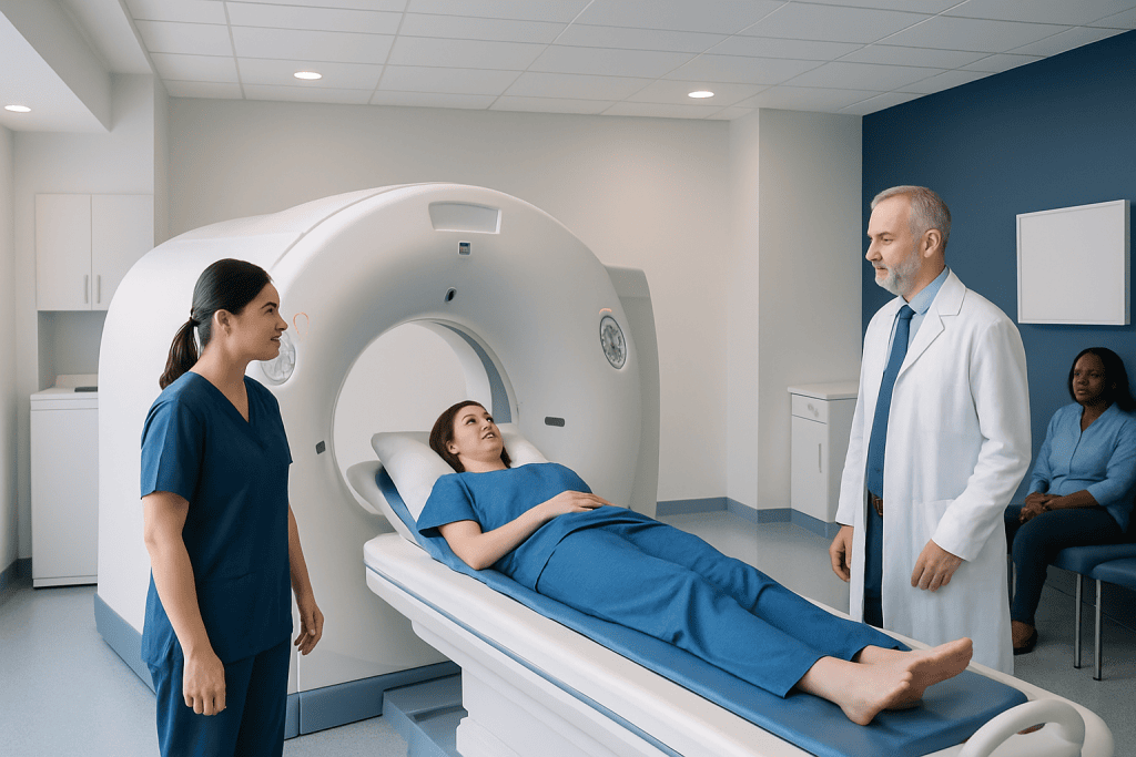
A vq scan measures ventilation (airflow) and perfusion (blood flow) in the lungs. It is commonly used to help assess suspected pulmonary embolism when appropriate.
Key Takeaways
- A vq scan measures ventilation (airflow) and perfusion (blood flow) in the lungs.
- It is commonly used to help assess suspected pulmonary embolism when appropriate.
- The test usually involves breathing in a small amount of tracer and receiving a small tracer injection.
- Results are interpreted together with symptoms, examination findings, and other tests.
- Preparation is usually simple, but patients should tell the care team about pregnancy, breastfeeding, allergies, and recent imaging tests.
A vq scan is a nuclear medicine imaging test that compares how air and blood move through the lungs. It is most often used when doctors need to evaluate a possible pulmonary embolism or better understand certain lung and circulation problems.
Overview: what a VQ scan is and why it is used
A vq scan, short for ventilation-perfusion scan, is an imaging test that shows how well air reaches different parts of the lungs and how well blood flows through them. It uses small amounts of radioactive tracer to create pictures that help doctors compare ventilation and perfusion. This comparison can reveal areas where airflow is normal but blood flow is reduced, a pattern that may suggest a blockage in the lung circulation.
The test is especially helpful when a clinician needs more information about a possible blood clot in the lungs, known as a pulmonary embolism. It may also be considered when other imaging tests are not suitable, such as in some people who should avoid iodinated contrast dye or when radiation exposure to certain organs should be minimized. The exact choice of test depends on the person’s symptoms, medical history, physical examination, and available imaging options.
Unlike a standard chest X-ray, a vq scan focuses on function rather than structure. It does not simply show what the lungs look like; it helps show how the lungs are working. That makes it useful in selected cases of unexplained shortness of breath, chronic clot-related lung disease, or preoperative lung assessment.
How the test works
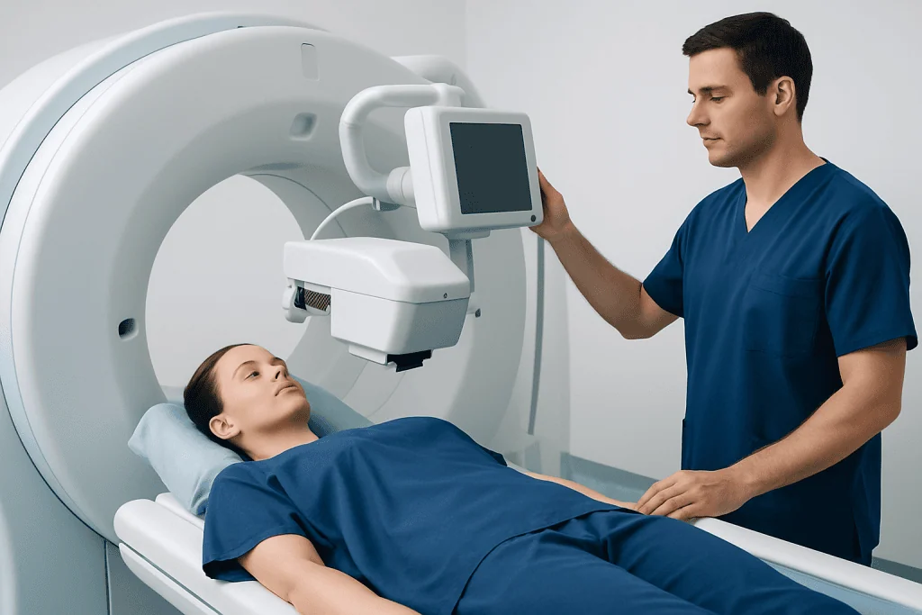
The name of the test describes its two parts. The ventilation part evaluates airflow. During this stage, the patient breathes in a small amount of tracer gas or aerosol through a mask or mouthpiece while images are taken. These images show whether air is reaching the lungs evenly or whether some areas are less well ventilated.
The perfusion part evaluates blood flow. A tracer is injected into a vein, usually in the arm, and a special camera detects how that tracer travels through the blood vessels of the lungs. Areas that receive less blood flow than expected may stand out on the scan.
Doctors then compare the two sets of images. If a region of the lung has normal ventilation but reduced perfusion, this mismatch can suggest a problem in the blood supply, including a possible pulmonary embolism. If both airflow and blood flow are reduced in the same area, the cause may be another lung condition rather than a clot alone.
The scan is usually performed in a nuclear medicine department by trained imaging professionals. It is noninvasive apart from the intravenous injection, and most people tolerate it well. In some centers, the study may be combined with other imaging, depending on the clinical question.
When doctors may recommend a VQ scan

A vq scan is most commonly requested when there is concern about a pulmonary embolism and the medical team needs functional imaging of the lungs. Symptoms that may lead to evaluation include sudden shortness of breath, chest pain that worsens with breathing, rapid heart rate, low oxygen levels, or unexplained fainting. The scan is not ordered based on one symptom alone; doctors consider the whole clinical picture.
It may also be used in people with chronic thromboembolic disease, in follow-up after certain clotting problems, or before some lung surgeries to estimate how much each lung contributes to breathing function. In selected patients with underlying lung disease, the scan can provide useful information that complements pulmonary function tests and structural imaging.
A vq scan can be particularly helpful when CT pulmonary angiography is not the best option, such as in some patients with contrast allergy, kidney concerns, or other reasons to avoid contrast-enhanced CT. If broader evaluation is needed, the medical team may also recommend tests such as CT scanning or MRI scanning for related conditions, depending on the symptoms being investigated.
- Suspected pulmonary embolism
- Assessment of chronic blood flow problems in the lungs
- Preoperative evaluation before some lung procedures
- When CT contrast is not suitable or should be avoided
What to expect before, during, and after the scan
Preparation is usually straightforward. Many patients do not need to fast, but local instructions may vary. The care team may ask about recent chest imaging, current medicines, prior lung disease, pregnancy, breastfeeding, allergies, or any history of blood clots. A chest X-ray is often reviewed alongside the scan because it can help interpret the images more accurately.
During the ventilation phase, the patient breathes the tracer while remaining still for imaging. During the perfusion phase, a small amount of tracer is injected into a vein and more images are taken. The camera moves around the chest, but the test is generally painless. Remaining still is important because movement can affect image quality.
The full appointment length varies by center and by whether one or both parts of the study are performed. Many scans are completed within about an hour, though timing can differ. Most people can return to normal activities afterward unless the doctor gives different instructions.
After the test, drinking fluids may be advised to help the body clear the tracer. The amount of radioactivity used is small and chosen for medical imaging purposes. A radiologist or nuclear medicine specialist reviews the images and sends a report to the referring clinician, who explains what the findings mean in context.
Understanding the results
Vq scan results are not usually described as simply normal or abnormal without explanation. Instead, the report looks at the pattern of airflow and blood flow. A normal scan makes a significant pulmonary embolism less likely. A mismatched defect, where ventilation is preserved but perfusion is reduced in one or more areas, may support the diagnosis of a clot.
Sometimes the result is indeterminate, meaning it does not clearly confirm or exclude the suspected diagnosis on its own. This can happen if there is underlying lung disease, if the image pattern is complex, or if the scan findings do not fit neatly into one category. In those situations, doctors may use blood tests, leg vein ultrasound, clinical risk scores, or other imaging to reach a diagnosis.
Interpretation is especially important in people with conditions such as chronic obstructive pulmonary disease, pneumonia, asthma, or prior lung surgery, because these can affect ventilation and perfusion patterns. The final decision is based on the person’s symptoms and risk factors, not only on the scan.
If a blood clot is confirmed or strongly suspected, treatment may involve interventional treatment in selected vascular situations or, more commonly, anticoagulation under specialist guidance depending on the overall diagnosis. The exact treatment plan is individualized and may also involve evaluation for related vascular conditions such as deep vein thrombosis.
Benefits, limitations, and safety
The main benefit of a vq scan is that it provides functional information about the lungs in a relatively gentle and well-established way. For the right patient, it can answer an important question while avoiding tests that may be less suitable because of contrast exposure or other concerns. It is also useful in situations where matching airflow and blood-flow data helps narrow the diagnosis.
Like any medical test, it has limitations. A vq scan may be less definitive in people with significant underlying lung disease or an abnormal chest X-ray. Results can sometimes be nondiagnostic, which means further tests may still be needed. It also does not replace a full medical assessment.
Radiation exposure from a vq scan is generally low and carefully controlled, but it is still a factor that doctors consider, especially during pregnancy and in younger patients. Patients should tell the care team if they are pregnant, could be pregnant, or are breastfeeding so the imaging team can discuss the safest approach.
Allergic reactions to the tracers used in nuclear medicine are uncommon. The intravenous injection may cause minor brief discomfort. Overall, serious complications are rare, and the test is considered safe when used appropriately and interpreted by experienced clinicians.
When to seek medical care
Medical care should be sought promptly for sudden shortness of breath, chest pain, coughing up blood, fainting, severe unexplained weakness, or a rapid heartbeat, especially if there is recent surgery, prolonged immobility, cancer treatment, pregnancy, or a history of blood clots. These symptoms do not always mean a pulmonary embolism, but they should be assessed without delay.
Non-urgent medical advice is also appropriate for ongoing breathlessness, reduced exercise tolerance, leg swelling, or unexplained chest discomfort. A doctor can decide whether a vq scan, blood tests, ultrasound, or another imaging test would be more helpful. Self-diagnosis is not reliable in this area because many heart and lung conditions can cause similar symptoms.
For international patients who need coordinated evaluation, Acibadem International’s multidisciplinary specialists and JCI-accredited hospitals diagnose and treat lung and vascular conditions using modern imaging and evidence-based care. The most suitable test is chosen after individual assessment rather than using one test for everyone.
Frequently asked questions
What does a vq scan check for?
A vq scan checks how air and blood move through the lungs. It is most often used to help evaluate a possible pulmonary embolism, but it can also help assess other lung circulation and function problems.
Is a vq scan the same as a CT scan?
No. A vq scan is a nuclear medicine test that looks at lung function, while a CT scan creates detailed structural images. Doctors choose between them based on the clinical question, the patient's health history, and whether contrast dye is appropriate.
How long does a vq scan take?
Many vq scans are completed within about an hour, but timing can vary by hospital and by whether both the ventilation and perfusion parts are performed. Extra time may be needed for preparation, waiting between steps, or review of related imaging such as a chest X-ray.
Is a vq scan painful?
The test is usually not painful. Most people only notice the brief discomfort of the intravenous injection and the need to stay still during imaging. Breathing in the tracer is generally well tolerated.
Is the radiation from a vq scan safe?
The scan uses a small amount of radioactive tracer, and the dose is carefully selected for medical imaging. For most patients, the test is considered safe when it is clearly indicated. The imaging team takes extra care in pregnancy and breastfeeding, so these should always be mentioned in advance.
Do patients need to prepare for a vq scan?
Preparation is often simple, and many people can eat and drink normally unless told otherwise. Patients should inform the care team about pregnancy, breastfeeding, lung disease, recent imaging tests, medicines, and any history of clotting problems.
References
- American College of Radiology
- Society of Nuclear Medicine and Molecular Imaging
- Radiological Society of North America
- National Heart, Lung, and Blood Institute
- Centers for Disease Control and Prevention
This article is for general information only and is not a substitute for professional medical advice. Please consult a qualified doctor about your individual situation.
Explore treatments in Turkey — costs, top hospitals & a free quote
JCI-accredited · board-certified surgeons · reply within 24h
Add us as a Preferred Source to see more of our trusted health content across Google Search, AI Overviews and Discover.
Check your numbers in seconds
BMI, calories, due date, blood pressure and 30+ more clinical calculators — free, instant, doctor-reviewed ranges.

More from the Health Library

Bezoar: A Complete Medical Overview

Al Beato — Explained by Medical Evidence, Not Myths

Lungs — Explained by Medical Evidence, Not Myths

Lump Under Chin: A Complete Medical Overview

Spank: A Complete Medical Overview