Intradermal Nevus: An Evidence-Based Guide for Patients

An intradermal nevus is usually a benign mole that sits within the dermis, the deeper layer of skin. These moles are often raised, smooth or slightly warty, and may be skin-colored, tan, or light brown.
Key Takeaways
- An intradermal nevus is usually a benign mole that sits within the dermis, the deeper layer of skin.
- These moles are often raised, smooth or slightly warty, and may be skin-colored, tan, or light brown.
- Doctors diagnose intradermal nevus mainly through a skin examination and may use dermoscopy or biopsy when needed.
- Removal is not always necessary, but it may be considered if the mole is irritated, cosmetically bothersome, or uncertain in diagnosis.
- A mole that changes in size, color, shape, or starts bleeding should be assessed promptly.
An intradermal nevus is a common, usually harmless mole formed by melanocyte cells located deeper in the skin. It often looks dome-shaped, soft, or flesh-colored, but any changing or unusual mole should be checked by a qualified doctor.
Overview
An intradermal nevus is a type of mole made up of melanocytes, the pigment-producing cells of the skin. In this type of mole, the cells are located mainly in the dermis rather than at the skin surface, which is why the lesion often appears raised, rounded, and lighter in color than some other moles. In most cases, it is benign and does not cause health problems.
Patients often notice an intradermal nevus as a long-standing bump on the face, neck, trunk, or scalp. It may be flesh-colored, pinkish, tan, or light brown, and some lesions have hairs growing from them. Because many people worry that any mole might be skin cancer, it is helpful to know that intradermal nevi are common and usually harmless when their appearance is stable.
Even so, not every raised mole is an intradermal nevus. Other benign growths and some skin cancers can sometimes look similar, especially to the untrained eye. That is why medical evaluation matters when a lesion is new, changing, repeatedly irritated, or different from a person’s other moles.
How an Intradermal Nevus Looks and Feels
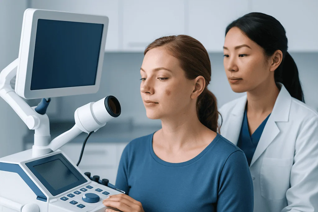
Intradermal nevi often have a characteristic appearance. They are commonly dome-shaped or slightly pedunculated, meaning they may sit on a small stalk. The surface may be smooth, soft, or mildly papillomatous, giving it a subtle uneven or wart-like texture. Unlike many flatter pigmented moles, they may have little visible pigment and blend closely with the surrounding skin tone.
These moles usually develop gradually over time and can remain unchanged for years. Some begin as more pigmented junctional or compound nevi and later become more elevated and less dark as the nevus cells move deeper into the skin. This pattern is one reason intradermal nevi are often seen in adults.
Most do not cause symptoms. However, a lesion may become tender, itchy, or irritated if it rubs against clothing, jewelry, razors, or combs. A mole on the scalp or beard area may be especially prone to repeated minor trauma. If a bump catches often or bleeds after shaving, a doctor can help determine whether it is simply irritated or needs closer assessment.
- Usually raised rather than flat
- Often flesh-colored, pink, tan, or light brown
- May have hair growing from it
- Common on the face, neck, scalp, and trunk
- Typically stable over time
Causes and Risk Factors
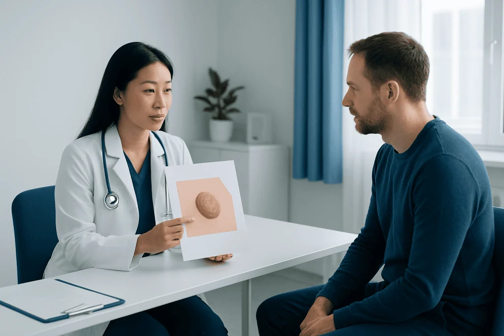
An intradermal nevus develops when melanocyte cells cluster within the skin. The exact reason a particular mole forms is not always clear, but genetics, natural skin development, and cumulative sun exposure may all play a role in the number and behavior of moles over a lifetime. Having moles is extremely common, and most are not a sign of disease.
Age can influence how a mole looks. Some nevi become more raised and less pigmented over time, which may reflect the normal maturation process of a benign mole. Hormonal changes, such as those that occur during adolescence or pregnancy, can also affect the appearance of existing moles, although any significant change still deserves medical review.
People with fair skin, a family tendency to develop numerous moles, or a history of significant sun exposure may notice more pigmented lesions overall. Still, an intradermal nevus itself is generally considered a benign entity. The main clinical question is often not why it formed, but whether the lesion is truly an intradermal nevus or another condition that needs treatment, such as skin cancer.
How Doctors Diagnose It
Diagnosis begins with a careful skin examination. A dermatologist or other trained doctor looks at the lesion’s size, shape, border, color, texture, and location, and asks whether it has changed over time. The patient’s personal and family history of atypical moles or skin cancer also helps guide the assessment.
Many specialists use dermoscopy, a noninvasive magnified examination of the skin surface that can reveal patterns not visible to the naked eye. Dermoscopy can improve the accuracy of distinguishing a benign mole from other lesions. In cases where the diagnosis remains uncertain, the doctor may recommend removing part or all of the lesion for laboratory analysis.
A biopsy is the most reliable way to confirm what a skin lesion is when there is any doubt. Under the microscope, pathologists can identify the typical features of an intradermal nevus and rule out other conditions. In practice, many stable lesions with classic features can be monitored, while suspicious or bothersome lesions are often removed through a minor office-based procedure such as mole removal or biopsy.
Treatment Options and When Removal May Help
Treatment is not always necessary for an intradermal nevus. If the lesion has a typical benign appearance, has remained stable, and causes no symptoms, a doctor may simply advise observation. Many patients choose no treatment once they understand that the mole is harmless.
Removal may be considered for several reasons. These include repeated irritation, bleeding from friction or shaving, cosmetic concerns, or uncertainty about the diagnosis. Depending on the lesion’s size, shape, and location, the doctor may use a shave excision, surgical excision, or another minor technique to remove it safely and send tissue for examination if needed.
Because the goal is both diagnosis and appropriate care, treatment planning should be individualized. A raised lesion on a visible area such as the face may need a different approach than one on the trunk. If there is concern that a lesion could represent another type of growth, a clinician may recommend further evaluation by specialists in dermatology and, if malignancy is suspected, more focused assessment related to skin cancer treatment.
Near the end of the care pathway, some patients seek support from centers with multidisciplinary expertise. Acibadem International’s multidisciplinary specialists and JCI-accredited hospitals evaluate and treat skin lesions, including suspicious moles, for international patients when further assessment or treatment is needed.
Self-Monitoring, Prevention, and Skin Care Habits
There is no guaranteed way to prevent every mole, including an intradermal nevus. However, healthy sun habits are important for overall skin protection and may help reduce damage that can complicate the evaluation of pigmented lesions. These habits include seeking shade when practical, wearing protective clothing, and using broad-spectrum sunscreen as advised by a healthcare professional.
Self-monitoring is also useful. Patients can become familiar with their usual moles and look for changes in size, shape, color, symmetry, surface, or symptoms such as itching and bleeding. Taking clear photographs at intervals may help some people notice changes more accurately, especially for lesions in hard-to-see areas.
It is best not to pick, scratch, or try to remove a mole at home. Home removal can cause bleeding, infection, scarring, and delayed diagnosis if the lesion is not actually benign. If a mole is frequently irritated by clothing, shaving, or hair care routines, medical assessment is safer than self-treatment.
When to Seek Medical Care
Medical care is advisable if a presumed intradermal nevus is new in adulthood, changes noticeably, or looks different from a person’s other moles. A lesion should also be checked if it bleeds without clear injury, develops a crust, becomes painful, or repeatedly gets inflamed. These features do not always mean cancer, but they should not be ignored.
Doctors often advise using the well-known ABCDE warning signs when considering pigmented lesions: asymmetry, irregular border, varied color, increasing diameter, and evolution over time. Another helpful clue is the “ugly duckling” sign, meaning one lesion appears unlike the rest. These tools are not diagnostic on their own, but they can help patients know when to arrange an examination.
Urgent evaluation is particularly important for anyone with a personal history of skin cancer, a strong family history of melanoma, or many atypical moles. In these situations, regular professional skin checks may be recommended. If there is any uncertainty, a qualified doctor can determine whether the lesion is a benign intradermal nevus or another condition such as melanoma.
Frequently asked questions
Is an intradermal nevus cancerous?
An intradermal nevus is usually a benign, noncancerous mole. However, a doctor should assess any lesion that is changing, bleeding, or looks unusual, because not every raised skin lesion is an intradermal nevus.
What does an intradermal nevus look like?
It often appears as a raised, rounded, flesh-colored, pink, tan, or light brown bump. The surface may be smooth or slightly warty, and some lesions have hair growing from them.
Does an intradermal nevus need to be removed?
Not always. If it is clearly benign, stable, and not bothersome, observation may be enough; removal is more often considered if it is irritated, cosmetically unwanted, or uncertain in diagnosis.
Can an intradermal nevus turn into melanoma?
Most intradermal nevi remain benign. Still, any mole can become a source of concern if it changes over time, so prompt evaluation is important when warning signs appear.
How is an intradermal nevus diagnosed?
Doctors diagnose it mainly through a clinical skin examination, often supported by dermoscopy. If the lesion does not have a clearly benign appearance, a biopsy may be recommended to confirm the diagnosis.
Is it safe to remove a raised mole at home?
No, home removal is not recommended. It can cause bleeding, infection, scarring, and may delay the diagnosis of a lesion that needs proper medical evaluation.
References
- American Academy of Dermatology
- National Cancer Institute
- DermNet
- Mayo Clinic
- British Association of Dermatologists
This article is for general information only and is not a substitute for professional medical advice. Please consult a qualified doctor about your individual situation.
Hair transplant in Turkey — costs, graft counts & a free assessment
JCI-accredited · board-certified surgeons · reply within 24h
Add us as a Preferred Source to see more of our trusted health content across Google Search, AI Overviews and Discover.

More from the Health Library

Straight Hair: What Patients Need to Know

Palinopsia: A Complete Medical Overview

Low Carb Meal Delivery — Explained by Medical Evidence, Not Myths

Largest Muscle in the Body — Explained by Medical Evidence, Not Myths

Trepanation: What Patients Need to Know