Brown Spot on Eye: A Complete Medical Overview

A brown spot on eye may be a harmless nevus, but it should be assessed if it is new or changing. The exact meaning depends on where the spot appears: on the white of the eye, the iris, or deeper inside the eye.
Key Takeaways
- A brown spot on eye may be a harmless nevus, but it should be assessed if it is new or changing.
- The exact meaning depends on where the spot appears: on the white of the eye, the iris, or deeper inside the eye.
- Warning signs include growth, changing color, blurred vision, eye pain, flashes, or loss of part of vision.
- Diagnosis usually involves a slit-lamp eye exam and sometimes retinal imaging or ultrasound.
- Many pigment spots only need monitoring, while suspicious lesions may require specialist treatment.
A brown spot on eye is commonly caused by a benign pigment spot, similar to a freckle or mole, on the colored part of the eye or its surface. Most are not dangerous, but any new, changing, raised, or symptomatic spot should be checked by an eye specialist.
Overview: what a brown spot on eye can mean
A brown spot on eye often turns out to be a small area of extra pigment, much like a freckle on the skin. These spots can appear on the iris, which is the colored part of the eye, on the conjunctiva that covers the white of the eye, or less commonly inside the eye where they cannot be seen without an eye examination.
In many people, the spot is benign and remains stable for years. Common explanations include an iris nevus, a conjunctival nevus, or racial melanosis, which is a benign increase in pigmentation on the eye surface. However, some pigmented lesions need closer attention because they can grow, affect vision, or rarely represent a cancerous change.
The main question is not simply whether the spot is brown, but where it is located, whether it is new, and whether it has changed in size, shape, or color. This is why eye specialists focus on careful examination and comparison over time rather than guessing from appearance alone.
A practical rule is reassuring but cautious: a long-standing, stable spot without symptoms is often not urgent, but a new or changing brown spot deserves professional assessment. Early evaluation helps distinguish harmless findings from conditions that may need monitoring or treatment.
Types of brown spots seen on or in the eye
Brown spots are not all the same. A spot on the iris may be an iris freckle or iris nevus. A freckle usually lies flat and is often linked to light exposure, while a nevus is a benign cluster of pigment cells that may be slightly thicker. On the eye surface, a conjunctival nevus is common and may look like a small brown or tan lesion near the cornea or on the white of the eye.
Some people have diffuse pigment rather than one distinct spot. This can occur with complexion-associated melanosis, often seen as a flat brown area on the conjunctiva. Another possibility is a deposit caused by prior inflammation, injury, or a foreign body, which may darken the surface of the eye and mimic a pigmented lesion.
Doctors also consider less common but important causes. Primary acquired melanosis is an abnormal pigmentation of the conjunctiva that may require close follow-up because some forms can progress. Uveal melanoma, including iris or choroidal melanoma, is rare but serious and may first be noticed as a dark spot or be found only during a detailed dilated eye exam. For related information, some patients may hear the term eye cancer during evaluation of suspicious lesions.
Because different lesions can look similar to a non-specialist, self-diagnosis is unreliable. A clear photo can help document change, but it cannot replace an examination by an ophthalmologist, especially if the spot is raised, irregular, or associated with symptoms.
Symptoms and warning signs to watch for

Many benign eye pigment spots cause no symptoms at all. People often notice them while looking in a mirror, seeing a photograph, or being told about them during a routine eye exam. A stable, painless spot with no effect on vision is often less concerning than a spot that changes over time.
Symptoms that deserve attention include blurred vision, a shadow in part of the visual field, flashes of light, new floaters, eye pain, redness that does not settle, or a feeling that something is in the eye. Changes in the pupil shape, a visibly growing spot, or a lesion that develops blood vessels or elevation should also be assessed.
Location matters. A spot on the conjunctiva may become irritated or more noticeable with dryness or inflammation, while a lesion inside the eye may not be visible at all but can affect vision. If a brown spot appears after trauma, surgery, or chemical exposure, the eye should be examined to rule out injury or retained foreign material.
- New brown spot appearing in adulthood
- Rapid growth or change in shape or color
- Raised, thickened, or irregular surface
- Blurred vision, flashes, floaters, or visual field loss
- Persistent redness, pain, or recurrent irritation
Causes and risk factors
The most common cause of a brown spot on eye is localized pigment from melanocytes, the cells that make melanin. Benign nevi may develop in childhood or early adulthood and stay unchanged for many years. Freckles and some iris pigmentation changes may become more noticeable with age or sun exposure, although the relationship is not always straightforward.
Inherited traits and natural eye color also play a role. Some people are simply more likely to have visible ocular pigmentation. Surface pigment can also become more obvious in people with chronic eye irritation, prior inflammation, or certain benign racial or complexion-associated pigmentation patterns.
Less commonly, a brown spot may be linked to disease processes such as primary acquired melanosis, dysplastic changes, or melanoma. Previous skin melanoma, a family history of ocular tumors, and certain genetic or pigmentation conditions may raise concern, although most eye spots are still not cancer.
Environmental and medical factors can occasionally contribute. Prior eye injury, surgery, inflammation, or retained material may leave a dark mark on the eye surface. A careful history helps the doctor decide whether the spot is likely to be a benign pigment change or something that needs imaging and specialist review.
How doctors diagnose a brown spot on eye
Diagnosis begins with a detailed eye history and examination. The ophthalmologist will ask when the spot was first noticed, whether it has changed, and whether there are any symptoms such as blurred vision, irritation, or pain. Past eye disease, injury, and family history may also be relevant.
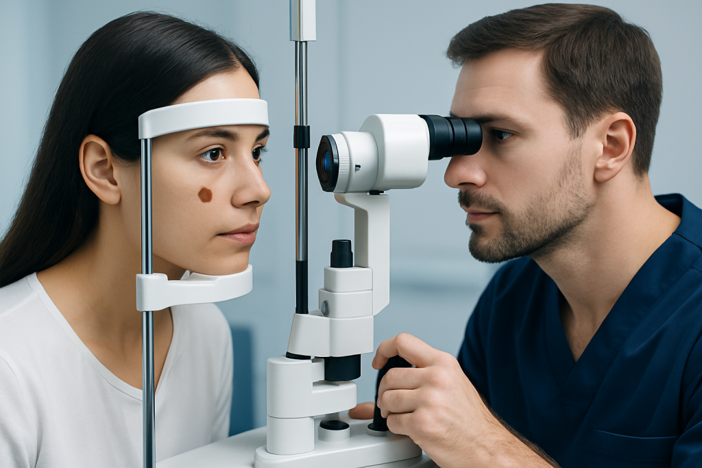
Most visible spots are examined with a slit lamp, which allows the doctor to see whether the lesion is flat or elevated, well-defined or irregular, and whether it involves the iris, conjunctiva, cornea, or nearby tissues. Photographs are often taken to document the lesion so future changes can be compared accurately.
If the lesion may be inside the eye, a dilated retinal examination is usually needed. Additional tests may include ocular ultrasound, optical coherence tomography, fundus photography, or other imaging to measure thickness and assess deeper structures. In some situations, referral for a comprehensive eye examination or subspecialty care is the next step.
Biopsy is not necessary for most simple, stable lesions, but it may be considered if the appearance is suspicious or if treatment is being planned. The goal of diagnosis is to classify the lesion correctly, identify any signs of malignancy, and decide whether observation alone is safe.
Treatment options and follow-up
Treatment depends entirely on the cause. Many benign spots, such as stable conjunctival or iris nevi, do not need removal. Instead, doctors usually recommend periodic observation with repeat photographs and eye exams to confirm that there is no growth or structural change.
If the spot causes irritation, treatment may focus on the surface problem rather than the pigment itself. Lubricating drops, management of dryness, or treatment of associated inflammation may help comfort. If there is uncertainty about the diagnosis, a specialist in ocular tumors or anterior segment disease may be involved.
Suspicious lesions may require removal, biopsy, laser treatment, or radiation-based approaches depending on location and findings. When a lesion affects the retina or deeper structures, management may involve advanced retinal disease treatment or coordination with oncology specialists. For patients with lesions strongly suggestive of malignancy, care may be linked to services related to cancer treatment.
Near the end of the evaluation process, some patients benefit from multidisciplinary care. Acibadem International’s specialists in JCI-accredited hospitals diagnose and treat eye conditions for international patients, including cases that need imaging, ophthalmic follow-up, or coordinated specialty care. If the brown spot is associated with broader eye diseases, follow-up planning is especially important.
Prevention and self-care
Not every brown spot can be prevented, because many are related to natural pigmentation or age. Still, sensible eye protection is helpful. Wearing UV-protective sunglasses and avoiding direct eye exposure to irritants may reduce cumulative stress on the eye surface and support overall eye health.
Self-care also means avoiding rubbing the eyes, managing dryness, and keeping regular eye appointments, especially for people who already know they have a pigmented lesion. Contact lens users should follow hygiene guidance carefully and seek advice if a spot is accompanied by redness or discomfort.
It can be useful to note the appearance of a visible spot with a dated photograph, but this should be done only as a record for discussion with a doctor. Home observation is not a substitute for an eye examination, especially if the spot is new, changes, or is linked to visual symptoms.
People who have had a brown spot identified by an ophthalmologist should keep the recommended review schedule. Long-term stability is reassuring, but regular monitoring is the best way to catch important change early if it occurs.
When to seek medical care
A prompt eye assessment is sensible if a brown spot on eye is new, appears to be growing, or has an irregular outline. The same is true if the spot is accompanied by blurred vision, flashes, floaters, partial loss of vision, persistent redness, pain, or a change in pupil shape.
Urgent care is particularly important after eye injury, chemical exposure, or sudden visual symptoms. These situations may reflect more than a pigment spot and should not be monitored at home. Children with a new eye spot should also be evaluated rather than assuming it is harmless.
If the spot has been present for a long time and has not changed, it is still reasonable to mention it at a routine eye check. An ophthalmologist can confirm whether it looks benign and decide if periodic follow-up is needed. Clear diagnosis provides reassurance and reduces the risk of missing a treatable condition.
Frequently asked questions
Is a brown spot on eye usually serious?
Usually not. Many brown spots are benign pigment changes such as a nevus or freckle. Even so, a new, changing, or raised spot should be checked by an ophthalmologist to make sure it is not a more significant lesion.
Can a brown spot on the white of the eye be normal?
Yes, it can be normal, especially if it is a stable conjunctival nevus or benign surface pigmentation. The doctor will look at its shape, thickness, borders, and whether it has changed over time. Surface irritation or redness around the spot may still need evaluation.
What is the difference between an eye freckle and eye melanoma?
An eye freckle or nevus is usually a benign cluster of pigment cells that remains stable. Melanoma is a cancerous growth that may enlarge, thicken, or affect vision. Only an eye specialist can reliably tell the difference through examination and sometimes imaging.
Should a child with a brown spot on eye see a doctor?
Yes. Although many spots in children are harmless, a proper eye examination helps identify what the lesion is and whether follow-up is needed. It is especially important if the spot is new, enlarging, or associated with redness or vision complaints.
Can a brown spot on eye go away on its own?
Some spots remain unchanged for years, while others may appear more or less noticeable over time. Most true nevi do not simply disappear. A doctor can explain whether the spot is likely to stay stable or needs monitoring.
How is a suspicious brown eye spot treated?
Treatment depends on the diagnosis and location. Some lesions are only observed with repeat photographs, while others may need biopsy, removal, laser treatment, or radiation-based therapy. The plan is individualized to protect eye health and vision as much as possible.
This article is for general information only and is not a substitute for professional medical advice. Please consult a qualified doctor about your individual situation.
Explore treatments in Turkey — costs, top hospitals & a free quote
JCI-accredited · board-certified surgeons · reply within 24h
Add us as a Preferred Source to see more of our trusted health content across Google Search, AI Overviews and Discover.

More from the Health Library

Raccoon Eyes: An Evidence-Based Guide for Patients

Mirapex: A Complete Medical Overview

Rosacea — Explained by Medical Evidence, Not Myths

Sophia Burns Md: What Patients Need to Know

Burning Sensation in Stomach: What Patients Need to Know