

Radiologist: A Complete Medical Overview

A radiologist is a physician who specializes in reading and interpreting medical images. Radiologists help diagnose injuries, infections, cancers, vascular problems, and many other conditions.
Key Takeaways
- A radiologist is a physician who specializes in reading and interpreting medical images.
- Radiologists help diagnose injuries, infections, cancers, vascular problems, and many other conditions.
- Some radiologists, called interventional radiologists, also perform minimally invasive image-guided treatments.
- Patients may not always meet the radiologist directly, but the radiologist’s report guides medical care.
- Imaging tests are chosen based on symptoms, medical history, and the clinical question being asked.
A radiologist is a medical doctor trained to diagnose, monitor, and sometimes treat disease using imaging tests such as X-rays, ultrasound, CT, MRI, and nuclear medicine scans. They usually work behind the scenes with referring doctors, but their interpretation plays a central role in accurate diagnosis and treatment planning.
Overview: what a radiologist does
A radiologist is a doctor who specializes in medical imaging. This physician reviews images from tests such as X-rays, ultrasound, computed tomography (CT), magnetic resonance imaging (MRI), mammography, and nuclear medicine studies to help identify the cause of symptoms or monitor a known condition. In many healthcare settings, the radiologist works closely with the referring doctor rather than serving as the main doctor for ongoing care.
The radiologist’s role goes beyond simply “looking at scans.” They choose the most appropriate imaging approach in some situations, compare current images with prior studies, assess image quality, and provide a structured medical interpretation called a radiology report. This report helps other specialists decide on next steps, such as additional testing, medication, surgery, or follow-up observation.
Some radiologists also perform procedures. Interventional radiologists use real-time imaging guidance to place needles, drains, stents, or catheters through small skin openings instead of large incisions. This can support diagnosis, relieve symptoms, or treat certain conditions in a minimally invasive way.
How radiologists fit into a patient’s care
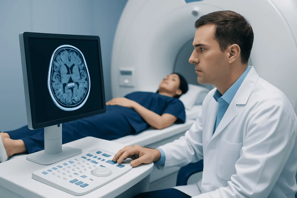
Many patients are referred for imaging by a primary care doctor, emergency physician, surgeon, neurologist, oncologist, or another specialist. The radiologist then reviews the clinical question, interprets the images, and communicates important findings to the referring team. In urgent situations, such as suspected stroke, bleeding, or a blocked blood vessel, the radiologist may contact the treating doctor immediately.
Radiologists are especially important because many illnesses cannot be fully evaluated through symptoms and examination alone. Imaging can reveal fractures, pneumonia, kidney stones, tumors, internal bleeding, organ inflammation, and vascular abnormalities. In some cases, imaging is also used to guide biopsies or other procedures so tissue samples can be taken accurately and safely.
Depending on the country, hospital, and type of test, a patient may or may not meet the radiologist face to face. For example, patients often meet the doctor during ultrasound, breast imaging, or image-guided procedures. For routine X-rays or CT scans, the radiologist may interpret the study remotely and send the report to the referring clinician.
Radiologists also contribute to ongoing disease monitoring. They may compare new imaging with previous scans to track healing after injury, assess response to cancer treatment, or monitor chronic disorders such as stroke-related brain changes or vascular conditions that need periodic review.
Types of radiologists and imaging they use

Radiology includes several subspecialties. Diagnostic radiologists focus on interpreting imaging studies to identify disease. Interventional radiologists perform minimally invasive procedures using imaging guidance. There are also neuroradiologists, pediatric radiologists, breast imaging specialists, musculoskeletal radiologists, and nuclear medicine physicians, among others.
Each imaging method has strengths. X-rays are commonly used for bones and chest conditions. Ultrasound uses sound waves and is often used for pregnancy, abdominal organs, blood vessels, and soft tissues. CT creates detailed cross-sectional images and is helpful in trauma, lung disease, abdominal pain, and many emergencies. MRI provides high-detail images of the brain, spine, joints, and soft tissues without using ionizing radiation. Nuclear medicine and PET imaging can show how tissues function as well as what they look like.
Interventional radiology is a distinct area because it combines imaging with treatment. These specialists may help manage blocked vessels, abscess drainage, certain bleeding problems, or targeted biopsies. In selected situations, patients may be referred for image-guided minimally invasive procedures as part of a broader care plan.
Some patients are also sent to highly focused imaging experts when symptoms point to a specific system. For example, neurological symptoms may lead to advanced brain or spine imaging reviewed by specialists in neuroradiology evaluation and imaging.
Common reasons someone may need a radiologist
Patients do not usually seek a radiologist for a “condition” in the same way they would seek a cardiologist or dermatologist. Instead, a radiologist becomes involved when imaging is needed to answer a medical question. This may happen after an injury, with unexplained pain, during cancer screening, or when a doctor needs more information about a symptom.
Common reasons include persistent headaches, chest pain, abdominal pain, shortness of breath, back pain, a suspected fracture, a lump, abnormal blood test results, or symptoms such as weakness, numbness, or dizziness. Imaging can also be used before surgery, after treatment, or to monitor chronic illness. Screening exams, such as mammography or some forms of vascular imaging, also rely on radiologists.
Radiologists are part of care pathways for many serious and routine conditions. For example, imaging may help evaluate possible brain tumor findings, detect internal causes of pain, or guide treatment decisions when cancer is suspected or already diagnosed. In selected cases, doctors may request MRI scanning or CT imaging because these tests provide different kinds of anatomical detail.
- Emergency care: trauma, suspected bleeding, stroke, pulmonary embolism
- Outpatient care: joint pain, abdominal symptoms, kidney stones, sinus disease
- Screening and monitoring: breast imaging, follow-up scans, treatment response
- Procedure support: biopsies, drain placement, vascular access, targeted treatment planning
What happens during imaging and how results are reported
The patient experience depends on the imaging test. A simple X-ray may take only a few minutes. Ultrasound usually involves a handheld probe on the skin with gel. CT scans are quick and may require a contrast dye to improve visibility of organs or blood vessels. MRI generally takes longer and can be noisy, but it is painless. Some nuclear medicine tests involve a small amount of tracer that helps show how organs or tissues function.
Before imaging, patients may be asked about pregnancy, kidney function, allergies, implanted devices, recent surgeries, or claustrophobia. These questions help the team choose the safest and most useful test. In some cases, fasting or avoiding certain medicines may be advised beforehand. Clear instructions are usually given by the imaging center or referring doctor.
After the images are obtained, the radiologist studies them carefully and prepares a report. This report usually describes the findings, notes anything abnormal, and may suggest follow-up tests or comparison with previous imaging. The referring doctor then explains how the results fit the patient’s symptoms, examination, and other test results.
It is important to remember that an imaging result is one part of the diagnostic process. A scan can be normal even when symptoms are real, and an abnormal finding does not always mean a serious disease. Radiologists interpret images in context, helping reduce uncertainty rather than replacing the broader medical evaluation.
Safety, benefits, and possible limitations of radiology
Medical imaging can provide valuable information that may not be available in any other way. It can confirm or rule out suspected disease, guide treatment, avoid unnecessary surgery, and detect problems early. In emergencies, radiology often helps doctors make rapid decisions that improve safety and outcomes.
At the same time, each test has limitations. Some findings are nonspecific, meaning they do not point to one single diagnosis. Small abnormalities may need follow-up imaging to clarify whether they are important. Different tests may show different aspects of the same problem, which is why a doctor might order ultrasound first and MRI or CT later.
Radiation exposure is a common concern. X-rays and CT scans use ionizing radiation, while ultrasound and MRI do not. When radiation-based tests are recommended, the medical team aims to use the lowest appropriate dose and only when the expected benefit outweighs the risk. Contrast materials also have potential risks, such as allergic reactions or kidney-related concerns in certain patients, so doctors screen for these issues in advance.
Patients who are anxious about scans, contrast dye, or enclosed spaces should tell the care team. Adjustments can often be made, and the team can explain the purpose of the test, what to expect, and how to prepare in a way that helps patients feel more comfortable.
How patients can prepare and make the most of an imaging visit
Good preparation helps imaging go smoothly and improves the usefulness of the results. Patients should bring their referral information, a list of current medications, details of allergies, and any relevant prior imaging if it was done at another facility. Sharing previous reports can help the radiologist compare old and new findings more accurately.
Following preparation instructions matters. Some tests require fasting, drinking water, avoiding metal items, or arriving early for paperwork and contrast screening. MRI patients should report pacemakers, aneurysm clips, metal fragments, cochlear implants, or other devices. CT or MRI with contrast may require information about kidney health or prior reactions to dye.
Patients can also ask practical questions, such as when results will be ready, whether a contrast agent is planned, and if normal activities can be resumed afterward. For procedures performed by interventional radiologists, it is reasonable to ask about sedation, recovery time, wound care, and follow-up arrangements.
Near the end of the care journey, some patients benefit from evaluation in centers where radiologists work alongside surgeons, oncologists, neurologists, and other specialists. Acibadem International’s multidisciplinary specialists and JCI-accredited hospitals diagnose and treat patients who need coordinated imaging and image-guided care, particularly for complex or cross-specialty conditions.
When to seek medical care
A person usually does not need to decide on their own whether to “see a radiologist.” Instead, they should seek medical care for concerning symptoms, and a doctor can decide if imaging is needed. Prompt evaluation is appropriate for sudden severe headache, chest pain, shortness of breath, new weakness or numbness, significant injury, unexplained abdominal pain, or symptoms that are severe, worsening, or not improving.
Urgent medical attention is especially important when symptoms suggest an emergency, such as signs of stroke, severe trauma, heavy bleeding, or sudden confusion. In these situations, radiology often helps confirm the diagnosis quickly so treatment can begin without delay.
For non-emergency concerns, a scheduled appointment is a good next step if there is a new lump, ongoing back or joint pain, recurrent headaches, abnormal screening results, or persistent symptoms that interfere with daily life. A qualified clinician can decide whether imaging is helpful, which type is most appropriate, and whether specialist review is needed.
Frequently asked questions
Is a radiologist a real medical doctor?
Yes. A radiologist is a fully qualified medical doctor who has completed medical school and then specialized training in interpreting medical images and, in some cases, performing image-guided procedures. Many also complete additional fellowship training in subspecialties such as neuroradiology, breast imaging, or interventional radiology.
Do patients usually meet the radiologist?
Not always. For many routine imaging tests, the radiologist reviews the images and sends a report to the referring doctor without seeing the patient in person. Patients are more likely to meet the radiologist during ultrasound exams, breast imaging assessments, or image-guided procedures.
What is the difference between a radiologist and an interventional radiologist?
A diagnostic radiologist mainly interprets imaging studies to help diagnose and monitor disease. An interventional radiologist also uses imaging in real time to perform minimally invasive procedures, such as biopsies, drain placement, or treatments involving blood vessels. Some doctors have training that overlaps both areas.
Can a radiologist diagnose cancer?
A radiologist can identify imaging findings that may suggest cancer and describe how suspicious an abnormality appears. However, imaging alone does not always provide a final diagnosis. In many cases, tissue sampling such as a biopsy is needed to confirm the diagnosis and determine the exact type.
Are radiology tests safe?
Most imaging tests are considered safe when used appropriately. Some tests, such as X-rays and CT scans, involve radiation, while MRI and ultrasound do not. Healthcare teams weigh the benefits and risks carefully and use safety measures to reduce unnecessary exposure.
Why would a doctor order MRI instead of CT, or the other way around?
MRI and CT provide different kinds of information. CT is often faster and especially useful in emergencies, trauma, lung imaging, and some abdominal problems, while MRI gives more detail for the brain, spine, joints, and soft tissues. The best test depends on the symptom, the body area being examined, and the specific clinical question.
References
- American College of Radiology
- Radiological Society of North America
- National Institute of Biomedical Imaging and Bioengineering
- U.S. Food and Drug Administration
- World Health Organization
This article is for general information only and is not a substitute for professional medical advice. Please consult a qualified doctor about your individual situation.
Explore treatments in Turkey — costs, top hospitals & a free quote
JCI-accredited · board-certified surgeons · reply within 24h
Add us as a Preferred Source to see more of our trusted health content across Google Search, AI Overviews and Discover.

More from the Health Library



Related Specialists

Dr. Nazlı Özcan
Radiology
Dr. Can Turan (m)
Intensive Care
Dr. Ömercan Yağız Öksüz
Aesthetic Plastic & Reconstructive Surgery
Assoc. Prof. Dr. Lütfiye Uygur
Gynecology & Obstetrics