How Many Ultrasounds During Pregnancy? Here Is What the Evidence Says

Most uncomplicated pregnancies do not need many ultrasounds; two routine scans are often standard. The exact number depends on medical history, symptoms, fetal growth, and whether the pregnancy is high risk.
Key Takeaways
- Most uncomplicated pregnancies do not need many ultrasounds; two routine scans are often standard.
- The exact number depends on medical history, symptoms, fetal growth, and whether the pregnancy is high risk.
- Ultrasound uses sound waves, not radiation, and is considered safe when used appropriately by trained professionals.
- Extra scans are usually done for a clear medical reason, such as bleeding, pain, twins, growth concerns, or placenta issues.
- Patients should contact a doctor promptly for heavy bleeding, severe pain, fluid leakage, reduced fetal movement, or signs of preterm labor.
In most low-risk pregnancies, two routine ultrasounds are commonly enough: one in early pregnancy to confirm dates and one around 18 to 22 weeks to check the baby’s development. More scans may be recommended when there are symptoms, uncertainty about dates, or maternal or fetal conditions that need closer follow-up.
Overview: How Many Ultrasounds Are Usually Needed?
For most people with a low-risk pregnancy, the answer to how many ultrasounds during pregnancy is usually two routine scans. One is often performed in the first trimester to confirm that the pregnancy is in the uterus, estimate the due date, and check whether there is one baby or more. The second is usually done around 18 to 22 weeks to examine the baby’s anatomy, growth, placenta, and amniotic fluid.
In many cases, needing more than two ultrasounds does not mean something is seriously wrong. Extra scans are often ordered because the pregnancy dates are unclear, symptoms such as bleeding need evaluation, or the doctor wants to follow growth or placenta position more closely. Ultrasound is a common imaging tool in obstetric care because it provides useful information without using ionizing radiation.
Recommendations can vary slightly between countries, hospitals, and individual pregnancies. A person carrying twins, living with diabetes or high blood pressure, or having a history of pregnancy complications may need more frequent monitoring. The best schedule is the one that fits the clinical situation rather than a fixed number for everyone.
Why Ultrasounds Are Done During Pregnancy
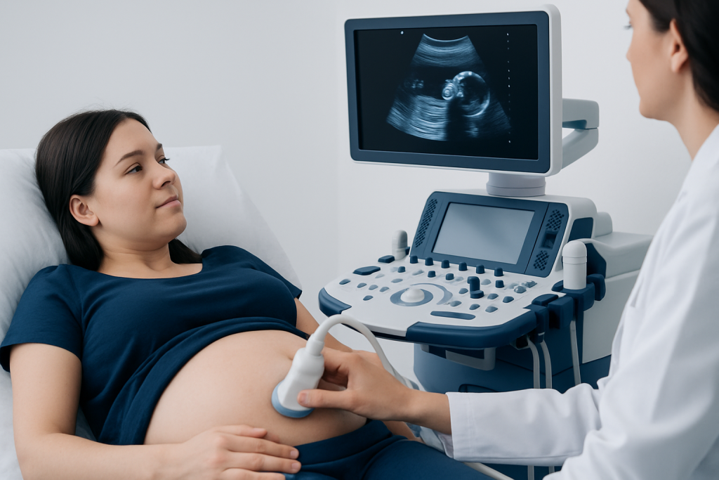
Pregnancy ultrasounds are used to answer specific medical questions. Early in pregnancy, they can confirm the location of the pregnancy, estimate gestational age, and assess early development. Later in pregnancy, they help doctors evaluate fetal anatomy, growth, movement, position, placenta location, and the amount of amniotic fluid.
Ultrasound can also support decisions when symptoms occur. If a pregnant person has bleeding, pelvic pain, or concerns about fetal movement, a scan may help the care team understand what is happening and what follow-up is needed. In this way, the value of ultrasound is not just counting scans, but using them thoughtfully to guide safe care.
Sometimes ultrasound is combined with other prenatal assessments rather than used alone. These may include blood tests, blood pressure checks, urine tests, and physical examination. If an early scan raises concern about pregnancy location or viability, the doctor may also discuss conditions such as ectopic pregnancy and the need for close follow-up.
Typical Pregnancy Ultrasound Schedule

Although care is individualized, many pregnancies follow a broad pattern. A first-trimester ultrasound may be done around 6 to 12 weeks if there is uncertainty about dates, a history of miscarriage, fertility treatment, pain, bleeding, or simply as part of local routine prenatal care. This scan can confirm the heartbeat, the number of fetuses, and the estimated due date.
The mid-pregnancy anatomy scan, often between 18 and 22 weeks, is the major routine ultrasound in many care pathways. It looks at the baby’s organs, head, spine, limbs, abdominal wall, placenta, cervix, and amniotic fluid. It can also help identify structural concerns that may need follow-up with maternal-fetal medicine or pediatric specialists.
Some people will have a later third-trimester scan, but this is not routine for every low-risk pregnancy. It may be recommended to assess growth, check fetal position, monitor placenta previa, or follow pregnancies affected by maternal health conditions. If a doctor suspects a structural issue, specialized imaging such as fetal echocardiography may be advised to look more closely at the baby’s heart.
- Early scan: often for dating, location, heartbeat, and number of fetuses
- Mid-pregnancy scan: detailed anatomy and placenta assessment
- Later scans: usually for a specific medical indication rather than routine screening
When More Ultrasounds May Be Recommended
Additional ultrasounds are commonly recommended when a pregnancy needs closer observation. This can happen with vaginal bleeding, pelvic pain, uncertain dating, suspected miscarriage, prior pregnancy complications, high blood pressure, diabetes, or autoimmune disease. Multiple pregnancy, such as twins or triplets, also usually requires more frequent scans because growth and complications need more careful monitoring.
Doctors may also order extra scans if the baby seems smaller or larger than expected, if there are concerns about amniotic fluid, or if the placenta is low-lying. A later scan can check whether placenta previa has resolved as the uterus grows, or whether the baby has moved into a head-down position near delivery. If a growth issue is suspected, serial ultrasounds may be arranged rather than a single additional scan.
In some cases, ultrasound is used to investigate specific concerns such as reduced movement or signs of preterm labor. Depending on the findings, the doctor may recommend additional tests or treatment. When structural findings suggest a wider health issue, they may also discuss related conditions such as preeclampsia or refer the patient for targeted fetal assessment and closer antenatal care.
Safety, Accuracy, and What Ultrasound Can and Cannot Show
Ultrasound uses sound waves and does not expose the pregnant person or baby to ionizing radiation. When performed by trained professionals for a medical reason, it is widely considered safe in pregnancy. This is why it is commonly used as a first-line imaging method in obstetrics.
Even so, more scans are not always better. Ultrasound is most useful when it is done at the right time and interpreted in the context of the whole pregnancy. Images can be affected by gestational age, fetal position, body habitus, scar tissue, and the specific question the scan is trying to answer.
It is also important to understand that ultrasound has limits. A normal scan cannot guarantee that every condition is absent, and an uncertain result does not automatically mean the baby has a serious problem. Sometimes a repeat scan, blood test, Doppler study, or consultation with a specialist is needed to clarify findings. If monitoring suggests a placenta or cord issue, doctors may use tests such as Doppler ultrasound to assess blood flow in more detail.
How Doctors Decide if Extra Scans Are Needed
The decision to do another ultrasound usually starts with a symptom, a physical finding, or a known risk factor. Doctors consider the pregnancy stage, previous scan results, fetal growth pattern, maternal medical history, and whether there are signs that the placenta, cervix, or amniotic fluid need reassessment. This individualized approach helps avoid unnecessary imaging while still identifying pregnancies that benefit from closer follow-up.
If symptoms arise, the evaluation often includes more than imaging alone. A doctor may review the timing and amount of bleeding, the location of pain, blood pressure, fetal heart rate, urine or blood test results, and whether contractions are present. Depending on the situation, pelvic examination or cardiotocography later in pregnancy may also be appropriate.
When findings need specialized review, the care team may recommend a detailed perinatology assessment. This can be especially helpful in high-risk pregnancy, suspected fetal anomalies, growth restriction, or maternal conditions that may affect the baby. Near the end of care, patients seeking international evaluation may wish to know that Acibadem International’s multidisciplinary specialists and JCI-accredited hospitals diagnose and treat pregnancy-related conditions for international patients.
When to Seek Medical Care
Most pregnancies progress normally, and many symptoms turn out to have a harmless explanation. Still, certain warning signs deserve prompt medical review rather than waiting for the next routine ultrasound. Medical attention is especially important because symptoms, not just scan timing, often determine whether an urgent assessment is needed.
A pregnant person should contact a doctor or maternity unit promptly for heavy vaginal bleeding, severe or one-sided abdominal pain, fainting, fever, painful contractions, fluid leaking from the vagina, or a sudden severe headache with vision changes. Later in pregnancy, reduced or absent fetal movement also deserves urgent evaluation. These symptoms do not always signal a serious problem, but they should be checked without delay.
During the visit, the doctor may ask about symptoms, examine the patient, listen to the fetal heartbeat, and decide whether ultrasound is needed. Depending on the pregnancy stage, they may also order blood tests, urine tests, blood pressure monitoring, or fetal monitoring. This step-by-step approach helps identify common causes, reassure when things are normal, and act quickly if treatment is needed.
Frequently asked questions
Is two ultrasounds really enough in pregnancy?
For many low-risk pregnancies, yes. One early scan and one anatomy scan are often enough when the pregnancy is progressing normally and there are no symptoms or risk factors that call for closer monitoring.
Do more ultrasounds mean something is wrong with the baby?
Not necessarily. Extra scans are often done for practical or precautionary reasons, such as uncertain dates, bleeding, a low-lying placenta, twins, or checking growth over time. They are commonly used to provide reassurance and guide care.
Are pregnancy ultrasounds safe for the baby?
Medical ultrasound is generally considered safe during pregnancy when used appropriately by trained professionals. It uses sound waves rather than radiation, which is one reason it is widely used in prenatal care.
Is a third-trimester ultrasound routine?
Not in every low-risk pregnancy. A third-trimester scan is usually recommended when there is a medical reason, such as growth concerns, placenta position, maternal health conditions, or checking the baby’s position later in pregnancy.
What if an ultrasound result is unclear?
An unclear result is fairly common and does not automatically mean there is a serious problem. The doctor may suggest repeating the scan later, using a more detailed ultrasound, or combining the findings with other tests and clinical follow-up.
Should someone ask for extra scans just for reassurance?
It is best to discuss this with a qualified maternity professional. In general, ultrasounds are most helpful when they are done for a clinical reason and at a time when they can answer a meaningful medical question.
References
- World Health Organization
- American College of Obstetricians and Gynecologists
- National Institute for Health and Care Excellence
- Royal College of Obstetricians and Gynaecologists
This article is for general information only and is not a substitute for professional medical advice. Please consult a qualified doctor about your individual situation.
Ultrasound in Turkey — costs, top hospitals & a free quote
JCI-accredited · board-certified surgeons · reply within 24h
Add us as a Preferred Source to see more of our trusted health content across Google Search, AI Overviews and Discover.
Check your numbers in seconds
BMI, calories, due date, blood pressure and 30+ more clinical calculators — free, instant, doctor-reviewed ranges.

Check-up & Preventive Medicine
Comprehensive health screenings designed for early detection and prevention.
3 specialists in this unit