Posterior Cul De Sac: A Complete Medical Overview

The posterior cul de sac is a normal space behind the uterus, also called the pouch of Douglas. A small amount of fluid in this area can be normal, especially around ovulation.
Key Takeaways
- The posterior cul de sac is a normal space behind the uterus, also called the pouch of Douglas.
- A small amount of fluid in this area can be normal, especially around ovulation.
- Symptoms usually come from the underlying cause, not from the space itself.
- Common related conditions include endometriosis, pelvic inflammatory disease, ovarian cyst problems, and internal bleeding.
- Ultrasound is often the first test used to assess posterior cul de sac findings.
- Treatment depends on the cause and may range from observation to medication or surgery.
The posterior cul de sac is a normal anatomical space in the female pelvis, located between the uterus and the rectum. It often becomes important in medicine when imaging shows fluid, scar tissue, cysts, or signs of conditions such as endometriosis or pelvic infection.
Overview: what the posterior cul de sac is
The posterior cul de sac is a small space in the female pelvis located behind the uterus and in front of the rectum. It is also known as the rectouterine pouch or pouch of Douglas. This space is part of normal pelvic anatomy and, by itself, is not a disease.
Doctors often mention the posterior cul de sac in ultrasound reports, gynecology exams, or surgical notes. Most often, this happens because the area can collect fluid or show changes related to nearby organs such as the uterus, ovaries, fallopian tubes, and pelvic lining. In many cases, these findings are mild or temporary.
Understanding the term helps patients read test results more confidently. The key point is that the posterior cul de sac is simply a location. What matters medically is what, if anything, is seen there, such as free fluid, adhesions, nodules, or masses.
Why this area matters in pelvic health
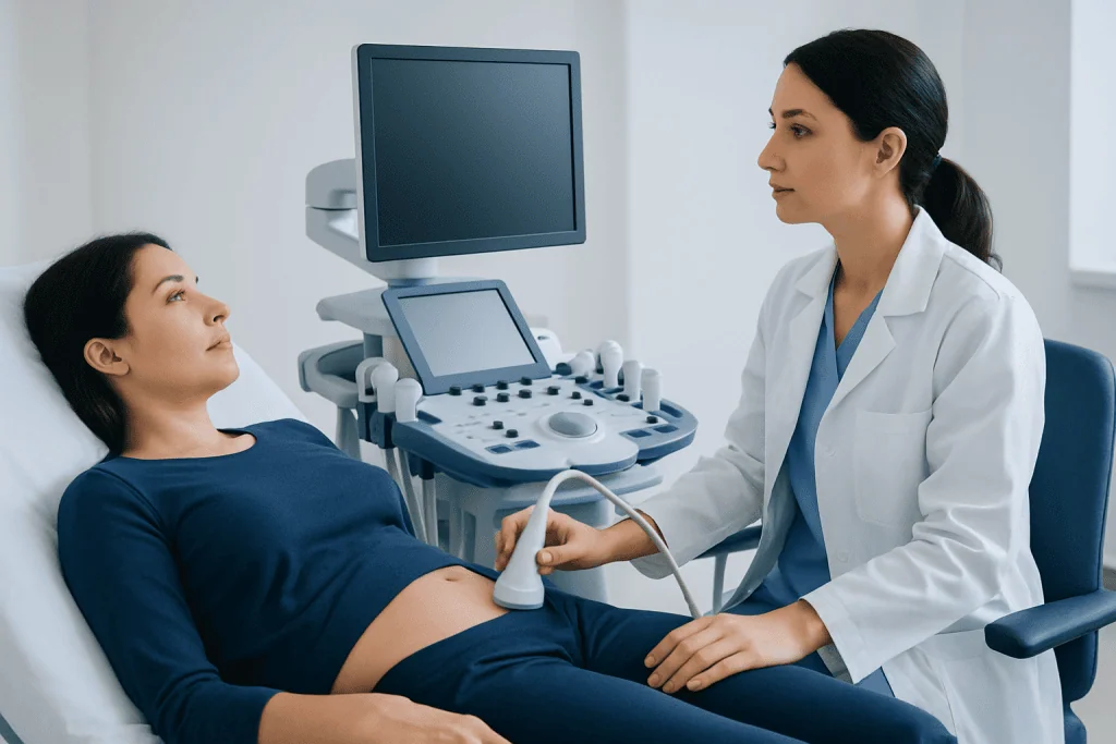
The posterior cul de sac is the lowest part of the pelvic cavity when a person is upright or lying on the back. Because of this, small amounts of fluid may settle there and become visible on imaging. This is one reason why sonographers and gynecologists pay close attention to it.
Structures near this space can affect it directly. Conditions involving the ovaries, uterus, fallopian tubes, peritoneum, or bowel may lead to inflammation, bleeding, fluid collection, or scar tissue in the posterior cul de sac. For example, endometriosis can form deposits or adhesions in this area, sometimes causing pain with periods, sex, or bowel movements.
This region may also be assessed when a person has pelvic pain, abnormal bleeding, suspected infection, or fertility concerns. In some cases, findings in the posterior cul de sac help doctors decide whether simple monitoring is enough or whether more detailed testing is needed, including evaluation for endometriosis or an ovarian cyst.
Possible symptoms linked to posterior cul de sac findings

The posterior cul de sac itself does not usually cause symptoms. Symptoms typically come from the underlying condition affecting this area. Some people have no symptoms at all, and a finding is discovered incidentally during a routine pelvic ultrasound.
When symptoms do occur, they can vary widely depending on the cause. Common complaints may include pelvic pain, cramping, pressure, painful periods, pain during sexual intercourse, or pain with bowel movements. Some people also notice bloating, abnormal vaginal bleeding, fever, nausea, or changes in urination or bowel habits.
Symptoms that need prompt medical attention include sudden severe pelvic pain, fainting, shoulder pain, fever, heavy bleeding, or signs of pregnancy with pain. These can suggest more urgent problems, such as bleeding from a ruptured cyst, ectopic pregnancy, or pelvic infection.
- Mild or moderate pelvic discomfort
- Pain during menstruation
- Pain during or after sex
- Pressure in the lower abdomen or pelvis
- Abnormal bleeding or spotting
- Fever or unusual discharge if infection is present
Common causes and risk factors
A small amount of free fluid in the posterior cul de sac can be a normal finding, especially around ovulation or just after a follicle releases an egg. In this setting, fluid is usually temporary and not a cause for concern if there are no troubling symptoms.
Other causes are linked to gynecologic or abdominal conditions. Endometriosis may create implants, inflammation, and scar tissue in this space. Pelvic inflammatory disease can lead to fluid, tenderness, or abscess formation. Bleeding from a ruptured ovarian cyst or internal pelvic bleeding may also collect there. In some cases, ectopic pregnancy, postoperative adhesions, or less commonly tumors can affect the area.
Risk factors depend on the condition involved. They may include a history of endometriosis, prior pelvic infections, sexually transmitted infections, pelvic surgery, fertility problems, ovarian cysts, or pregnancy-related complications. A doctor considers both symptoms and medical history before deciding how significant a posterior cul de sac finding may be.
Some people being evaluated for chronic pelvic pain may also need assessment for related concerns such as uterine fibroids, even though fibroids do not arise in the posterior cul de sac itself.
How doctors diagnose problems in the posterior cul de sac
Diagnosis starts with a medical history and pelvic examination. The doctor asks about pain, menstrual patterns, vaginal bleeding, sexual health, digestive symptoms, pregnancy status, and any previous surgeries or known gynecologic conditions. This helps narrow the list of likely causes.
Ultrasound, especially transvaginal ultrasound, is usually the first imaging test. It can show free fluid, cysts, masses, abscesses, or signs of deep endometriosis and adhesions. In more complex cases, magnetic resonance imaging may provide additional detail, particularly when surgery is being considered or when deep pelvic disease is suspected.
Lab tests may include a pregnancy test, blood tests, or infection screening. If the diagnosis remains uncertain, or if symptoms are severe, minimally invasive surgery may be recommended to look directly inside the pelvis. Procedures such as laparoscopy can help diagnose and sometimes treat the problem at the same time.
Treatment options and what recovery may involve
Treatment is based on the underlying cause rather than on the posterior cul de sac itself. If imaging shows only a small amount of simple fluid and the person feels well, the doctor may recommend observation and follow-up. Many temporary findings resolve without intervention.
When infection is the cause, antibiotics are commonly used. If symptoms suggest hormonal conditions such as endometriosis, treatment may include pain relief, hormonal therapy, or planned surgery depending on severity, age, fertility goals, and response to prior treatment. Management for endometriosis may involve endometriosis treatment when disease in the posterior cul de sac contributes to chronic pain.
Ovarian cyst complications, abscesses, internal bleeding, or suspected ectopic pregnancy may require urgent treatment. In selected cases, surgery is needed to stop bleeding, remove a cyst, release adhesions, drain infection, or confirm a diagnosis. If fertility is part of the discussion, doctors may tailor treatment carefully to protect reproductive health.
For some patients, a multidisciplinary approach is helpful, especially when pain is ongoing or symptoms overlap with bowel or urinary problems. Near the end of the care pathway, centers such as Acibadem International can coordinate evaluation through multidisciplinary specialists in JCI-accredited hospitals for international patients, including advanced pelvic imaging and gynecology care when appropriate.
Self-care, monitoring, and prevention
There is no specific way to prevent all posterior cul de sac findings because this area is simply an anatomical space. Prevention focuses on reducing the risk of the conditions that can affect it. Regular gynecologic care, prompt treatment of infections, and follow-up for known ovarian cysts or endometriosis are sensible steps.
At home, symptom tracking can be useful. Keeping a record of menstrual cycles, pelvic pain, bleeding, bowel symptoms, and triggers helps doctors identify patterns. People who are advised to watch and wait should understand when to return for repeat imaging or reassessment.
General pelvic health measures may include safer sex practices, completing prescribed treatment for infections, staying hydrated, and avoiding delay if new pelvic pain develops. Self-care should never replace medical evaluation when symptoms are severe, persistent, or associated with pregnancy.
When to seek medical care
Medical care is recommended if pelvic pain is persistent, worsening, or interfering with daily life. A person should also speak with a doctor if they have abnormal vaginal bleeding, painful intercourse, fever, unusual discharge, difficulty with bowel movements, or fertility concerns.
Urgent assessment is important for sudden severe pain, fainting, dizziness, heavy bleeding, vomiting, or symptoms during early pregnancy. These can point to emergencies that need prompt diagnosis and treatment. It is especially important not to ignore pain that feels very different from usual menstrual discomfort.
Even when imaging mentions the posterior cul de sac without obvious danger signs, follow-up matters if symptoms continue. A qualified doctor can explain whether the finding is normal, temporary, or a clue to a treatable pelvic condition.
Frequently asked questions
Is the posterior cul de sac a disease?
No. The posterior cul de sac is a normal space in the female pelvis behind the uterus. Doctors focus on it because fluid or signs of disease can appear there on imaging.
What does fluid in the posterior cul de sac mean?
A small amount of fluid can be normal, especially around ovulation. Larger amounts, blood, or fluid seen with pain may suggest a ruptured cyst, infection, endometriosis, or another pelvic problem.
Can posterior cul de sac findings affect fertility?
Sometimes they can, depending on the cause. For example, endometriosis, pelvic inflammatory disease, or scar tissue in this area may affect reproductive organs and fertility, while simple temporary fluid often does not.
How is a posterior cul de sac problem found?
It is often identified during a pelvic ultrasound, especially a transvaginal ultrasound. Doctors may also use a pelvic exam, lab tests, MRI, or laparoscopy if more information is needed.
Does every posterior cul de sac finding need treatment?
No. Treatment is only needed when the underlying cause requires it. Some findings are normal or resolve on their own, while others need medication, monitoring, or surgery.
Can endometriosis occur in the posterior cul de sac?
Yes. The posterior cul de sac is a common location for endometriosis implants, scar tissue, and deep pelvic disease. This may contribute to painful periods, pain during sex, or bowel-related pelvic pain.
References
- American College of Obstetricians and Gynecologists
- Office on Women's Health
- National Institutes of Health
- Radiological Society of North America
- World Health Organization
This article is for general information only and is not a substitute for professional medical advice. Please consult a qualified doctor about your individual situation.
Explore treatments in Turkey — costs, top hospitals & a free quote
JCI-accredited · board-certified surgeons · reply within 24h
Add us as a Preferred Source to see more of our trusted health content across Google Search, AI Overviews and Discover.
Check your numbers in seconds
BMI, calories, due date, blood pressure and 30+ more clinical calculators — free, instant, doctor-reviewed ranges.

More from the Health Library



Related Specialists

Prof. Dr. Nail Suat Ünver
Internal Medicine
Embriyolog Feray Farsak
Vitro Fertilization and Reproductive Medicine Center
Dt. Kerim Muhammed Aktaş
Oral & Dental Health
Dr. Zeynep Sağırkaya
Occupational Medicine