Calf Muscle Anatomy: A Complete Medical Overview
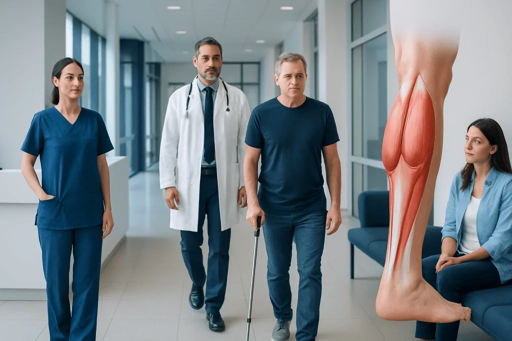
The main calf muscles are the gastrocnemius and soleus, with the plantaris present in some people as a smaller supporting muscle. These muscles join the Achilles tendon and are essential for walking, climbing stairs, standing on tiptoe, and balance.
Key Takeaways
- The main calf muscles are the gastrocnemius and soleus, with the plantaris present in some people as a smaller supporting muscle.
- These muscles join the Achilles tendon and are essential for walking, climbing stairs, standing on tiptoe, and balance.
- Calf symptoms can come from muscle strain, overuse, cramps, tendon problems, circulation issues, or nerve-related conditions.
- A physical examination is often enough to assess many calf problems, but imaging may be needed when injury or other causes are suspected.
- Self-care may help mild overuse symptoms, but sudden swelling, severe pain, weakness, or shortness of breath need prompt medical attention.
Calf muscle anatomy refers to the muscles at the back of the lower leg, mainly the gastrocnemius and soleus, which work together to point the foot downward and support walking, running, and balance. Understanding how these muscles are built and how they function can also help explain common symptoms such as tightness, strain, cramps, and Achilles-related pain.
Overview of calf muscle anatomy
Calf muscle anatomy describes the group of muscles at the back of the lower leg that create strength, stability, and movement at the ankle and knee. The two main muscles are the gastrocnemius and the soleus. Together, they form most of the visible calf and connect to the heel bone through the Achilles tendon.
The calf is not only important for sports. It is active during everyday tasks such as walking, standing, climbing stairs, and pushing off the ground. These muscles also help the body stay upright and steady, especially when standing for long periods or changing direction.
A complete medical overview of calf muscle anatomy also includes nearby tendons, nerves, blood vessels, and connective tissue. This broader view matters because pain or tightness in the calf does not always come from the muscle itself. Problems involving the Achilles tendon, veins, nerves, or nearby joints can create similar symptoms.
Main muscles of the calf and how they are arranged
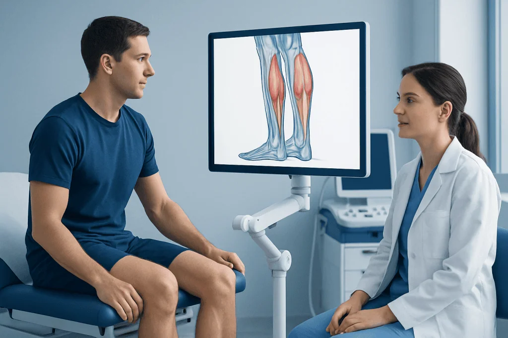
The gastrocnemius is the larger, more superficial calf muscle. It has two heads, called the medial and lateral heads, which start above the knee from the lower end of the thigh bone. Because it crosses both the knee and ankle joints, it helps bend the knee and point the foot downward, a movement known as plantarflexion.
Underneath the gastrocnemius lies the soleus, a broad and powerful muscle that starts below the knee from the tibia and fibula. The soleus does not cross the knee joint, so its main role is plantarflexion at the ankle. It is especially important for posture and endurance activities such as walking and standing.
A smaller muscle called the plantaris may also be present. It has a small muscle belly and a long thin tendon, and some people do not have it at all. It usually plays only a minor role in movement, but it can sometimes be involved in calf pain or be mistaken for a muscle injury during assessment.
The gastrocnemius and soleus join together to form the Achilles tendon, the thick tendon that attaches to the heel bone. This arrangement allows the calf to transmit force efficiently from muscle to foot, making strong push-off movements possible.
How the calf muscles work in movement and circulation
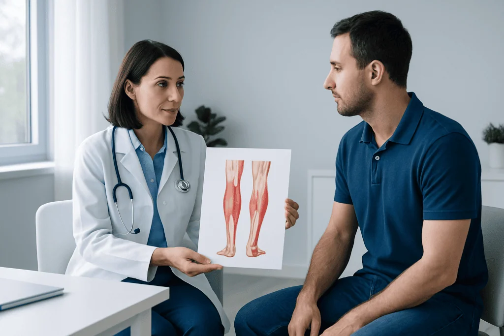
The calf muscles generate plantarflexion, which means pointing the toes downward. This action is needed for walking, running, jumping, and rising onto the toes. During gait, the calf helps control the body as it moves over the foot, then provides the final push that moves the body forward.
The gastrocnemius is more active during faster and more forceful activities, especially when the knee is straight. The soleus is particularly important during slower, steady movements and postural control. Because the soleus contains many fatigue-resistant muscle fibers, it is well suited to long periods of standing and walking.
The calf also supports circulation. As the muscles contract, they help squeeze deep veins and assist the return of blood from the legs to the heart. This function is sometimes called the muscle pump. Reduced movement, prolonged sitting, or certain vascular conditions can interfere with this mechanism and contribute to swelling or discomfort.
Healthy calf function depends on coordinated work between muscles, the Achilles tendon, bones, joints, and nerves. If one part is irritated or injured, movement patterns may change and symptoms may spread to the ankle, heel, or knee. In some cases, calf symptoms may overlap with Achilles tendonitis.
Blood supply, nerves, and nearby structures
The calf receives blood from branches of the popliteal and tibial arteries. Good blood flow is important for oxygen delivery, muscle performance, and healing after activity or injury. Reduced circulation can cause cramping, fatigue, or pain with walking, especially in people with vascular disease.
Nerve supply to the main calf muscles comes primarily from the tibial nerve, a branch of the sciatic nerve. This nerve allows the muscles to contract and also contributes to sensation in nearby areas of the lower leg and foot. Nerve irritation or compression higher up in the leg or lower back may sometimes be felt as calf discomfort or weakness.
Several structures around the calf can contribute to symptoms that people may think are purely muscular. These include the Achilles tendon, deep veins, fascial compartments, the knee joint, and the ankle joint. Because of this, a careful medical assessment is often needed if pain is persistent, severe, or unexplained.
Some conditions require urgent attention because they may mimic a muscle problem. For example, calf swelling and tenderness can sometimes be related to a blood clot such as deep vein thrombosis. This is one reason sudden or one-sided calf symptoms should not be ignored.
Common symptoms and what they may suggest
Symptoms related to calf muscle anatomy can include pain, tightness, cramping, weakness, tenderness, swelling, or difficulty pushing off when walking. Athletes may notice sharp pain during sprinting or jumping, while other people may feel aching after long periods of standing or walking. The exact pattern often helps guide the cause.
A calf strain commonly causes sudden pain, tenderness, and difficulty bearing weight or standing on tiptoe. Overuse can lead to aching, stiffness, or a sense of heaviness that develops more gradually. Muscle cramps tend to come on suddenly and may be linked to dehydration, prolonged exertion, or nighttime tightening of the muscle.
Not all calf pain starts in the muscle itself. Problems involving the Achilles tendon, nerves, or blood vessels may feel similar. A clinician may also consider conditions such as peripheral artery disease when calf pain occurs repeatedly with walking and improves with rest.
- Sudden sharp pain may suggest a strain or tendon injury.
- Cramping may be linked to fatigue, hydration, medications, or circulation issues.
- Swelling, warmth, or redness may point to inflammation or a vascular cause.
- Weakness or numbness may raise concern for nerve involvement.
Causes of calf problems and who is at risk
Calf problems can develop from sports injuries, sudden increases in activity, poor conditioning, tight muscles, or incomplete warm-up. Running, tennis, football, hiking, and activities that involve repeated push-off can place significant stress on the gastrocnemius and soleus. The gastrocnemius is especially prone to strain during explosive movements.
Age, previous injury, reduced flexibility, poor footwear, and altered walking mechanics can increase risk. Tendon disorders, arthritis, and muscle imbalances may place extra load on the calf. Inactive periods followed by sudden exercise can also trigger pain because the muscle is not prepared for the demand.
Medical causes should also be considered. Circulation problems, nerve compression, electrolyte imbalance, and medication side effects may all affect the calf. In some people, Achilles problems are part of the picture, and treatment may include targeted assessment of the tendon and surrounding structures, sometimes including Achilles tendon repair when significant structural injury is present.
Less commonly, persistent calf symptoms may be related to compartment syndrome, inflammatory conditions, or referred pain from the lower back. For this reason, ongoing or unusual symptoms should be evaluated rather than assumed to be a simple strain.
How doctors evaluate calf pain or injury
Assessment begins with a medical history and physical examination. A doctor usually asks when the symptoms began, whether there was a sudden injury, what activities make the symptoms worse, and whether swelling, redness, numbness, or weakness are present. The pattern of pain during walking, standing, or exercise can provide important clues.
On examination, the clinician may check tenderness, bruising, swelling, range of motion, strength, and the ability to walk or stand on tiptoe. They may compare both legs and assess the Achilles tendon, ankle, knee, pulses, and sensation. This helps distinguish muscle strain from tendon injury, vascular problems, or nerve-related causes.
Imaging is not always needed for mild muscle symptoms, but it may be recommended when a tear, tendon rupture, or another condition is suspected. Ultrasound can help assess soft tissue injuries, while MRI gives more detailed images of muscle and tendon structures. If a blood clot is a concern, urgent vascular ultrasound may be needed. In selected cases of serious tendon damage, treatment may involve orthopedic surgery.
Blood tests or additional vascular or neurological studies may be used when the history suggests a non-muscular cause. The goal is to identify the exact source of the symptom so care can be matched to the condition.
Treatment, self-care, and when to seek medical care
Treatment depends on the cause. Mild calf strain or overuse symptoms often improve with relative rest, gentle movement, activity modification, and gradual return to exercise. Some people benefit from supervised stretching and strengthening through physical therapy and rehabilitation, especially when tightness, weakness, or recurring injury is present.
If the problem involves the Achilles tendon, a more structured treatment plan may be needed, including tendon-loading exercises, footwear advice, and medical follow-up. Severe tears, major weakness, or loss of function may require specialist treatment. Near the end of the care pathway, Acibadem International’s multidisciplinary specialists in JCI-accredited hospitals diagnose and treat calf, tendon, and lower-limb conditions for international patients.
Self-care is most appropriate when symptoms are mild and clearly related to exercise or temporary overuse. General measures include warming up before activity, increasing training gradually, wearing supportive footwear, staying active during long travel or sitting periods, and addressing flexibility and strength imbalances.
Medical care should be sought promptly for sudden severe calf pain, a popping sensation, marked swelling, one-sided redness or warmth, inability to bear weight, new numbness, or shortness of breath. These symptoms can suggest a significant muscle or tendon injury, a vascular problem, or another condition that needs urgent assessment.
Frequently asked questions
What muscles make up the calf?
The calf is mainly formed by the gastrocnemius and soleus muscles. A smaller muscle called the plantaris may also be present, but it is less important for overall calf strength and function.
What is the difference between the gastrocnemius and soleus?
The gastrocnemius is the larger, more visible calf muscle and crosses both the knee and ankle joints. The soleus sits underneath it, crosses only the ankle, and plays a major role in posture, standing, and endurance walking.
Why does calf pain sometimes happen during walking?
Calf pain during walking can come from muscle overuse, strain, tightness, tendon irritation, or poor conditioning. In some cases, it may also be related to circulation problems, so repeated pain with walking should be assessed by a doctor.
Can calf muscle anatomy explain Achilles tendon pain?
Yes. The gastrocnemius and soleus join to form the Achilles tendon, so tightness or overload in the calf can increase stress on the tendon. This is one reason calf strength and flexibility are often considered when evaluating Achilles symptoms.
How do doctors tell if calf pain is a strain or something more serious?
Doctors look at how the pain started, whether there was a sudden injury, and whether swelling, weakness, redness, or numbness are present. A physical examination is often very helpful, and imaging or vascular tests may be used if a tear, clot, or other condition is suspected.
When should someone worry about calf swelling?
Calf swelling deserves prompt attention if it is sudden, affects one leg, or comes with warmth, redness, tenderness, chest pain, or shortness of breath. These features can point to a vascular problem and should be checked urgently.
References
- American Academy of Orthopaedic Surgeons
- National Library of Medicine
- Cleveland Clinic
- MedlinePlus
- American College of Sports Medicine
This article is for general information only and is not a substitute for professional medical advice. Please consult a qualified doctor about your individual situation.
Joint and spine care in Turkey — expert assessment & treatment
JCI-accredited · board-certified surgeons · reply within 24h
Add us as a Preferred Source to see more of our trusted health content across Google Search, AI Overviews and Discover.