Liver Ultrasound Photos: A Complete Medical Overview

Liver ultrasound is a painless imaging test that uses sound waves and does not involve ionizing radiation. A single ultrasound image rarely provides a diagnosis; interpretation depends on the complete scan, measurements and clinical context.
Key Takeaways
- Liver ultrasound is a painless imaging test that uses sound waves and does not involve ionizing radiation.
- A single ultrasound image rarely provides a diagnosis; interpretation depends on the complete scan, measurements and clinical context.
- Ultrasound may identify fatty change, cysts, enlargement, fluid, bile duct changes or focal areas that need further assessment.
- Not every finding is serious, and many liver ultrasound changes are common or benign.
- Further tests such as blood work, elastography, CT or MRI may be recommended when ultrasound findings are unclear or need closer evaluation.
Liver ultrasound photos are grayscale images created by sound waves, not ordinary photographs. They provide useful information about the liver’s size, texture, blood vessels and surrounding structures, but a radiologist or qualified clinician must interpret them alongside symptoms, medical history and, when needed, blood tests.
Overview: What Liver Ultrasound Photos Show
Liver ultrasound photos are still images captured during an ultrasound examination of the upper abdomen. The scan uses a handheld device called a transducer to send high-frequency sound waves through the skin. Reflected sound waves are converted into moving images and saved photographs that show the liver and nearby organs.
These images can help clinicians evaluate liver size, overall appearance, tissue texture, blood vessels, bile ducts, the gallbladder and, in many cases, the spleen and kidneys. A liver ultrasound may be requested after abnormal liver blood tests, upper abdominal symptoms, an injury, or as follow-up for a known liver condition.
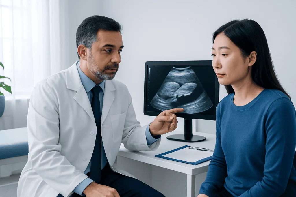
It is important to view liver ultrasound photos as part of a medical assessment rather than as standalone pictures to interpret at home. The appearance of a structure can vary with the machine settings, scanning angle, body shape and preparation for the test. A formal radiology report explains what was seen and whether additional evaluation is appropriate.
How to Read the Main Features in Ultrasound Images
Most liver ultrasound photos appear in shades of gray, black and white. Fluid usually appears black because sound travels through it with few echoes. Solid tissues create different shades of gray depending on how strongly they reflect sound. The liver normally has a relatively uniform, medium-gray appearance, although normal appearance varies from person to person.
The sonographer captures images from several angles rather than relying on one view. The report may include measurements of the liver and spleen, images of the liver’s right and left lobes, views of the portal vein and hepatic veins, and images of the gallbladder and bile ducts. Doppler ultrasound may add color or waveforms to show the direction and speed of blood flow.
Words such as echogenicity, echotexture, hyperechoic and hypoechoic describe how tissues reflect sound. For example, increased echogenicity means the liver looks brighter than expected. These are imaging descriptions, not diagnoses by themselves, and their significance depends on the full pattern and the person’s health information.
- Homogeneous: a generally even tissue appearance.
- Diffuse change: an appearance affecting much of the liver rather than one small area.
- Focal lesion: a localized area that looks different from surrounding tissue.
- Simple cyst: a fluid-filled area with typical benign imaging features.
- Vascular flow: blood movement assessed with Doppler imaging.
Common Findings Seen on Liver Ultrasound Photos
One frequent finding is increased liver echogenicity, sometimes described as a “bright liver.” This can be consistent with fatty liver, also called hepatic steatosis, particularly when the liver appears brighter than the kidney on the same scan. Ultrasound can suggest moderate or more marked fatty change, but it is less reliable for detecting mild fat and cannot precisely measure the amount of fat present.
Ultrasound may also show an enlarged liver, known as hepatomegaly, or an enlarged spleen. These findings have several possible causes and need interpretation with blood tests, symptoms and medical history. A coarse or uneven texture can occur in chronic liver disease, but ultrasound alone cannot confirm the stage of fibrosis or cirrhosis in every case.
Small simple liver cysts are commonly found incidentally. They often look like round or oval black areas with a thin wall and are usually harmless. Other focal findings, including benign blood-vessel growths such as hemangiomas, may have recognizable features, but some areas cannot be fully characterized by ultrasound. In that situation, a clinician may recommend contrast-enhanced imaging such as CT or MRI.
The scan can also identify fluid around the liver or in the abdomen, widening of bile ducts, gallstones, and changes in major blood vessels. These observations may help explain symptoms such as abdominal discomfort or jaundice, but they must be correlated carefully with laboratory results and examination findings.
What Ultrasound Cannot Tell You Alone
Liver ultrasound is valuable, but it has limits. It does not directly measure liver function, so normal-looking liver ultrasound photos do not necessarily mean liver blood tests are normal. Conversely, a mildly abnormal appearance does not always mean there is significant liver damage.
Image quality can be reduced by bowel gas, excess body tissue, inability to hold a breath briefly, or limited visibility of a deep area of the liver. Very small lesions may not be visible, and ultrasound can sometimes have difficulty distinguishing among different types of focal liver abnormalities. The test is therefore often one step in a broader diagnostic process.
If there is concern about liver scarring, clinicians may request elastography, which estimates tissue stiffness. Blood tests can assess inflammation and liver function, while CT or MRI can provide more detailed information about anatomy and certain lesions. In selected circumstances, specialists may discuss biopsy, although it is not required for every person with an abnormal ultrasound result.
People should avoid drawing conclusions by comparing their images with examples online. Similar-looking ultrasound patterns can have different causes, and the written report is designed to summarize the clinically meaningful findings. Asking the ordering clinician to explain the report is the safest and most useful approach.
Why a Liver Ultrasound May Be Requested
A clinician may arrange a liver ultrasound because of persistently abnormal liver enzymes, a feeling of fullness in the upper right abdomen, unexplained abdominal pain, nausea, jaundice, itching, or a history of viral hepatitis, alcohol-related liver injury, metabolic conditions or certain medicines. It may also be used to monitor a previously identified cyst, fatty liver changes or chronic liver disease.
People with metabolic risk factors, including excess weight, type 2 diabetes, high cholesterol or high triglycerides, may be assessed for fatty liver disease. The ultrasound result can support a care plan that addresses overall metabolic health, but it should not be used as the only measure of risk or treatment response.
In urgent settings, ultrasound can help evaluate suspected bile duct blockage, gallbladder disease, abdominal fluid or blood-flow problems. However, the choice of imaging test depends on the clinical question. A normal ultrasound does not rule out every cause of abdominal symptoms, and the clinician may recommend other tests if symptoms continue.
Preparing for the Scan and Understanding the Report
Preparation instructions vary by facility and reason for the scan. For many abdominal ultrasounds, patients are asked not to eat for several hours beforehand. Fasting can reduce bowel gas and keep the gallbladder fuller, which may improve visibility. Water and regular medicines may often be allowed, but the imaging center’s instructions should always be followed.
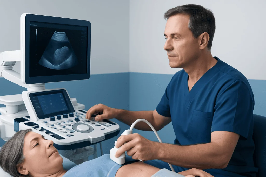
During the examination, gel is placed on the upper abdomen and the transducer is moved over the skin. Gentle pressure may be applied, and the person may be asked to change position or hold their breath for a few seconds. The procedure is usually comfortable, takes a relatively short time, and does not use radiation.
A report commonly includes the liver’s size and echotexture, whether a focal lesion was seen, the appearance of the portal vein and bile ducts, and relevant findings in nearby structures. Phrases such as “no focal lesion identified” or “no biliary dilatation” are generally reassuring, but the report should still be reviewed in the context of the reason for testing.
If the report recommends follow-up, this does not necessarily indicate a serious problem. It may simply mean that another imaging method can provide a clearer answer. Keeping copies of previous reports can help clinicians compare whether a finding is stable over time.
Supporting Liver Health After an Ultrasound
The appropriate next step depends on the results. When fatty liver is suspected, clinicians commonly focus on sustainable lifestyle measures such as balanced eating patterns, regular physical activity, gradual weight management when appropriate, diabetes and cholesterol management, and avoiding unnecessary alcohol exposure. Individual advice is especially important for people who have existing liver disease or take regular medicines.
People should not stop prescribed medicines or begin supplements based on ultrasound photos alone. Some supplements and herbal products can affect the liver or interact with medication. A doctor or pharmacist can help review what is safe, particularly when liver blood tests are abnormal.
Follow-up may involve repeat blood tests, an elastography examination, or more detailed imaging. Acibadem International’s multidisciplinary specialists and JCI-accredited hospitals evaluate liver and digestive health concerns for international patients, using imaging results alongside clinical assessment rather than relying on a single image.
When to Seek Medical Care
People should arrange a medical appointment to discuss liver ultrasound photos if the report shows an unexpected finding, if liver blood tests are abnormal, or if symptoms persist despite a normal scan. Timely review is also sensible for a newly detected liver lesion, enlarged liver, bile duct change, abdominal fluid or a recommendation for additional imaging.
Urgent medical assessment is needed for yellowing of the skin or eyes, severe or worsening abdominal pain, vomiting that prevents fluid intake, fever with abdominal pain, confusion, fainting, vomiting blood, or black stools. These symptoms can have many causes, but they should not be managed by waiting for an outpatient imaging result.
For less urgent questions, patients can ask their clinician what the finding means, whether it is likely related to their symptoms, whether blood tests are needed, and when follow-up should occur. Clear communication helps turn a technical imaging report into an individualized care plan.
Frequently asked questions
Are liver ultrasound photos accurate?
Liver ultrasound photos can accurately show many structural features, including liver size, fluid-filled cysts, gallstones, bile duct widening and moderate to marked fatty change. Their accuracy depends on the question being asked, image quality and the experience of the imaging team. Some findings require blood tests, elastography, CT or MRI for confirmation or more detail.
What does a bright liver on ultrasound mean?
A bright liver means the liver reflects more ultrasound waves than expected and therefore appears whiter or more echogenic on the image. This pattern is often associated with fatty change, but it can also be influenced by other diffuse liver changes and technical factors. A clinician should interpret it with liver blood tests and relevant health history.
Can an ultrasound show liver cancer?
Ultrasound can detect some liver masses and may be used for surveillance in selected people at increased risk of liver cancer. However, it cannot confirm the nature of every lesion or rule out all small abnormalities. If a suspicious area is seen, contrast-enhanced CT or MRI is often used to investigate it further.
Can liver ultrasound photos show cirrhosis?
Ultrasound may show features that raise concern for advanced chronic liver disease, such as an irregular liver surface, altered shape, enlarged spleen or abdominal fluid. However, it cannot reliably stage all degrees of fibrosis or confirm cirrhosis by itself. Elastography, blood tests and specialist assessment can provide additional information.
Do people need to fast before a liver ultrasound?
Many abdominal ultrasound appointments require fasting for several hours because food can increase bowel gas and affect gallbladder appearance. The exact instructions vary according to the facility and the type of scan. Patients should follow the instructions provided by their imaging center and ask about regular medicines if unsure.
Is a normal liver ultrasound enough to rule out liver disease?
No. A normal ultrasound is reassuring for many structural abnormalities, but it does not directly assess liver function or identify every type of liver disease. If symptoms, risk factors or abnormal blood tests remain present, a clinician may recommend further evaluation.
References
- American College of Radiology
- Radiological Society of North America
- National Institute of Diabetes and Digestive and Kidney Diseases
- European Association for the Study of the Liver
- World Gastroenterology Organisation
This article is for general information only and is not a substitute for professional medical advice. Please consult a qualified doctor about your individual situation.
Ultrasound in Turkey — costs, top hospitals & a free quote
JCI-accredited · board-certified surgeons · reply within 24h
Add us as a Preferred Source to see more of our trusted health content across Google Search, AI Overviews and Discover.

Check-up & Preventive Medicine
Comprehensive health screenings designed for early detection and prevention.
1 specialists in this unit