Bleeding Mole: An Evidence-Based Guide for Patients
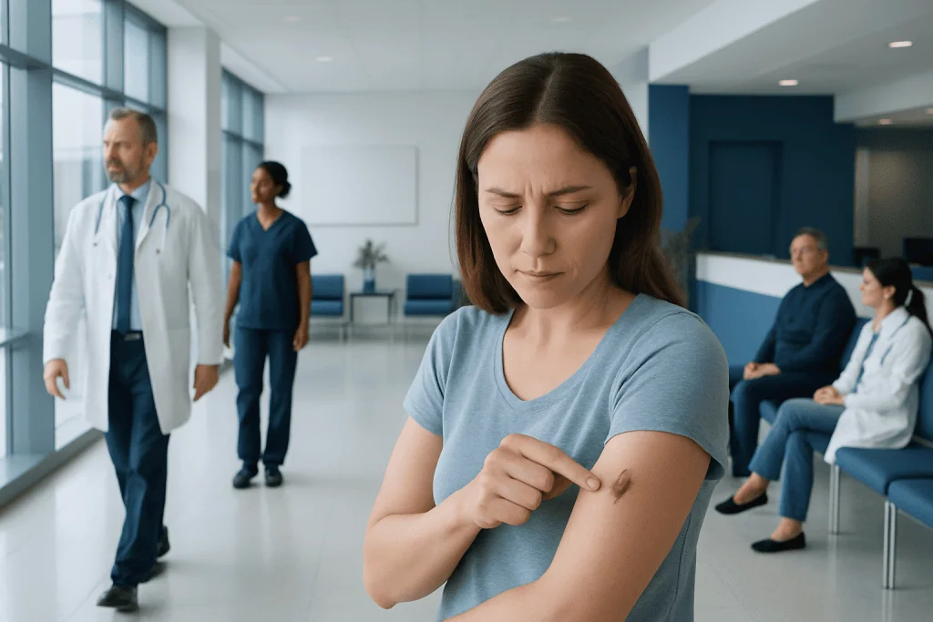
Most bleeding moles result from trauma or friction, especially in areas affected by clothing, shaving, or jewelry. Bleeding alone does not diagnose melanoma, but unexplained, recurrent, or persistent bleeding needs medical review.
Key Takeaways
- Most bleeding moles result from trauma or friction, especially in areas affected by clothing, shaving, or jewelry.
- Bleeding alone does not diagnose melanoma, but unexplained, recurrent, or persistent bleeding needs medical review.
- The ABCDE pattern and the “ugly duckling” sign can help people notice moles that look different or are changing.
- A dermatologist can examine a concerning mole with dermoscopy and may recommend a biopsy or removal if needed.
- Avoid picking, cutting, or attempting to remove a mole at home; protect it and arrange an evaluation when appropriate.
A bleeding mole is commonly caused by rubbing, scratching, shaving, or another minor injury. However, a mole that bleeds without a clear cause, bleeds repeatedly, or changes in appearance should be assessed by a qualified clinician to rule out skin cancer and other conditions.
Bleeding Mole: What It Usually Means
A bleeding mole is a mole that has broken open or released blood from its surface. In many cases, this happens because the mole has been accidentally scratched, caught by clothing, nicked while shaving, rubbed by a bra strap, or irritated by sports equipment. Because moles may be raised and contain small blood vessels, even minor trauma can make them bleed.
Still, a mole should not be assumed harmless simply because an injury seems possible. A mole that begins bleeding without being scratched or injured, continues to bleed, forms a recurring scab, or changes in color, shape, size, or sensation deserves professional assessment. These changes can occur with benign skin growths, but they can also be signs that the lesion needs closer examination.
Bleeding is not, by itself, proof of melanoma or another skin cancer. The most useful question is whether the bleeding is part of a broader change. Looking at the mole’s history, its appearance, and whether it differs from a person’s other moles helps a clinician decide what should happen next.
Why Can a Mole Bleed?
The most frequent explanation is direct physical irritation. Moles on the neck, scalp, face, underarms, waistline, groin, feet, or areas where clothing fits tightly are more likely to be rubbed or injured. Shaving over a raised mole can cut its surface, and scratching due to dry skin, eczema, insect bites, or irritation can also cause bleeding.
Some benign lesions are naturally more delicate than ordinary flat moles. For example, raised intradermal nevi, skin tags, seborrheic keratoses, and blood-vessel growths may catch on clothing or bleed after small injuries. It is not always possible to identify a lesion accurately by appearance alone, particularly after it has become inflamed or scabbed.
Less commonly, bleeding may be associated with an abnormal or cancerous skin lesion. Melanoma can sometimes bleed, ooze, crust, itch, or become tender, usually alongside a new or evolving appearance. Non-melanoma skin cancers, including basal cell carcinoma and squamous cell carcinoma, can also form a sore that repeatedly crusts or bleeds and does not heal as expected.
People who take blood-thinning medicines or have a bleeding disorder may notice more prolonged bleeding after a small skin injury. These factors do not explain why a mole changed, so a new or unusual lesion should still be checked when it is persistent or concerning.
Changes That Deserve Closer Attention
A useful way to observe pigmented moles is the ABCDE guide. “A” refers to asymmetry, meaning one half does not resemble the other. “B” means an irregular, blurred, scalloped, or uneven border. “C” refers to uneven color, such as a mixture of brown, black, tan, red, white, or blue. “D” means a diameter that is growing or is larger than about 6 millimeters, although melanomas may be smaller. “E” stands for evolving: any meaningful change over time.
For a bleeding mole, evolving is especially important. Warning features include spontaneous bleeding, repeated bleeding after minimal contact, a scab that returns after healing, a sore that does not heal, new elevation, darkening, loss of color, or changes in texture. Persistent itch, pain, tenderness, or oozing may also warrant an examination, especially when they are new.
The “ugly duckling” approach is another practical tool. Most people’s ordinary moles tend to share a general pattern. A mole that looks noticeably different from the others in color, size, shape, or behavior may need attention even if it does not match every ABCDE feature.
Not every changing spot is dangerous, and moles may change gradually during childhood, adolescence, pregnancy, or with age. Nevertheless, a clinician should evaluate a new, changing, or bleeding pigmented lesion rather than relying on self-diagnosis. This is particularly important for people with a personal or family history of melanoma, many moles, fair skin that burns easily, or substantial past ultraviolet exposure.
What to Do if a Mole Starts Bleeding
If a mole has been accidentally injured, gentle first aid is usually appropriate. Wash hands, apply steady direct pressure with clean gauze or a clean cloth for several minutes, and cover the area with a simple dressing once bleeding has stopped. Avoid repeatedly lifting the cloth to check, as this can disturb the forming clot.
It is best not to pick at a scab, use harsh antiseptics repeatedly, or try to burn, cut, tie off, or remove the mole at home. Home removal can cause infection, scarring, and incomplete removal. It may also make it harder for a pathologist to evaluate the lesion if there is a concern about skin cancer.
After an obvious injury, note whether the area heals normally. Taking a clear photograph after bleeding has stopped can be helpful for tracking changes and for discussing the lesion with a clinician. The photograph should supplement, not replace, an in-person assessment if the mole is unusual or keeps bleeding.
Seek urgent medical help if bleeding is heavy, does not stop with continuous firm pressure, or is associated with a deep wound. People using anticoagulant medicines should follow their clinician’s instructions about prolonged bleeding and should not stop prescribed medication without medical advice.
How a Bleeding Mole Is Evaluated
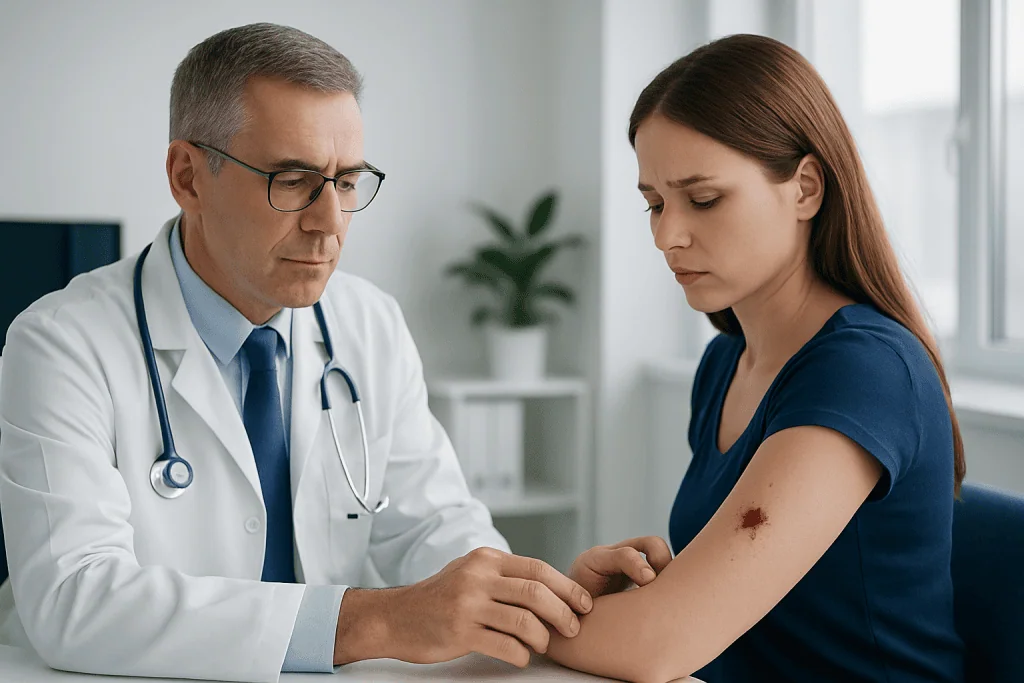
A primary care clinician or dermatologist will usually begin by asking when the mole first appeared, whether it has changed, whether there was an injury, and whether there is a personal or family history of skin cancer. They will examine the lesion and may inspect the surrounding skin and other moles as well.
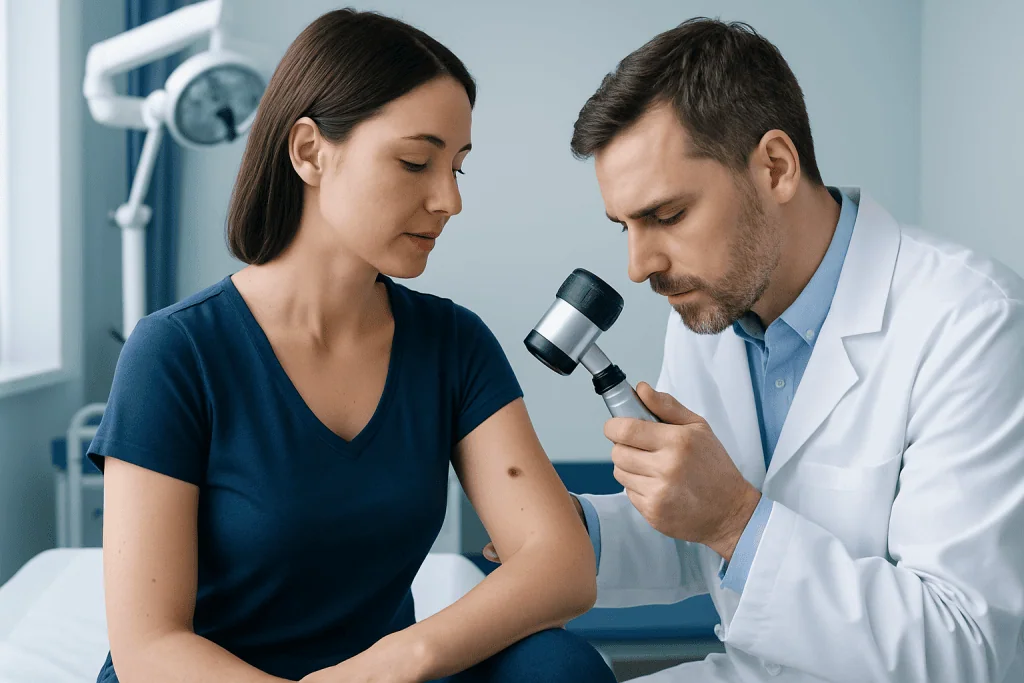
Dermatologists often use dermoscopy, a handheld lighted instrument that magnifies skin structures not visible to the unaided eye. Dermoscopy is painless and can help distinguish patterns associated with common benign moles from patterns that require further assessment. It does not involve cutting the skin.
If a lesion is suspicious or cannot be confidently identified, the clinician may recommend a skin biopsy. In this procedure, all or part of the lesion is removed under local anesthetic and examined in a laboratory by a pathologist. A biopsy is the reliable method for confirming whether melanoma, another skin cancer, or a benign condition is present.
Management depends on the result. A benign but frequently irritated mole may be removed for comfort or to prevent repeated injury. If skin cancer is diagnosed, treatment is planned according to the specific type, depth, location, and other clinical findings. Early evaluation generally allows clearer decision-making and appropriate care.
Skin Protection and Self-Care
Protecting the skin from ultraviolet radiation is an important part of reducing the risk of new sun-related skin damage and skin cancers. This includes seeking shade when practical, wearing protective clothing and a wide-brimmed hat, and using broad-spectrum sunscreen with an SPF of 30 or higher on exposed skin. Sunscreen should be reapplied according to its label, particularly after swimming, sweating, or towel drying.
It is also sensible to avoid intentional tanning and tanning beds. Ultraviolet radiation can damage skin cells and may contribute to melanoma and other skin cancers. Sun protection is useful year-round, including on cloudy days, because ultraviolet exposure can still occur.
A regular skin self-check can help people notice change. Once a month, examine the front and back of the body, arms, hands, legs, soles, scalp, and areas that are difficult to see using mirrors or help from someone trusted. The aim is not to diagnose a mole, but to notice spots that are new, different, or evolving.
People who have many atypical moles, previous melanoma, a strong family history of melanoma, or immune suppression may need personalized surveillance advice from a dermatologist. A clinician can recommend how often professional skin examinations are appropriate for the individual.
When to Seek Medical Care
Arrange a medical appointment promptly for a bleeding mole that bled without a clear injury, bleeds more than once, does not heal, or develops a recurring crust or ulcer. An appointment is also appropriate when a mole is growing, changing color or border, becoming asymmetrical, or looking distinctly different from other moles.
Medical review is particularly important if the lesion is new in adulthood, is dark or multi-colored, or is associated with persistent itching, pain, tenderness, or discharge. These features often have non-cancerous explanations, but they should be assessed rather than monitored indefinitely at home.
Emergency care may be needed when skin bleeding is severe or does not stop after sustained direct pressure, especially after a significant injury or in someone taking blood-thinning medication. For most minor shaving or scratching injuries, a routine appointment is sufficient if the mole heals fully and has no concerning changes.
Acibadem International’s multidisciplinary specialists and JCI-accredited hospitals can assess suspicious skin lesions and coordinate diagnostic and treatment planning for international patients. A qualified dermatologist can provide the most appropriate advice based on the lesion’s appearance, medical history, and examination findings.
Frequently asked questions
Is a bleeding mole always cancer?
No. Many moles bleed because they have been scratched, shaved, rubbed, or otherwise injured. However, bleeding that occurs without an obvious cause, keeps coming back, or accompanies changes in the mole should be evaluated by a clinician.
Can a normal mole bleed after shaving?
Yes. Raised moles can be cut easily during shaving and may bleed more than expected because the skin is disrupted. Apply direct pressure and let the area heal, but arrange an assessment if it repeatedly bleeds or has other changes.
How long should a bleeding mole take to heal?
A minor surface injury often begins to improve over several days and should continue healing over the following weeks. A spot that remains open, crusted, bleeding, or unhealed should be examined, particularly if there was no clear injury.
Should a person remove a mole that keeps catching on clothing?
A clinician can assess whether removal is appropriate for a repeatedly irritated mole. It should not be removed at home, as this can lead to bleeding, infection, scarring, and loss of tissue needed for an accurate diagnosis.
What does melanoma bleeding look like?
There is no single appearance that reliably identifies melanoma. A suspicious lesion may bleed or crust repeatedly, but melanoma can also appear as a new or changing pigmented spot without bleeding. A professional examination is needed to determine the cause.
Can a photograph help monitor a bleeding mole?
A well-lit photograph can help document the appearance of a mole after bleeding has stopped and may show whether it changes over time. It cannot diagnose the lesion or replace a dermatology appointment when warning signs are present.
References
- American Academy of Dermatology
- American Cancer Society
- National Cancer Institute
- NHS
- World Health Organization
This article is for general information only and is not a substitute for professional medical advice. Please consult a qualified doctor about your individual situation.
Oncology care in Turkey — second opinion and treatment plan
JCI-accredited · board-certified surgeons · reply within 24h
Add us as a Preferred Source to see more of our trusted health content across Google Search, AI Overviews and Discover.