Bubble Study: What Patients Need to Know

A bubble study uses sterile saline mixed with a small amount of air to create visible microbubbles during an echocardiogram. The test can help detect a right-to-left shunt, including a patent foramen ovale (PFO) or an abnormal blood-vessel connection in the lungs.
Key Takeaways
- A bubble study uses sterile saline mixed with a small amount of air to create visible microbubbles during an echocardiogram.
- The test can help detect a right-to-left shunt, including a patent foramen ovale (PFO) or an abnormal blood-vessel connection in the lungs.
- Most people need little preparation and can return to normal activities shortly after a standard transthoracic study.
- A positive bubble study does not always mean treatment is needed; results must be interpreted alongside symptoms, medical history, and other tests.
- Complications are uncommon, but patients should tell the care team about allergies, breathing concerns, pregnancy, or prior reactions to medical tests.
A bubble study is a specialized ultrasound test of the heart, usually performed during an echocardiogram, that helps clinicians identify an abnormal connection or passageway in the heart or lungs. It is minimally invasive, does not use radiation, and is commonly used to investigate unexplained stroke, low oxygen levels, or certain heart findings.
What Is a Bubble Study?
A bubble study, also called an agitated saline contrast study or bubble echocardiogram, is an ultrasound-based test used to look for unusual blood flow through the heart or lungs. A clinician injects a small amount of sterile saltwater solution that has been gently mixed to form tiny bubbles into a vein, usually in the arm. An echocardiogram then tracks the bubbles as they travel through the right side of the heart.
Normally, these microbubbles are filtered out by the lungs and do not appear in the left side of the heart. If bubbles appear there unusually quickly, it may suggest a pathway that allows blood to pass from the right side of the circulation to the left without following the usual route through the lungs. This is called a right-to-left shunt.
The bubble solution is not the same as the iodine-containing contrast used for some CT scans, and the study does not involve radiation. It is generally an added part of a standard heart ultrasound rather than a separate major procedure.
Why a Bubble Study May Be Recommended
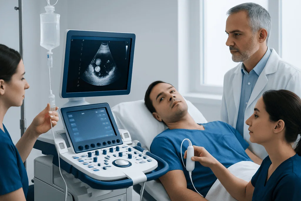
Clinicians may recommend a bubble study when they need more information about a possible opening between the upper heart chambers, particularly a patent foramen ovale (PFO). Before birth, the foramen ovale is a normal opening that helps blood circulate. It closes after birth in most people, but it remains partly open in some adults.
A bubble study may be part of an evaluation after a stroke or transient ischemic attack when no clear cause has been found, especially in a younger person or someone without typical vascular risk factors. It may also be considered for unexplained low blood oxygen levels, shortness of breath in certain positions, suspected congenital heart conditions, or evaluation of an abnormal connection between blood vessels in the lungs.
The test does not diagnose every cause of neurological symptoms, breathing symptoms, or stroke. Instead, it provides one piece of information that helps the clinical team decide whether further assessment is appropriate. Findings are considered together with a physical examination, medical history, ECG, standard echocardiography, brain imaging, and blood-vessel studies when needed.
How the Test Is Performed
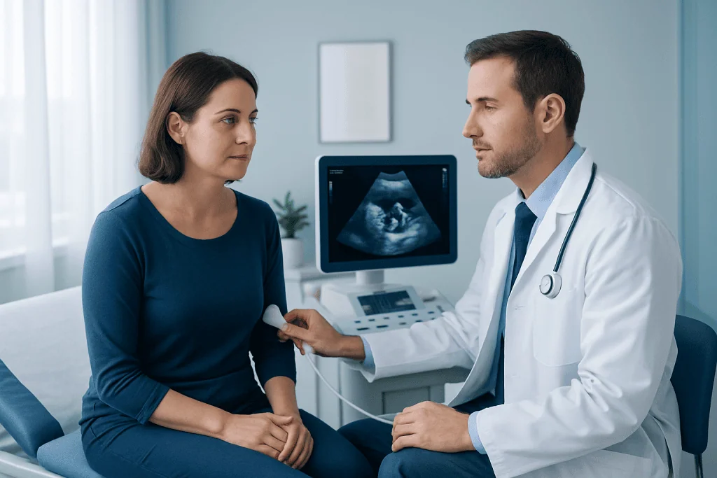
Most bubble studies are performed as part of a transthoracic echocardiogram, meaning the ultrasound probe is placed on the outside of the chest. The patient usually lies on an examination couch, often on the left side. Small adhesive electrodes may be placed on the chest to monitor the heart rhythm, and a small intravenous line is inserted into a vein in the arm or hand.
The sonographer or physician first records standard ultrasound images. Sterile saline is then mixed between syringes to create temporary microbubbles and injected through the IV while the heart is imaged. The patient may be asked to breathe normally, cough, or briefly perform a Valsalva maneuver, which involves bearing down or forcefully exhaling against a closed airway. These actions can briefly change pressure inside the heart and make a small shunt easier to detect.
In some circumstances, a transesophageal echocardiogram may provide a clearer view. For this test, an ultrasound probe is passed into the esophagus after the throat is numbed and sedation may be given. The care team will explain fasting instructions, transport arrangements, and recovery guidance if this approach is planned. A standard transthoracic bubble study often takes about 30 to 60 minutes, although appointment times vary.
Understanding Bubble Study Results
A negative bubble study means that bubbles were not seen crossing into the left side of the heart during the images obtained. This makes a significant right-to-left shunt less likely, although no single test is perfect. Image quality, the timing of breathing maneuvers, and the size or intermittent nature of a shunt can affect what is seen.
If bubbles appear in the left atrium or left ventricle within a few heartbeats, this can support the presence of an intracardiac shunt, such as a PFO or, less commonly, an atrial septal defect. If bubbles appear later, a clinician may consider a shunt in the lungs, such as a pulmonary arteriovenous malformation. The exact timing and pattern are assessed by an experienced clinician rather than interpreted in isolation.
A positive result does not automatically mean that an opening caused a stroke, symptoms, or another health concern. PFOs are relatively common and many people never experience related problems. Depending on the reason for testing, further imaging, a consultation with cardiology or neurology, or monitoring may be recommended before decisions about treatment are made.
Safety, Risks, and Preparation
For most patients, a bubble study is well tolerated. The main sensations are usually the brief IV insertion and the pressure of the ultrasound probe on the chest. Some people may feel temporary warmth, notice the saline injection, or find the breathing maneuver mildly uncomfortable. The microbubbles dissolve quickly and are cleared naturally by the body.
Serious complications from a standard agitated saline study are uncommon. As with any IV procedure, there can be minor bruising, discomfort, bleeding, or rarely infection at the injection site. Staff monitor patients during the test and are trained to respond to unexpected symptoms. People should tell the team before the examination if they have significant heart or lung disease, a known congenital heart condition, pregnancy, difficulty lying flat, or concerns about IV access.
Preparation is usually simple for a transthoracic bubble study. Patients should follow the instructions provided by their clinic, wear comfortable clothing, and bring a medication list. Medicines are often continued as usual unless a doctor advises otherwise. If a transesophageal echocardiogram is planned, fasting and sedation-related instructions are important and should be followed carefully.
What Happens After the Study?
After a standard bubble study, most people can leave soon after the examination and return to usual daily activities. The IV is removed, and a small dressing may be placed over the site. Drinking fluids and keeping the area clean can help with minor tenderness or bruising.
The images are reviewed by a cardiologist or another qualified clinician, and results may not be available immediately. Patients should ask when and how they will receive their results, what the finding may mean in their specific situation, and whether additional tests are needed. If a PFO or other shunt is found, management can range from no treatment and follow-up to medicines, treatment of another underlying condition, or selected catheter-based procedures.
Decisions are individualized. For example, closure of a PFO may be considered in carefully selected people with certain types of stroke after a thorough evaluation has excluded other likely causes. The potential benefits and risks should be discussed with a specialist. Acibadem International’s multidisciplinary specialists in JCI-accredited hospitals can assess heart and neurological findings and coordinate care for international patients.
When to Seek Medical Care
A bubble study itself rarely causes concerning symptoms. However, patients should seek urgent medical care for chest pain, severe shortness of breath, fainting, sudden weakness or numbness on one side of the body, facial drooping, new difficulty speaking, sudden confusion, or a sudden severe headache. These symptoms may indicate a time-sensitive heart, lung, or neurological condition and should not be attributed to a recent test without medical assessment.
Patients should contact the testing clinic or their doctor if the IV site becomes increasingly red, swollen, painful, warm, or starts draining fluid, or if fever develops. Mild bruising is common and usually settles without treatment, but worsening local symptoms should be checked.
It is also appropriate to arrange a non-urgent appointment to discuss persistent unexplained breathlessness, low oxygen readings, recurrent migraines with neurological symptoms, or questions about an abnormal test result. A qualified clinician can explain whether these symptoms may be related to a heart or lung shunt and what next steps are suitable.
Frequently asked questions
Is a bubble study painful?
A bubble study is usually not painful. Most people only feel a brief pinch when the IV is placed and may feel mild pressure from the ultrasound probe. The saline microbubble injection is generally well tolerated.
Does a bubble study use radiation or dye?
No, a standard bubble study does not use radiation. It uses sterile saline mixed to form tiny air microbubbles, rather than the contrast dyes commonly associated with CT scans or some X-ray procedures.
What does a positive bubble study mean?
A positive result means bubbles were seen reaching the left side of the heart or circulation when they normally would not. This may suggest a PFO, another opening in the heart, or an abnormal blood-vessel connection in the lungs. Further interpretation depends on the timing of the bubbles and the reason the test was ordered.
Can a bubble study detect a PFO?
Yes. A bubble study is commonly used to help identify a PFO, particularly when combined with a breathing or straining maneuver. A transesophageal echocardiogram or other tests may sometimes be needed to define the anatomy more clearly.
Do patients need to fast before a bubble study?
Fasting is not usually required for a standard transthoracic bubble study performed through the chest wall. If the test is being done with transesophageal echocardiography and sedation, fasting is typically required. The testing center will provide specific instructions.
Can patients drive home after a bubble study?
Patients can usually drive after a standard transthoracic bubble study because no sedation is used. If sedation is given for a transesophageal echocardiogram, patients will need someone else to take them home and should follow the recovery instructions provided.
This article is for general information only and is not a substitute for professional medical advice. Please consult a qualified doctor about your individual situation.
Explore treatments in Turkey — costs, top hospitals & a free quote
JCI-accredited · board-certified surgeons · reply within 24h
Add us as a Preferred Source to see more of our trusted health content across Google Search, AI Overviews and Discover.

More from the Health Library

Extra Virgin Olive Oil Benefits — Explained by Medical Evidence, Not Myths
Back Extensions: A Complete Medical Overview

Anadrol: An Evidence-Based Guide for Patients

Moxibustion: An Evidence-Based Guide for Patients

Terminal Agitation: An Evidence-Based Guide for Patients