Liver Ultrasound: Uses, Preparation and Results
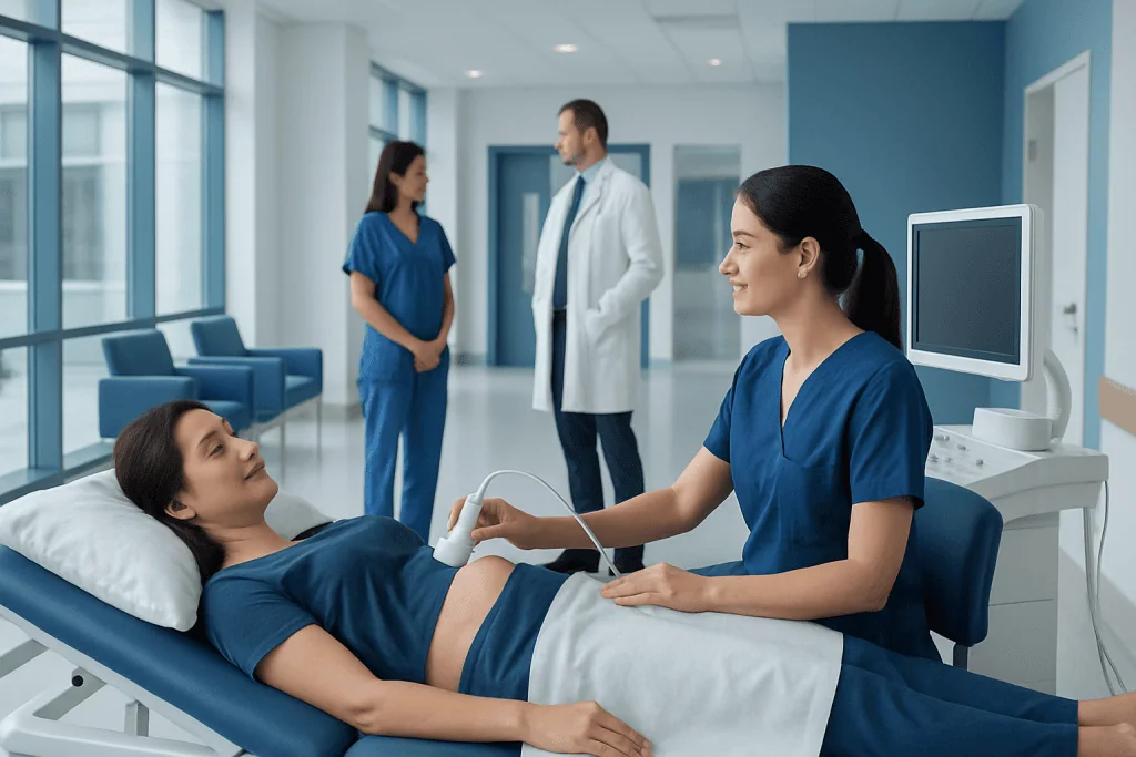
Liver ultrasound does not use radiation and is generally painless. It may assess liver size, texture, blood flow, focal lesions, bile ducts and nearby organs.
Key Takeaways
- Liver ultrasound does not use radiation and is generally painless.
- It may assess liver size, texture, blood flow, focal lesions, bile ducts and nearby organs.
- Fasting for several hours may be requested to improve visibility of the liver and gallbladder.
- Ultrasound findings are interpreted alongside symptoms, examination findings and blood test results.
- An abnormal result does not always mean a serious condition; further imaging or tests may be needed for clarification.
A liver ultrasound is a noninvasive imaging examination that uses sound waves to create pictures of the liver and surrounding abdominal organs. It can help clinicians investigate symptoms, assess known liver conditions and guide follow-up testing when needed.
Overview: What Is a Liver Ultrasound?
A liver ultrasound, also called a liver sonogram or upper abdominal ultrasound, is an imaging test that uses high-frequency sound waves to produce real-time images of the liver. A handheld device called a transducer is moved over the skin of the upper abdomen. The sound waves reflect differently from tissues and organs, allowing a computer to create images for review.
The examination is commonly used because it is noninvasive, does not expose the patient to ionizing radiation and is widely available. It may be performed as part of a broader abdominal ultrasound, which can also examine the gallbladder, bile ducts, pancreas, spleen, kidneys and major abdominal blood vessels.
Doctors may request liver ultrasound to investigate abnormal liver blood tests, abdominal symptoms or a previously identified liver finding. It can also be used to monitor some chronic liver diseases and to help evaluate changes seen on other imaging tests.
What Can a Liver Ultrasound Show?
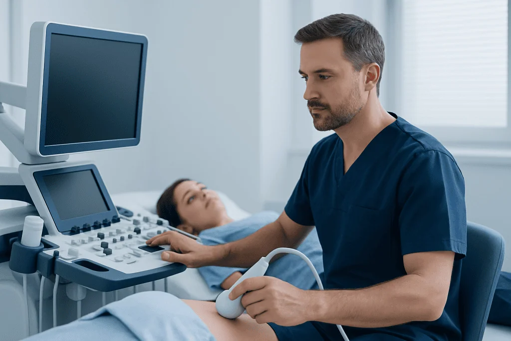
Ultrasound can provide important information about the liver’s size, outline and general tissue appearance. For example, it may show an enlarged liver, a fatty appearance consistent with steatosis, changes that can occur with long-standing liver disease, fluid around the liver, or widening of the bile ducts.
The scan may also identify focal changes, meaning an area that looks different from surrounding liver tissue. These can include cysts, benign lesions such as hemangiomas, or areas that require further assessment. Ultrasound can detect many findings, but it cannot always determine exactly what a lesion is. A doctor may therefore recommend contrast-enhanced imaging, such as CT or MRI, when more detail is needed.
When a Doppler ultrasound is included, it assesses blood flow in vessels such as the portal vein and hepatic veins. This can be useful when evaluating suspected vascular problems or signs related to portal hypertension. Ultrasound may also help assess conditions related to fatty liver disease or gallbladder and bile duct concerns.
Why Might a Doctor Recommend It?
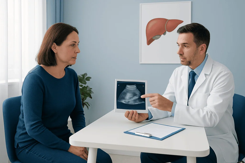
A clinician may recommend a liver ultrasound when a person has symptoms that could involve the liver or biliary system. These may include persistent discomfort in the upper right abdomen, unexplained abdominal swelling, jaundice, nausea, itching, or changes in liver function blood tests. Many of these symptoms have causes other than liver disease, and ultrasound is one way to investigate them carefully.
The test may also be used for people with known liver conditions, including chronic viral hepatitis, metabolic dysfunction-associated steatotic liver disease, cirrhosis or certain inherited liver disorders. In selected people at increased risk of liver cancer, ultrasound may be part of an ongoing surveillance plan determined by a liver specialist.
In addition, ultrasound can help evaluate suspected gallstones, bile duct blockage, abdominal fluid called ascites, or enlargement of the spleen. The exact purpose of the examination should be discussed with the referring clinician, who can explain what question the scan is intended to answer.
How to Prepare for a Liver Ultrasound
Preparation instructions can vary according to the type of ultrasound and the area being examined. Many imaging centers ask patients not to eat or drink for several hours before an upper abdominal or liver ultrasound. Fasting can reduce gas in the digestive tract and help keep the gallbladder full, which may improve the quality of the images.
Patients should follow the instructions provided by their healthcare team. It is important to ask whether regular medicines can be taken before the appointment; in many cases they can be taken with a small sip of water, but individual advice may differ. People with diabetes, pregnant individuals and those with other medical needs may require tailored fasting guidance.
Comfortable, loose clothing can make the appointment easier. The patient may be asked to change into a gown and remove jewelry or clothing around the upper abdomen. Previous imaging reports and relevant medical information can also be useful if the imaging department requests them.
What Happens During the Examination?
A liver ultrasound is usually performed by a trained sonographer or radiology professional. The patient lies on an examination table, often on their back and sometimes partly turned to one side. A clear gel is applied to the skin to help sound waves travel effectively between the transducer and the body.
The clinician gently moves the transducer over the upper abdomen and may ask the patient to take a deep breath, hold their breath briefly or change position. These steps can improve the view of the liver beneath the ribs. Mild pressure may be felt, particularly if the area is tender, but the test is not generally painful.
The examination often takes about 15 to 30 minutes, although timing varies with the reason for the scan and whether Doppler assessment is needed. Afterward, the gel is wiped away and most people can return to normal activities immediately unless they have been given different instructions.
In some circumstances, a specialist may use ultrasound to guide a procedure, such as draining fluid or obtaining a tissue sample. This is different from a routine diagnostic ultrasound and involves separate preparation, consent and aftercare guidance.
Understanding Liver Ultrasound Results
A radiologist or another qualified imaging physician reviews the images and prepares a report for the referring doctor. The report may describe liver size, echogenicity (how the liver reflects sound waves), texture, blood flow and any focal finding. It may also comment on the gallbladder, bile ducts, spleen and fluid in the abdomen when these structures are included in the scan.
Terms such as “increased echogenicity” can be seen with fatty change in the liver, while a nodular contour or other structural changes may suggest chronic liver disease. These descriptions are not a diagnosis on their own. The meaning depends on the person’s health history, symptoms, examination and laboratory results.
If a scan shows an unclear lesion or finding, the doctor may recommend follow-up imaging, blood tests or referral to a gastroenterologist or hepatologist. Further evaluation may include MRI scanning or CT scanning when these tests are appropriate. Many ultrasound findings are benign, but timely follow-up helps ensure that any necessary care is planned appropriately.
Benefits, Limitations and Safety
Liver ultrasound is considered very safe because it uses sound waves rather than radiation. It is suitable for many people, including children and pregnant patients when clinically indicated. It is also relatively quick and can show movement and blood flow in real time when Doppler techniques are used.
However, ultrasound has limitations. Image quality can be affected by bowel gas, body shape, the position of the liver and the patient’s ability to hold a breath briefly. Some areas of the liver may be difficult to see clearly, and small or deeply located lesions may require another imaging method for better characterization.
Ultrasound can suggest fatty liver and identify some features of advanced chronic liver disease, but it may not accurately measure the amount of fat, inflammation or scarring in every person. Depending on the clinical question, a clinician may recommend blood-based assessments, elastography, CT, MRI or occasionally biopsy. These complementary tests are selected based on individual needs rather than used routinely for everyone.
When to Seek Medical Advice
A liver ultrasound should be arranged when recommended by a qualified clinician, particularly after abnormal liver blood tests or ongoing abdominal symptoms. It is helpful to attend scheduled follow-up appointments so that the results can be explained in context and any next steps can be discussed.
Prompt medical assessment is appropriate for symptoms such as yellowing of the skin or eyes, severe or worsening abdominal pain, vomiting that prevents hydration, fever with abdominal pain, confusion, black stools, vomiting blood, or rapid abdominal swelling. These symptoms can have different causes and should not be self-diagnosed.
Healthy lifestyle measures can support overall liver health, including limiting alcohol where appropriate, maintaining a balanced diet, staying physically active and discussing medicines or supplements with a doctor. Acibadem International’s multidisciplinary specialists and JCI-accredited hospitals diagnose and treat liver-related conditions for international patients, with care plans based on individual clinical findings.
Frequently asked questions
Is a liver ultrasound painful?
A routine liver ultrasound is usually painless. The transducer may apply gentle pressure to the upper abdomen, and this can feel uncomfortable if the area is already tender. The examination does not involve needles or radiation unless it is being used to guide a separate procedure.
Do patients need to fast before a liver ultrasound?
Many patients are asked to fast for several hours before an upper abdominal ultrasound. This can improve visualization by reducing bowel gas and helping keep the gallbladder full. The imaging center’s instructions should always be followed, especially for people with diabetes or other health conditions.
Can a liver ultrasound diagnose fatty liver?
Ultrasound can often detect changes that are consistent with fatty liver, particularly when fat accumulation is moderate or marked. It may be less sensitive for mild fatty change and cannot reliably assess inflammation or the exact degree of fibrosis. A doctor may use blood tests or additional imaging to clarify the diagnosis and assess liver health.
Can liver ultrasound detect cancer?
Ultrasound can identify a liver lesion or an area that needs closer assessment, but it does not always confirm whether a lesion is cancerous. CT, MRI, contrast-enhanced ultrasound or other tests may be needed to characterize an abnormality. Results should be reviewed promptly with the referring doctor.
How long does it take to receive liver ultrasound results?
The timing varies by healthcare setting. Images are usually reviewed by a radiologist, who sends a report to the referring clinician. The clinician can then explain what the findings mean and whether any follow-up is needed.
What does an enlarged liver on ultrasound mean?
An enlarged liver, also called hepatomegaly, can occur for several reasons, including fatty liver disease, inflammation, congestion, infection or other conditions. It is a descriptive imaging finding rather than a diagnosis. Further assessment depends on symptoms, medical history, blood tests and the rest of the ultrasound report.
References
- National Institute of Diabetes and Digestive and Kidney Diseases
- American College of Radiology
- Radiological Society of North America
- National Health Service
- World Health Organization
This article is for general information only and is not a substitute for professional medical advice. Please consult a qualified doctor about your individual situation.
Ultrasound in Turkey — costs, top hospitals & a free quote
JCI-accredited · board-certified surgeons · reply within 24h
Add us as a Preferred Source to see more of our trusted health content across Google Search, AI Overviews and Discover.

Check-up & Preventive Medicine
Comprehensive health screenings designed for early detection and prevention.
1 specialists in this unit