Transvag Ultrasound Ovarian Cancer: Preparation, Procedure and Results

Transvaginal ultrasound produces detailed images of the ovaries and nearby pelvic organs using sound waves. The scan may show an ovarian cyst or mass, but further tests are needed to determine whether a mass is cancerous.
Key Takeaways
- Transvaginal ultrasound produces detailed images of the ovaries and nearby pelvic organs using sound waves.
- The scan may show an ovarian cyst or mass, but further tests are needed to determine whether a mass is cancerous.
- The procedure is usually brief, does not use radiation, and causes at most mild temporary pressure for most people.
- Preparation is simple; instructions about bladder filling depend on whether an abdominal ultrasound is also planned.
- Persistent bloating, pelvic pain, early fullness, or changes in urinary habits should be discussed with a clinician.
A transvaginal ultrasound is commonly used to examine the ovaries when symptoms, examination findings, or other tests raise concern. It can identify an ovarian mass and describe its features, but it cannot confirm or rule out ovarian cancer on its own.
Overview: transvag ultrasound ovarian cancer assessment
A transvag ultrasound ovarian cancer assessment, also called a transvaginal ultrasound, is an imaging examination that uses high-frequency sound waves to create pictures of the ovaries, uterus, fallopian tube region, and surrounding pelvic structures. It is often requested when a person has pelvic symptoms, an abnormal pelvic examination, a raised risk of ovarian cancer, or an ovarian finding on another scan.
The test is useful for identifying and describing ovarian cysts or masses. However, ultrasound findings alone cannot confirm that a mass is ovarian cancer, and a normal scan does not exclude every possible ovarian cancer. Doctors interpret the images alongside symptoms, medical history, examination findings, blood tests when appropriate, and sometimes additional imaging or surgery.
Many ovarian findings are benign, particularly in people who are still having periods. If a scan identifies a concerning mass, referral to a gynecologist or gynecologic oncology team may be recommended for timely, careful evaluation.
How transvaginal ultrasound works and who may need it
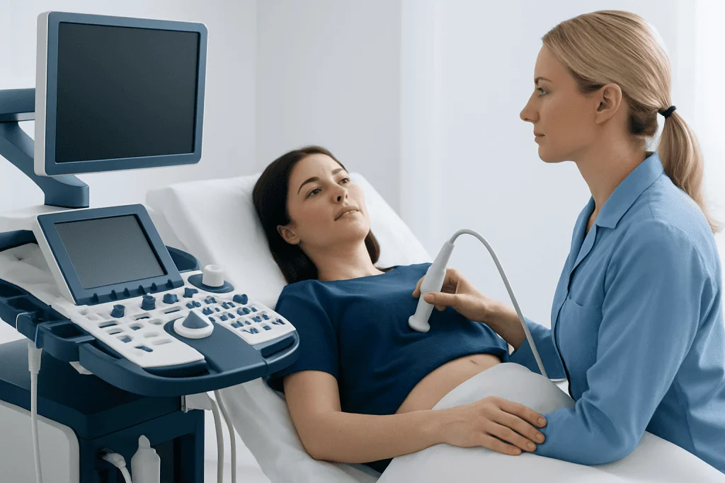
During transvaginal ultrasound, a slim ultrasound probe is placed a short distance into the vagina. The probe sends out sound waves and receives their echoes, which a computer converts into real-time images. Because the probe is close to the ovaries and uterus, it can usually provide more detail than an ultrasound performed only across the abdomen.
A clinician may recommend this examination for persistent bloating, pelvic or lower abdominal pain, a feeling of fullness soon after eating, unexplained urinary urgency, abnormal vaginal bleeding, or a pelvic mass. It may also be used to monitor a known ovarian cyst, evaluate inherited ovarian cancer risk, or clarify an unusual finding on a CT scan, MRI, or abdominal ultrasound.
Transvaginal ultrasound is not a routine screening test for ovarian cancer in people at average risk and without symptoms. In people with a strong family history or a known inherited gene change, a specialist can discuss individualized risk assessment and surveillance options. Related concerns may also be considered in an evaluation for ovarian cancer.
How should I prepare for an ovarian ultrasound?

Preparation for an ovarian ultrasound is usually straightforward. The imaging center will provide instructions in advance, and these should be followed because preparation can differ depending on the exact examination planned. No fasting is usually needed for a transvaginal ultrasound.
If an abdominal pelvic ultrasound is planned before the transvaginal part, the person may be asked to arrive with a comfortably full bladder. A full bladder can help move bowel loops out of the way and improve images obtained through the lower abdomen. After the abdominal images are completed, the person can usually empty the bladder before the internal scan.
It can help to wear comfortable clothing and bring prior imaging reports or relevant medical records if requested. The person should tell the care team about possible pregnancy, active vaginal bleeding, pelvic infection symptoms, previous pelvic surgery, or any concern about the examination. A transvaginal scan is performed only with consent, and the person may ask questions or request that the examination stop at any time.
Step-by-step: what happens during the procedure
The examination is usually performed by a trained sonographer, radiologist, or gynecology clinician in a private imaging room. After changing clothing as needed, the person lies on an examination couch, generally on their back with knees bent. A sheet is used to support privacy and comfort.
The probe is covered with a protective sheath and lubricating gel. It is then gently inserted into the vagina by the patient or clinician, depending on local practice and the person’s preference. The probe does not need to pass through the cervix or enter the uterus. It is moved slightly to obtain views of each ovary, the uterus, and the pelvis.
The operator may use Doppler imaging to assess blood flow within or around an ovarian mass. Images and measurements are recorded, including the size, shape, internal contents, wall appearance, and any solid areas or fluid in the pelvis. The scan itself does not spread cancer, disturb a cyst, or expose the patient to radiation.
Is a transvaginal ultrasound painful?
A transvaginal ultrasound is not usually painful, although some people experience pressure or short-lived discomfort during insertion or when the probe is positioned to obtain images. The probe is narrow, and the examination is performed gently. The clinician can adjust the position, pause, or stop if discomfort occurs.
People may be more sensitive if they have pelvic pain, endometriosis, a vaginal infection, recent surgery, trauma-related concerns, or anxiety about internal examinations. Sharing these concerns beforehand allows the team to explain each step, proceed more slowly, and discuss comfort measures. In many settings, the person may be able to insert the probe themselves.
Severe pain is not expected. If significant pain develops during or after the test, the person should tell the clinician promptly, particularly if it is accompanied by fever, fainting, heavy bleeding, or worsening abdominal pain.
How long does a transvaginal ultrasound take?
A transvaginal ultrasound commonly takes about 15 to 30 minutes, although timing varies with the reason for the scan and whether an abdominal ultrasound is performed as well. A more detailed assessment may take longer if an ovarian mass needs careful measurement and characterization.
Most people can return to normal daily activities immediately afterward. There is no sedation, recovery room stay, or radiation exposure. A small amount of gel may remain after the scan, and light spotting can occasionally occur, especially in people with a sensitive cervix or existing vaginal bleeding.
Preliminary information may sometimes be shared after the examination, but the formal result is generally issued after the images have been reviewed. The referring clinician explains what the findings mean in the context of the person’s overall health and next steps.
Understanding results, benefits, limitations and next steps
Ultrasound reports may describe a normal ovary, a simple cyst, a complex cyst, or an adnexal mass. Features such as size, solid components, thick walls, internal partitions, irregular surfaces, blood flow patterns, and fluid in the pelvis help doctors estimate whether a finding is more likely benign or needs further assessment. These descriptions are not a cancer diagnosis by themselves.
The main benefits are that the test is widely available, does not use ionizing radiation, and provides detailed pelvic images in real time. Its limitations include difficulty seeing some structures in certain body types or when bowel gas is present, and overlap between the appearance of some benign and malignant conditions. Small or early cancers may not always be visible.
Depending on the result, next steps may include watchful follow-up imaging, blood tests such as CA-125 in selected patients, MRI or CT imaging, or referral to a gynecologist or gynecologic oncologist. When cancer is suspected or confirmed, care may involve ovarian cancer treatment planning that combines appropriate surgery and systemic treatments. Acibadem International’s multidisciplinary specialists and JCI-accredited hospitals support diagnosis and treatment planning for international patients.
When to seek medical care
A person should arrange medical assessment if bloating, pelvic or abdominal pain, early fullness while eating, loss of appetite, urinary urgency or frequency, unexplained fatigue, or changes in bowel habits are new, persistent, or becoming more frequent. These symptoms are common and often have non-cancer causes, but they deserve evaluation when they do not settle.
Urgent medical care is appropriate for sudden severe pelvic or abdominal pain, pain with vomiting, fainting, severe dizziness, fever with pelvic pain, or heavy vaginal bleeding. Such symptoms may be related to an ovarian cyst complication, infection, ectopic pregnancy, or another condition that needs prompt assessment.
People with a close family history of ovarian, breast, pancreatic, prostate, or colorectal cancer should discuss their history with a doctor. Genetic counseling or specialist assessment may be helpful, particularly when several relatives have had related cancers or cancer occurred at a young age.
Frequently asked questions
Can a transvaginal ultrasound detect ovarian cancer?
A transvaginal ultrasound can detect an ovarian mass and identify features that may need further investigation. It cannot reliably confirm ovarian cancer on its own, because benign cysts and other non-cancerous conditions can sometimes look complex. Doctors use the scan together with clinical assessment and, when needed, additional tests.
Does a normal transvaginal ultrasound rule out ovarian cancer?
No single imaging test can rule out every ovarian cancer, especially at an early stage. A normal scan can be reassuring, but persistent or worsening symptoms should still be reviewed by a clinician. Further assessment may be appropriate based on symptoms, personal risk, and examination findings.
Is a transvaginal ultrasound painful?
Most people describe pressure or mild discomfort rather than pain. The examination should be performed gently, and the clinician can pause or stop if it is uncomfortable. It is helpful to mention pelvic pain, anxiety, previous trauma, or other concerns before the scan.
How long does a transvaginal ultrasound take?
The internal ultrasound portion often takes around 15 to 30 minutes. It may take longer when both abdominal and transvaginal imaging are required or when a finding needs detailed assessment. Normal activities can usually resume immediately after the examination.
How should I prepare for an ovarian ultrasound?
Most transvaginal ultrasounds do not require fasting. If an abdominal pelvic scan is also scheduled, the center may ask the patient to drink water and keep a full bladder until the first part of the examination is complete. The imaging provider’s instructions should always take priority.
What happens if an ovarian mass is found on ultrasound?
Many ovarian masses are benign and may only need observation or repeat imaging. If the appearance is complex, large, persistent, or concerning, a doctor may recommend blood tests, MRI or CT imaging, and referral to a gynecologist or gynecologic oncologist. The next step depends on the person’s age, symptoms, medical history, and scan findings.
References
- World Health Organization
- American College of Obstetricians and Gynecologists
- American Cancer Society
- National Cancer Institute
- Royal College of Obstetricians and Gynaecologists
This article is for general information only and is not a substitute for professional medical advice. Please consult a qualified doctor about your individual situation.
Ovarian Cancer in Turkey — costs, top hospitals & a free quote
JCI-accredited · board-certified surgeons · reply within 24h
Add us as a Preferred Source to see more of our trusted health content across Google Search, AI Overviews and Discover.

Check-up & Preventive Medicine
Comprehensive health screenings designed for early detection and prevention.
2 specialists in this unit