Monoka Stent: An Evidence-Based Patient Guide

Biliary stents can relieve blockage-related symptoms and help restore bile flow. Most bile duct stents are placed during ERCP, an endoscopic procedure performed with sedation or anesthesia.
Key Takeaways
- Biliary stents can relieve blockage-related symptoms and help restore bile flow.
- Most bile duct stents are placed during ERCP, an endoscopic procedure performed with sedation or anesthesia.
- Stent type determines whether and when removal or exchange is needed; plastic stents commonly require planned follow-up.
- Fever, worsening abdominal pain, vomiting, jaundice, or chills after placement require urgent medical assessment.
- MRI safety depends on the specific stent model and should always be confirmed with the treating team or radiology department.
A monoka stent is generally understood as a small drainage-supporting stent placed to keep a narrowed or blocked bile duct open and allow bile to flow. The exact device, material, and planned duration vary, so patients should receive individualized instructions from the endoscopy and hepatobiliary team that placed it.
Overview: What Is a Monoka Stent?
A monoka stent is a term that may be used in patient discussions for a small stent placed to support drainage through a bile duct. In biliary care, a stent is a narrow tube that helps keep a duct open when bile flow is reduced by a narrowing, stone, inflammation, scar tissue, a surgical change, or a tumor. Restoring drainage can reduce jaundice, itching, infection risk, and discomfort associated with obstruction.
It is important to confirm the exact name and type of stent with the clinician who placed it. “Monoka” can also refer to a type of monocanalicular stent used in tear-duct procedures, which is different from a bile duct stent. This guide addresses questions about bile duct stents, including placement, removal, follow-up, and recovery.
A biliary stent may be made of plastic or metal. Plastic stents are often temporary and usually need scheduled removal or replacement. Some metal stents are designed to remain in place for longer periods, while others can be removed in selected circumstances. The intended plan depends on the underlying reason for the blockage and the device used.
How Bile Duct Stents Work and Who May Need One
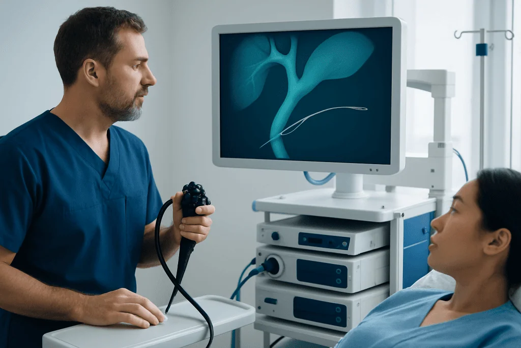
The liver produces bile, which travels through bile ducts to the small intestine, where it helps digest fats. If a duct becomes blocked or significantly narrowed, bile may build up. This can cause yellowing of the skin or eyes, dark urine, pale stools, itching, nausea, abdominal pain, or infection of the bile ducts.
A stent creates a channel through the narrowed area so bile can pass into the intestine. It may be used after a stone has been removed, to treat a benign narrowing, to support drainage during evaluation of an unexplained blockage, or to provide drainage when a cancer-related obstruction cannot be immediately removed surgically. Related digestive conditions may also be assessed during the same care pathway, including pancreatic cancer when imaging suggests an obstruction near the pancreas.
Clinicians consider symptoms, blood tests, imaging findings, anatomy, prior surgery, and the suspected cause of obstruction before recommending monoka stent placement or another biliary drainage approach. Patients who are pregnant, use blood-thinning medicine, have heart or lung conditions, or have had complex gastrointestinal surgery should tell the care team in advance so the procedure can be planned safely.
How Is a Bile Duct Stent Placed?

Most bile duct stents are placed during endoscopic retrograde cholangiopancreatography, known as ERCP. During this procedure, a specialist guides a flexible endoscope through the mouth, stomach, and into the first part of the small intestine. A small opening where the bile duct drains into the intestine is then accessed, and contrast imaging helps show the location of a narrowing or blockage.
If appropriate, the specialist may remove stones, widen a narrowed area, collect samples, and place the stent across the obstruction. Fluoroscopy, a type of real-time X-ray imaging, helps confirm positioning. Sedation or anesthesia is used, and patients are monitored closely throughout the procedure. ERCP for bile duct drainage is the primary endoscopic treatment used for many biliary stent placements.
After the procedure, people are observed until the effects of sedation have reduced. Some return home the same day, while others remain in hospital for monitoring, treatment of an infection, management of jaundice, or care of the condition causing the blockage. The team should provide clear written details about the stent, expected follow-up, and the date for any planned removal or exchange.
Benefits, Risks, and MRI Safety
The main benefit of a biliary stent is improved bile drainage. This can relieve jaundice and itching, improve liver test results, help control cholangitis, and allow further treatment or surgery to proceed more safely when needed. A stent does not by itself cure the condition causing the blockage, so diagnosis and treatment of the underlying cause remain essential.
ERCP and stent placement are established procedures, but they carry risks. These include inflammation of the pancreas (pancreatitis), bleeding, infection, perforation, reaction to sedation, stent migration, and stent blockage. The likelihood of particular complications varies with the reason for treatment, anatomy, and procedure complexity. The clinical team discusses individual risks before obtaining consent.
Monoka stent MRI safety cannot be assumed from the name alone. Many modern biliary stents may be MRI-conditional, meaning scanning is permitted only under stated conditions, but materials and manufacturer instructions differ. Before any MRI, patients should tell radiology staff that they have a bile duct stent and provide the device card or procedure report if available. The radiology department can verify compatibility and scan conditions.
Recovery Timeline and What to Avoid After Bile Duct Stent Placement
After ERCP, mild throat discomfort, bloating, or temporary tiredness from sedation can occur. On the day of the procedure, patients generally need a responsible adult to take them home and should not drive, drink alcohol, sign important documents, or make major decisions until the sedative has fully worn off. The treating team will advise when to restart food, medicines, and usual activities.
In many cases, gentle activity can resume within a day, depending on overall health and the reason for admission. Patients should follow instructions about blood thinners, diabetes medicines, and antibiotics carefully. There is usually no universal long-term “bile duct stent diet,” but a clinician may advise smaller, lighter meals initially if nausea or bloating occurs, and may recommend dietary changes based on gallbladder, pancreas, liver, or cancer treatment needs.
What should patients avoid after bile duct stent placement? They should avoid driving and alcohol for the period advised after sedation, avoid ignoring new symptoms, and avoid changing prescribed medication without medical advice. Until recovery is clear, strenuous activity may be postponed if it worsens pain or fatigue. A planned follow-up appointment is particularly important for temporary plastic stents, because a forgotten stent can become blocked or lead to infection.
- Take medicines only as instructed by the treating team.
- Keep the stent procedure report and follow-up schedule accessible.
- Attend blood tests, imaging, and planned endoscopy appointments.
- Contact the care team promptly if symptoms of obstruction or infection return.
How Is a Mini Monoka Stent Removed?
A bile duct stent is commonly removed or exchanged during another ERCP. The patient receives sedation or anesthesia, and the endoscopist reaches the bile duct opening with an endoscope. Small endoscopic instruments, such as a snare, forceps, or basket, can grasp the stent and withdraw it through the endoscope. If ongoing drainage is needed, a new stent may be placed during the same procedure.
The removal plan depends on the stent type and the reason it was inserted. Plastic stents are frequently removed or exchanged after a limited interval determined by the treating clinician, because they can clog over time. Some metal stents are not intended for routine removal, particularly when they are placed for long-term drainage; however, removable covered metal stents may be used in specific situations.
Patients should not attempt any form of monoka stent removal themselves. If a stent migrates or passes spontaneously, medical review is still needed to confirm that the bile duct is draining properly and that no replacement is required. The endoscopy team will explain the expected schedule before discharge and should be contacted if that date has not been arranged.
How Long Can a Stent Remain in the Bile Duct?
The answer depends on the device and the clinical purpose. A plastic bile duct stent is usually a temporary device and commonly requires removal or exchange on a schedule set by the endoscopist. It should not be left in place beyond the recommended follow-up period, because blockage, stone formation, and infection become more likely over time.
Metal stents may remain in place longer than plastic stents. In some people, particularly those receiving long-term palliation for a non-removable malignant obstruction, the stent may be intended to stay in place unless symptoms or testing suggest blockage or another complication. In other settings, such as some benign strictures, a removable covered metal stent may be placed with a planned removal date.
The safest approach is not to rely on a general timeline found online. Patients should ask their endoscopist whether the stent is plastic or metal, whether it is temporary or permanent, and exactly when follow-up is due. Recording this plan is an important part of safe long-term care.
When to Seek Medical Care
Patients should seek urgent medical assessment after biliary stent placement if they develop fever, chills, worsening or severe upper abdominal pain, persistent vomiting, fainting, confusion, black stools, vomiting blood, or difficulty breathing. These symptoms can indicate infection, pancreatitis, bleeding, or another complication that needs prompt evaluation.
Medical advice is also needed if jaundice, dark urine, pale stools, itching, or nausea return after initial improvement. These changes may indicate that bile flow has become reduced again. Early review helps the team decide whether blood tests, imaging, antibiotics, or repeat endoscopy are needed.
For people traveling for specialist digestive care, Acibadem International’s multidisciplinary specialists and JCI-accredited hospitals can assess bile duct obstruction and coordinate endoscopic and surgical treatment plans for international patients. Ongoing care may also involve gallbladder removal surgery when gallstones are the underlying concern, or coordinated oncology care when an obstruction is cancer-related.
Frequently asked questions
What is monoka?
The term monoka may refer to a small stent, but its precise meaning depends on the specialty and device being discussed. In eye care, Monoka is often associated with a monocanalicular tear-duct stent. If the discussion concerns bile drainage, patients should ask the endoscopy team for the exact stent brand, material, and intended duration.
How is a mini monoka stent removed?
A bile duct stent is usually removed during an ERCP procedure using endoscopic instruments passed through an endoscope. Sedation or anesthesia is used, and a replacement stent may be inserted at the same time if drainage is still needed. Removal should only be performed by an appropriately trained specialist.
How long can a stent remain in the bile duct?
The duration depends on whether the stent is plastic or metal and why it was placed. Plastic stents usually need planned removal or exchange, while some metal stents can remain longer. The treating endoscopist should provide the exact follow-up schedule.
What should I avoid after bile duct stent placement?
After sedation, patients should avoid driving, alcohol, and important decisions for the period specified by their care team. They should also avoid missing scheduled stent follow-up and should not stop or restart prescribed medicines without advice. New fever, pain, vomiting, or recurrent jaundice should be reported promptly.
Is a monoka stent safe for MRI?
MRI compatibility depends on the specific stent model and its manufacturer instructions. Many devices have MRI conditions, but not every device has the same requirements. Patients should tell the MRI department about the stent and provide their procedure documentation whenever possible.
What is a monoka stent CPT code?
A CPT code is a billing and procedure-coding term rather than a clinical description of the stent itself. The appropriate code can vary according to the procedure performed, such as ERCP with stent placement or removal, the indication, and local coding rules. A hospital billing team or treating clinician can clarify coding questions for an individual case.
References
- American Society for Gastrointestinal Endoscopy
- National Institute of Diabetes and Digestive and Kidney Diseases
- National Health Service
- European Society of Gastrointestinal Endoscopy
This article is for general information only and is not a substitute for professional medical advice. Please consult a qualified doctor about your individual situation.
Heart care in Turkey — expert evaluation and treatment
JCI-accredited · board-certified surgeons · reply within 24h
Add us as a Preferred Source to see more of our trusted health content across Google Search, AI Overviews and Discover.