Will Brain MRI Show Eye Problems: Preparation, Procedure and Results
Brain MRI can identify some causes of visual symptoms, including optic nerve inflammation, tumors, stroke and certain orbital abnormalities. Many common eye conditions, such as refractive errors, dry eye and early glaucoma, are not diagnosed with a brain MRI.
Key Takeaways
- Brain MRI can identify some causes of visual symptoms, including optic nerve inflammation, tumors, stroke and certain orbital abnormalities.
- Many common eye conditions, such as refractive errors, dry eye and early glaucoma, are not diagnosed with a brain MRI.
- The scan may need dedicated orbital images and contrast dye to assess the optic nerves and tissues behind the eyes clearly.
- MRI does not use ionizing radiation, but people should tell the imaging team about implants, kidney disease, pregnancy or possible pregnancy.
- Sudden vision loss, double vision, a severe new headache or eye pain with visual changes needs urgent medical assessment.
A brain MRI can show some eye problems, particularly those involving the optic nerves, eye sockets, brain pathways for vision, or nearby tissues. However, it does not replace a full eye examination, and a dedicated orbit MRI may be needed when an eye-specific condition is suspected.
Overview: Will Brain MRI Show Eye Problems?
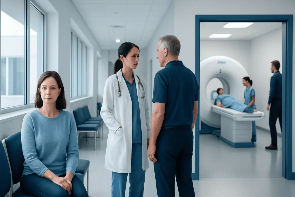
Will brain MRI show eye problems? A brain MRI may show eye-related problems when they involve the optic nerves, tissues behind the eyes, visual pathways in the brain, or structures around the eye sockets. It is particularly useful when symptoms such as unexplained vision loss, double vision, abnormal pupil responses, visual-field changes, or suspected neurological disease raise concern for a cause beyond the surface of the eye.
A routine brain MRI is not designed to detect every eye condition. For concerns focused on the eyeball, optic nerve, eye muscles or orbit, a clinician may request an MRI of the brain and orbits with specialized image sequences, sometimes using contrast material. An ophthalmologist or neuro-ophthalmologist combines imaging results with vision testing and an examination of the eye.
MRI uses a strong magnetic field and radio waves to create detailed images of soft tissues. It does not use X-rays or other ionizing radiation. The result is one part of the diagnostic process rather than a standalone test that can explain all visual symptoms.
How MRI Evaluates the Eyes, Optic Nerves and Visual Pathways
The visual system extends from the front of the eye to the back of the brain. Light is detected by the retina, and signals travel through the optic nerves, optic chiasm and optic tracts to areas of the brain that process vision. A brain MRI can assess many of these deeper structures, especially the optic pathways and the brain regions involved in sight.
When the clinical question concerns the eye sockets, radiologists use thin, high-resolution images through the orbits. These can show the optic nerves, eye muscles, tear glands, orbital fat and tissues surrounding the eyes. Contrast, usually gadolinium-based, may help identify inflammation, changes in the blood-brain barrier, infection, or some tumors.
Brain MRI may also help investigate visual symptoms caused by a neurological condition, such as a stroke, inflammation, pressure on the optic chiasm from a pituitary-area mass, or changes associated with demyelinating disease. Imaging findings must always be interpreted in the context of the person’s symptoms, examination and other tests.
What Eye Problems Can Be Diagnosed With an MRI Scan?
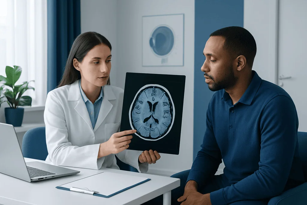
MRI can help diagnose or assess conditions affecting the optic nerve and structures behind the eye. Examples include optic neuritis, which is inflammation of the optic nerve; optic nerve compression from a mass or enlarged nearby structure; some orbital tumors; inflammation of eye muscles or other orbital tissues; and changes linked to thyroid eye disease. It can also help assess injury, infection, or an abscess in selected situations.
The scan may identify brain-related causes of visual symptoms, including certain strokes, tumors, inflammation, multiple sclerosis-related lesions and pituitary-region abnormalities that affect the optic chiasm. In people with double vision, MRI may be considered when the cause could involve the brainstem, cranial nerves, eye muscles, or the orbit.
For suspected optic nerve inflammation or other neuro-ophthalmic concerns, clinicians may coordinate assessment with specialists in ophthalmology and neurology. A complete evaluation can include visual acuity, color vision, eye pressure measurement, pupil assessment, slit-lamp examination, dilated retinal examination and visual-field testing alongside imaging.
- Optic neuritis and other optic nerve abnormalities
- Orbital inflammation, infection or selected masses
- Compression of the optic pathways
- Brain lesions affecting vision, including selected strokes
- Some causes of double vision or abnormal eye movement
How Accurate Is a Brain MRI for Eye Issues?
The accuracy of a brain MRI for eye issues depends on the suspected condition and the type of MRI ordered. MRI provides excellent detail of the brain, optic nerves and soft tissues around the eyes, making it valuable for identifying many structural, inflammatory and neurological causes of vision symptoms. Its usefulness is greater when the scan is tailored to the clinical question, such as an MRI of the brain and orbits with contrast when appropriate.
A normal brain MRI does not exclude every eye disorder. Some conditions are diagnosed mainly through examination, eye pressure testing, retinal imaging, optical coherence tomography, laboratory testing or functional vision tests. Small or early changes may not be visible, and MRI findings can occasionally be nonspecific, so a specialist interprets them carefully.
In short, MRI is highly informative for deeper structures and neurological causes, but it is not a universal eye test. If symptoms point to a problem on the cornea, lens, retina or within the eye itself, an ophthalmic examination may be more direct and more sensitive.
What Does Not Show Up on Brain MRI?
A brain MRI usually cannot diagnose common surface and focusing problems of the eye. It does not measure whether a person needs glasses or contact lenses, and it does not diagnose dry eye disease, most corneal conditions, cataracts or routine conjunctivitis. These conditions are generally assessed through an eye examination rather than brain imaging.
Early glaucoma may not appear on a brain MRI because glaucoma diagnosis relies on eye pressure, optic nerve examination, visual-field testing and detailed retinal nerve-fiber imaging. MRI can sometimes show advanced optic nerve changes or investigate an unusual presentation, but it is not a standard glaucoma screening tool.
Many retinal disorders also require specialized testing such as dilated retinal examination, retinal photography, optical coherence tomography or fluorescein angiography. MRI may occasionally reveal a large eye mass or deeper orbital involvement, but it does not replace tests designed to evaluate the retina and fine structures inside the eye.
Candidacy, Preparation and Step-by-Step Procedure
A doctor may recommend MRI when visual symptoms are unexplained, persistent, progressive, or associated with neurological signs. It can be especially relevant for reduced vision in one eye, pain with eye movement, new double vision, an unexplained visual-field defect, suspected optic nerve disease, or concern about structures near the optic pathways. The decision is individualized after a medical and eye assessment.
Before the scan, the person should tell the imaging team about a pacemaker, cochlear implant, aneurysm clip, implanted pump, metal fragments, prior surgery, pregnancy or possible pregnancy, kidney problems, and previous contrast reactions. Many modern implants are MRI-compatible under specific conditions, but the team must confirm this. If contrast is planned, kidney health and other relevant factors may be reviewed.
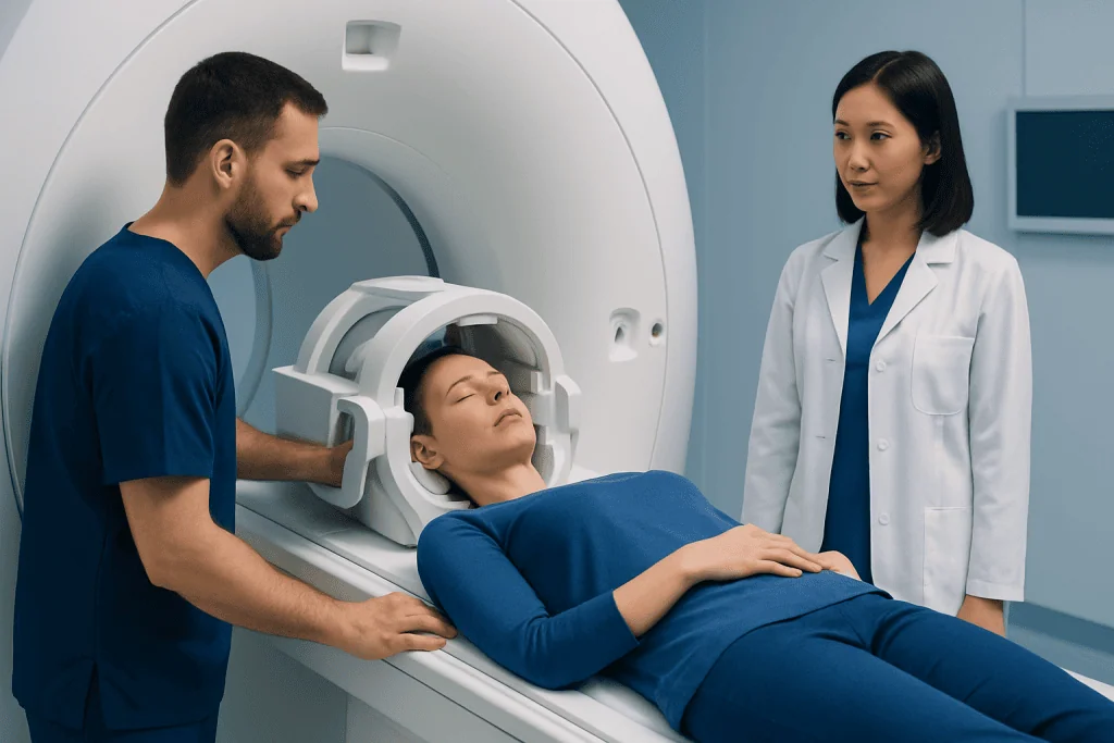
During the procedure, the person changes into clothing without metal and lies on a padded table that moves into the scanner. A head coil is placed around the head to obtain detailed images. The machine makes loud tapping sounds, so ear protection is provided. The person needs to remain still and can communicate with staff through an intercom; depending on the protocol, the scan often takes between 20 and 60 minutes.
Some scans involve an injection of contrast into a vein partway through the appointment. Sedation may be considered for severe claustrophobia or for individuals unable to remain still, with advance planning and appropriate supervision. The radiologist reviews the images and sends a report to the referring clinician, who explains what the findings mean in relation to the person’s symptoms.
Benefits, Risks and Recovery Timeline
The main benefit of MRI is its ability to produce detailed images of soft tissues without ionizing radiation. It can help clinicians distinguish between inflammation, compression, vascular changes and other causes of visual symptoms, guiding whether follow-up may involve ophthalmology, neurology, endocrinology, neurosurgery or another specialty.
MRI is generally considered safe when standard screening procedures are followed. Some people find the scanner noisy or enclosed, and lying still can be uncomfortable. Rarely, contrast material can cause an allergic-type reaction. Gadolinium contrast is used thoughtfully, particularly in people with significant kidney disease, and the imaging team will discuss whether it is appropriate.
Most people can return to normal activities immediately after a standard MRI. If sedating medication was used, they should arrange for an adult to take them home and follow the care instructions provided by the facility. Results may be available quickly in urgent settings, while non-urgent reporting timelines vary by service and the complexity of the images.
For coordinated evaluation of visual symptoms with possible neurological causes, Acibadem International’s multidisciplinary specialists and JCI-accredited hospitals can assess international patients and arrange appropriate imaging and follow-up care.
What Is the Most Common Finding on a Brain MRI?
For many people who have a brain MRI because of headaches, dizziness or nonspecific symptoms, the most common outcome is no significant abnormality related to the symptoms. MRI can also identify incidental findings, meaning changes that were not expected and may not require treatment, such as mild age-related white matter changes or harmless anatomical variations.
There is no single “most common” finding for people having MRI specifically because of eye symptoms. The likely findings depend on why the test was requested, the person’s age, symptoms and examination results. A radiology report may mention minor changes that are unrelated to vision, so the referring clinician is best placed to explain their importance.
If an MRI shows an abnormality, this does not automatically indicate a serious condition. Further tests or a follow-up scan may be recommended to clarify the finding, monitor it over time, or decide whether treatment is needed.
When to Seek Medical Care
Anyone with ongoing blurred vision, visual-field loss, new double vision, eye pain with movement, changes in color vision, or progressive changes in sight should arrange a timely assessment with an eye-care professional or doctor. The appropriate test may be an eye examination, MRI, another imaging study, blood tests, or a combination of these.
Urgent medical care is important for sudden loss of vision, sudden persistent double vision, a new drooping eyelid, severe headache with visual changes, weakness or numbness on one side of the body, trouble speaking, confusion, or severe eye pain with nausea. These symptoms can have several causes and need prompt evaluation.
It is helpful to note when symptoms began, whether they affect one or both eyes, whether they are constant or intermittent, and whether there are associated headaches, pain, weakness or balance changes. This information supports accurate assessment and helps clinicians determine whether brain MRI, dedicated orbital imaging or a different investigation is most appropriate.
Frequently asked questions
Will a brain MRI show optic neuritis?
A brain MRI can show signs of optic neuritis, especially when dedicated images of the orbits and optic nerves are included. Contrast may help reveal active inflammation. A normal scan does not completely rule it out, so eye and neurological examination findings remain important.
Can a brain MRI show glaucoma?
Brain MRI is not a standard test for diagnosing glaucoma. Glaucoma is usually assessed with eye pressure measurement, optic nerve evaluation, visual-field testing and retinal imaging. MRI may be used only when symptoms or findings suggest another problem involving the optic nerve or brain.
Can MRI detect a tumor behind the eye?
MRI is useful for detecting and characterizing many tumors or masses in the orbit and near the optic pathways. It can show their size, location and relationship to nearby nerves, muscles and brain structures. Further tests may still be needed to determine the exact type of lesion.
Do I need contrast for an MRI because of eye symptoms?
Not everyone needs contrast. It may be recommended when doctors are looking for inflammation, infection, abnormal blood-brain barrier changes, certain tumors or active optic nerve disease. The imaging team and referring clinician decide based on the clinical question and individual health factors.
Can a brain MRI explain blurry vision?
It can explain blurry vision when the cause involves the optic nerves, visual pathways or brain, but many causes of blurred vision begin within the eye and are better identified through an eye examination. Refractive errors, dry eye, cataracts and many retinal problems may not be explained by a routine brain MRI.
How long does it take to get brain MRI results?
In urgent situations, preliminary results may be reviewed rapidly. For routine scans, the radiologist usually prepares a report for the referring clinician within a timeframe set by the imaging service. The clinician then discusses how the findings relate to symptoms and whether further evaluation is needed.
References
- American Academy of Ophthalmology
- Radiological Society of North America
- National Institute of Neurological Disorders and Stroke
- American College of Radiology
This article is for general information only and is not a substitute for professional medical advice. Please consult a qualified doctor about your individual situation.
Explore treatments in Turkey — costs, top hospitals & a free quote
JCI-accredited · board-certified surgeons · reply within 24h
Add us as a Preferred Source to see more of our trusted health content across Google Search, AI Overviews and Discover.