Osteoid Osteoma on MRI: Preparation, Procedure and Results
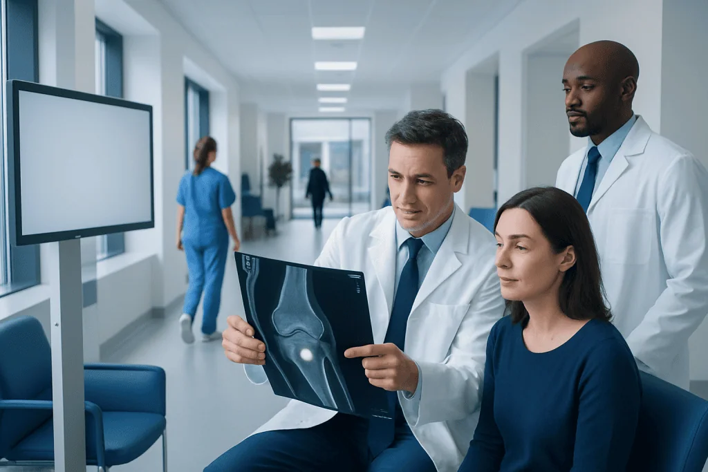
Osteoid osteoma is a small, benign bone-forming tumor that does not spread to other parts of the body. MRI commonly shows bone marrow and soft-tissue swelling, while CT may better reveal the central nidus.
Key Takeaways
- Osteoid osteoma is a small, benign bone-forming tumor that does not spread to other parts of the body.
- MRI commonly shows bone marrow and soft-tissue swelling, while CT may better reveal the central nidus.
- Night pain that improves with anti-inflammatory medicine is a typical symptom, although symptoms vary.
- The condition is not cancer and does not transform into a malignant tumor.
- Many symptomatic cases can be treated with minimally invasive image-guided ablation or, less commonly, surgery.
Osteoid osteoma on MRI can appear as extensive inflammation around a very small bone lesion, sometimes making the lesion itself difficult to see. MRI is valuable for assessing nearby bone and soft tissues, while CT is often used to identify the characteristic nidus and plan treatment when needed.
Overview: What does osteoid osteoma on MRI mean?
Osteoid osteoma on MRI usually refers to an MRI examination performed to investigate persistent, often night-time bone pain and identify features of this small benign bone tumor. MRI may show prominent inflammation in and around the affected bone, but the tiny central lesion, called a nidus, can be less distinct than it is on CT.
Osteoid osteoma is a non-cancerous tumor made of bone-forming tissue. It is usually small, commonly measuring less than 1.5 to 2 centimeters, but it can cause disproportionate pain because it produces substances that promote inflammation. Imaging findings, symptoms and clinical examination are considered together before a diagnosis and treatment plan are made.
The condition most often develops in children, adolescents and young adults, although it can occur at other ages. It can affect any bone. When symptoms are troublesome or the diagnosis remains uncertain, an orthopedic specialist and musculoskeletal radiologist may recommend further imaging, often including CT.
What are the MRI findings of osteoid osteoma?
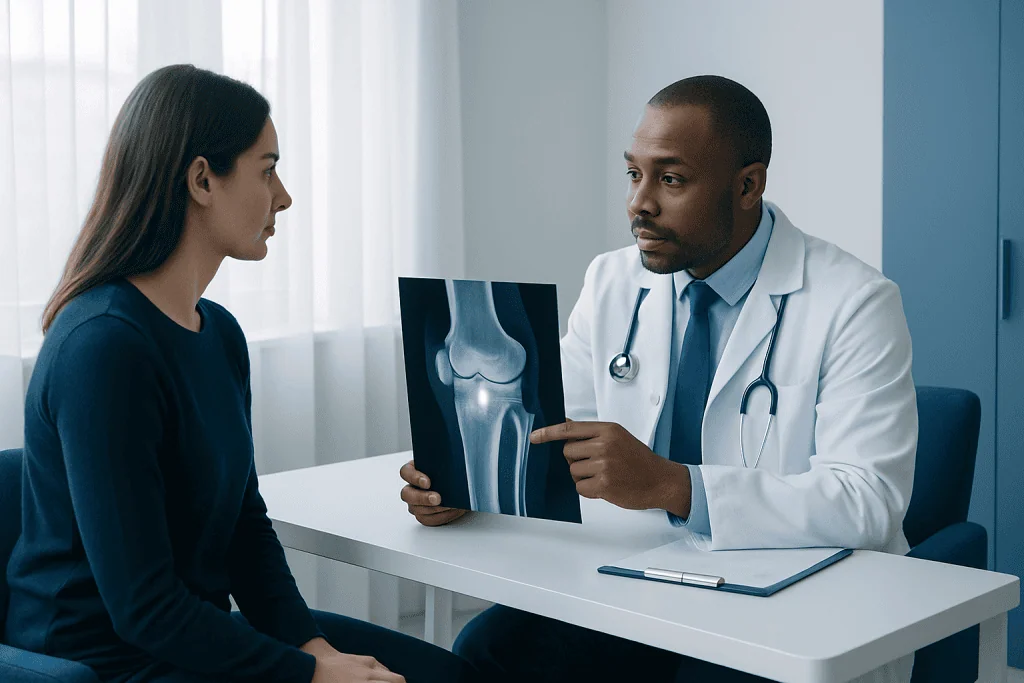
The classic lesion is a small nidus: a round or oval focus of vascular bone-forming tissue that may contain central mineralization. On MRI, the nidus may have variable signal characteristics because its appearance depends on the amount of mineralization, vascular tissue and surrounding reaction. After contrast material is given, the nidus often enhances because it has a rich blood supply.
More conspicuous MRI findings may occur around the nidus rather than within it. These can include bone marrow edema, cortical thickening, inflammation of nearby soft tissues and fluid in an adjacent joint. In a lesion near the spine, MRI may also show muscle edema or reactive changes around the posterior spinal elements.
These reactive changes can occasionally resemble infection, stress injury, inflammatory arthritis or another bone condition. CT provides detailed images of bone mineralization and is frequently the most useful test for locating a small nidus. A doctor may use MRI and CT as complementary examinations rather than viewing either test in isolation.
- MRI is especially useful for: showing marrow, joint and soft-tissue effects of the lesion.
- CT is especially useful for: demonstrating the nidus and any central calcification.
- Contrast MRI may help: when the nidus is difficult to distinguish from surrounding inflammatory change.
How MRI works, who may need it and how to prepare
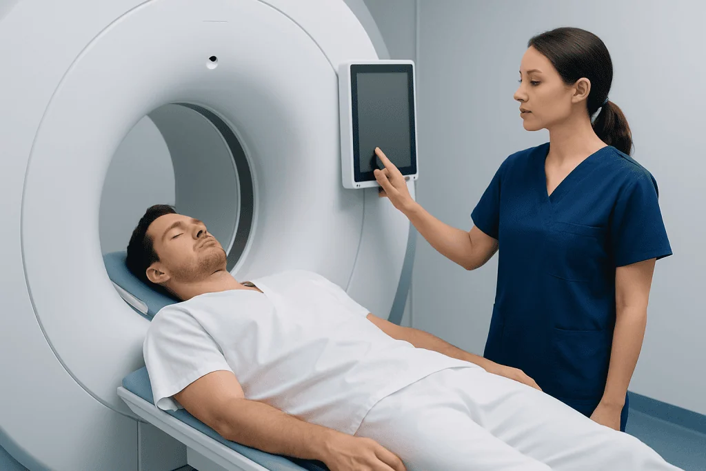
MRI uses a strong magnetic field and radiofrequency waves to create detailed images of bones, bone marrow, joints, muscles and other soft tissues. It does not use ionizing radiation. For a person with unexplained localized bone pain, MRI may be requested early because it can detect inflammation and other possible causes of symptoms.
People may be candidates for MRI when pain persists, is worse at night, limits movement, or is associated with a limp, localized tenderness or swelling. It can also help evaluate a suspected lesion near a joint, in the spine, or in a location where soft-tissue involvement is important. However, MRI alone may not always provide a definite diagnosis of osteoid osteoma.
Before the scan, patients should tell the imaging team about implanted devices, prior surgery, metal fragments, pregnancy, kidney disease, allergies and claustrophobia. Some implants are MRI-compatible and others require special assessment. If contrast is planned, the team will explain why it may be useful and review relevant safety information.
Most people can eat and drink normally before a standard MRI unless they are given different instructions. Metal objects, including jewelry, watches, hearing aids and removable dental appliances, must be removed. Comfortable clothing without metal fasteners may be helpful.
MRI procedure: what happens step by step?
At the appointment, a radiographer reviews safety questions and helps position the patient on a padded scanning table. A coil, which is a device that receives MRI signals, may be placed around the body part being examined. The table then moves slowly into the scanner. For many bone examinations, only the relevant area needs to be centered in the machine.
The scan itself is painless, although the MRI system makes rhythmic tapping and knocking sounds. Ear protection is provided. Remaining still is important because movement can blur images, and the radiographer can communicate with the patient throughout the examination. Scan time varies according to the body area and whether contrast sequences are needed.
If contrast is used, it is generally injected through a small intravenous line partway through the examination. This can help define vascular tissue and distinguish a possible nidus from edema. Following the scan, a radiologist reviews the images and prepares a report for the referring clinician.
When MRI findings suggest osteoid osteoma but do not clearly show the nidus, thin-section CT may be recommended. CT can also be used to plan image-guided radiofrequency ablation when treatment is appropriate.
How serious is osteoid osteoma?
Osteoid osteoma is benign, meaning it is not cancer and does not spread through the body. In this sense, it is not considered life-threatening. Nevertheless, the pain can be significant and may interfere with sleep, school, work, exercise and everyday activities.
Its impact depends partly on location. A lesion close to a joint may cause stiffness, swelling or reduced movement. In children and adolescents, persistent limping or reduced use of a limb should be assessed, as long-standing symptoms can affect function. Spinal lesions may lead to painful muscle spasm or, in some cases, a developing spinal curve caused by pain.
Symptoms may sometimes be controlled with clinician-guided anti-inflammatory medication while the lesion is monitored. If pain remains limiting, if medication is not suitable, or if the location creates functional concerns, minimally invasive treatment can usually be considered. The best approach depends on the lesion site, imaging findings, age, overall health and personal preferences.
What is the most common location for osteoid osteoma?
Osteoid osteoma most often occurs in the long bones of the legs, particularly the femur and tibia. It commonly arises in the cortex, the dense outer layer of bone. A lesion in the upper part of the femur can cause hip, thigh or knee pain, and symptoms may be mistaken for a joint problem because pain can be felt away from its source.
It can also occur in the bones of the spine, the foot, hand, pelvis or upper limb. Lesions within or close to a joint may produce joint fluid, stiffness and inflammation, and their symptoms can be less typical than the usual night pain pattern. This variation is one reason careful imaging review is important.
Wherever it occurs, precise localization matters. It helps the clinical team distinguish osteoid osteoma from conditions such as stress fracture, infection and other benign bone lesions, and it supports safe planning if an intervention is recommended.
Treatment options, benefits, risks and recovery
Management may include observation with pain control, particularly when symptoms are manageable and follow-up is appropriate. Non-steroidal anti-inflammatory medicines can be effective for some people because they reduce the inflammatory substances associated with the nidus. A clinician should advise on the safest medicine and duration, especially for people with kidney disease, stomach ulcers, cardiovascular conditions or medication interactions.
For persistent or disabling symptoms, percutaneous thermal ablation is often considered. Under CT guidance, a specialist places a probe into the nidus and uses controlled heat, such as radiofrequency energy, to destroy the lesion. Depending on its location and local practice, this may be performed with sedation or anesthesia. Conventional surgery is less common but may be necessary when image-guided ablation is unsuitable or when the diagnosis needs further clarification.
The potential benefit of ablation is targeted treatment with a small skin entry point and relatively rapid relief of pain. Risks are uncommon but can include bleeding, infection, skin or soft-tissue burn, fracture, nerve injury, injury to nearby structures, incomplete treatment or recurrence. Risk depends strongly on the lesion’s location, especially near nerves, joints or growth areas in children.
Recovery after a straightforward image-guided procedure is often measured in days to weeks rather than months, though activity restrictions vary. The care team may recommend a brief period of reduced weight-bearing or avoiding impact exercise. Follow-up focuses on symptom improvement, wound healing and return of function; repeat imaging is individualized rather than automatically required for every patient.
Can osteoid osteoma turn malignant?
No. Osteoid osteoma does not turn into cancer and is not known to transform into a malignant bone tumor. It is a benign lesion with a very different biological behavior from bone cancers.
However, a painful bone lesion should still be assessed carefully. Several conditions can produce overlapping symptoms or imaging changes, and a clear diagnosis helps ensure that the appropriate treatment is offered. If images are not typical, the specialist may request further imaging or, less often, a biopsy.
A specialist team may include orthopedic surgeons, interventional radiologists, musculoskeletal radiologists, anesthesiologists and rehabilitation professionals. Acibadem International’s multidisciplinary specialists and JCI-accredited hospitals evaluate and treat osteoid osteoma for international patients when specialist assessment or image-guided care is needed.
When to seek medical care
Medical assessment is appropriate for persistent focal bone pain, pain that regularly wakes a person at night, a limp, reduced movement or pain that does not improve as expected after a minor injury. Parents and caregivers should seek assessment for a child with ongoing limb pain, avoidance of activity or an unexplained change in walking pattern.
More prompt evaluation is important if pain is accompanied by fever, marked redness or warmth, unexplained weight loss, new weakness, numbness, loss of bladder or bowel control, or severe pain after an injury. These features do not necessarily indicate osteoid osteoma, but they need timely clinical review.
Patients should bring previous imaging reports and scans, if available, to their appointment. A qualified doctor can determine whether MRI, CT, blood tests, specialist referral or another next step is appropriate for the individual situation.
Frequently asked questions
Can MRI diagnose osteoid osteoma by itself?
MRI can strongly suggest osteoid osteoma by showing a small lesion and the surrounding bone marrow or soft-tissue inflammation. However, the nidus may be difficult to see on MRI because the inflammatory reaction can be extensive. CT is often used to confirm and precisely locate the nidus.
Why can osteoid osteoma be missed on MRI?
The nidus is very small and may have signal features similar to surrounding bone. MRI can make the surrounding edema more noticeable than the nidus itself. A thin-section CT scan can often show the lesion more clearly.
Is contrast always needed for an MRI of suspected osteoid osteoma?
No, contrast is not required in every case. It may be used when clinicians need more information about a suspected nidus, inflammation or nearby soft tissues. The decision depends on the body area, initial images and the patient's medical circumstances.
Does osteoid osteoma pain always get worse at night?
Night pain that responds to anti-inflammatory medication is a well-known pattern, but not every person has this exact experience. Pain can vary according to the lesion's location, particularly when it is close to a joint or in the spine. Persistent localized pain should be assessed even if it is not mainly nocturnal.
Will osteoid osteoma go away without a procedure?
Symptoms can sometimes be managed with anti-inflammatory medication, and osteoid osteoma may eventually resolve over time. This approach may not be suitable for everyone because pain can persist for a long period or medication may cause side effects. A clinician can discuss observation and intervention based on symptoms, location and overall health.
How quickly does pain improve after radiofrequency ablation?
Many patients experience meaningful pain improvement soon after a successful procedure, although recovery differs between individuals. Soreness at the procedure site and temporary activity limits are possible. The treating team provides tailored guidance on pain control, mobility and return to sport or work.
References
- American Academy of Orthopaedic Surgeons
- RadiologyInfo.org
- National Cancer Institute
- Orphanet
- Merck Manual Consumer Version
This article is for general information only and is not a substitute for professional medical advice. Please consult a qualified doctor about your individual situation.
Oncology care in Turkey — second opinion and treatment plan
JCI-accredited · board-certified surgeons · reply within 24h
Add us as a Preferred Source to see more of our trusted health content across Google Search, AI Overviews and Discover.