Choroidal Melanoma Treatment: How It Works, Results and What to Expect

Choroidal melanoma is the most common primary cancer inside the eye in adults, arising from pigment-producing cells in the choroid. Focused radiation is a common eye-preserving treatment; surgery may be needed for selected tumors or complications.
Key Takeaways
- Choroidal melanoma is the most common primary cancer inside the eye in adults, arising from pigment-producing cells in the choroid.
- Focused radiation is a common eye-preserving treatment; surgery may be needed for selected tumors or complications.
- Treatment planning considers tumor characteristics, vision in the affected eye, overall health and possible metastatic disease.
- The outlook varies substantially between individuals and depends on tumor features and whether cancer has spread.
- Regular examinations and liver-focused surveillance are important after treatment because metastases can occur years later.
Choroidal melanoma treatment is tailored to the tumor’s size, location, growth pattern and whether there is evidence of spread. Most care aims to control the tumor while preserving the eye and useful vision whenever safely possible, with regular lifelong follow-up remaining essential.
Overview: How choroidal melanoma treatment works
Choroidal melanoma treatment controls a cancer that begins in the choroid, the blood-vessel-rich layer beneath the retina at the back of the eye. It is a type of uveal melanoma. Treatment is planned by an ocular oncologist and aims to destroy or remove the tumor, protect the eye where possible, preserve vision when feasible, and identify any spread beyond the eye.
For many patients, focused radiation can treat the tumor without removing the eye. Other options include carefully selected laser-based treatments, surgery, or active surveillance for a very small lesion that is not clearly cancerous. The most appropriate choice depends on the tumor’s size and location, its behavior over time, vision in the eye, and the person’s general health and preferences.
Choroidal melanoma is different from melanoma of the skin. Although both originate in pigment-producing cells, their genetic features, patterns of spread and treatment approaches can differ. A specialist assessment is therefore important before any treatment decision is made.
Assessment and candidacy for treatment
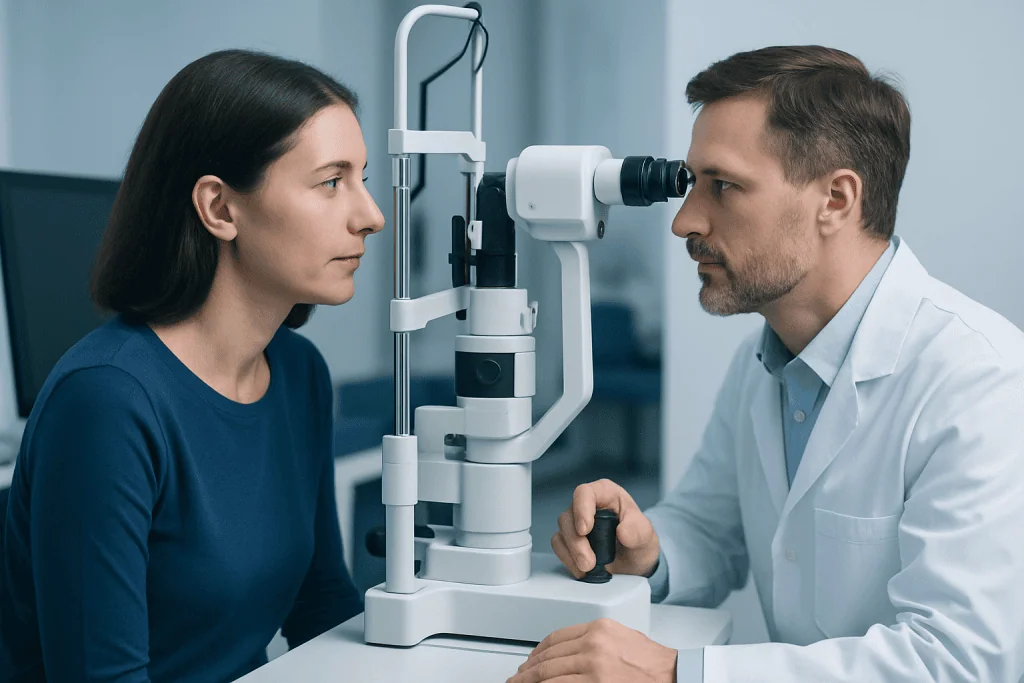
Not every pigmented spot in the choroid is a melanoma. Some are benign choroidal nevi, often called eye freckles, which may only need observation. A lesion is more concerning when it grows, has certain ultrasound or retinal features, causes fluid beneath the retina, or is associated with symptoms such as blurred vision or flashes of light.
Before recommending treatment, the care team establishes the diagnosis and maps the tumor precisely. Candidates for eye-preserving treatment commonly include people with small to medium tumors that can be targeted while limiting injury to critical structures. Larger tumors, tumors causing severe eye complications, or tumors with little possibility of retaining a comfortable, seeing eye may sometimes be treated with removal of the eye.
Testing also evaluates whether cancer has spread. This usually includes a medical history and physical examination, liver blood tests and imaging selected by the clinical team. Genetic testing of tumor tissue may be offered in some settings because it can help estimate metastatic risk and guide the intensity of follow-up, but it does not by itself determine a person’s outcome.
The treatment pathway: step by step

After detailed eye imaging, the ocular oncology team discusses the likely benefits and limitations of each approach. Radiation planning may involve measurements, photographs and imaging to define the tumor’s exact boundaries. In plaque brachytherapy, a small radioactive device is temporarily positioned on the outside of the eye directly over the tumor; it delivers radiation locally and is removed after the planned treatment period.
Some centers use proton beam radiation, which directs an external beam of radiation to the tumor with a highly controlled dose distribution. This may be considered according to tumor size, location and local expertise. Radiation does not make the patient radioactive after treatment is complete, and its effects on tumor cells develop gradually over time.
Laser approaches may be used in selected small tumors or as an additional treatment in particular circumstances. Surgical removal of the eye, called enucleation, can be recommended when it offers the safest local control or when the eye is painful or has very limited visual potential. An artificial eye can usually be fitted after healing to support appearance and comfort.
Care is often coordinated between ocular oncology, radiation oncology, medical oncology, radiology, pathology and supportive-care professionals. Patients considering care abroad can also discuss coordinated assessment and follow-up planning; Acibadem International’s multidisciplinary specialists and JCI-accredited hospitals provide diagnosis and treatment for international patients.
Benefits, risks and recovery timeline
The main benefit of treatment is local tumor control, meaning the tumor stops growing or becomes smaller over time. Eye-preserving approaches may retain the eye and, in some cases, useful vision. However, vision outcomes cannot be guaranteed because they depend strongly on whether the tumor or treatment is close to the macula, optic nerve or other sensitive structures.
Recovery varies with the treatment used. After plaque treatment or surgery, there may be temporary redness, soreness, light sensitivity or blurred vision. People commonly need prescribed eye drops, activity guidance and follow-up visits in the days and weeks after treatment. Imaging over subsequent months checks that the tumor is responding and that the retina and optic nerve remain healthy.
Potential late effects of radiation include cataract, dry eye, retinal swelling, radiation retinopathy, optic nerve damage, glaucoma and reduced vision. Some complications can be managed with eye injections, laser treatment, surgery or medicines, although not all visual loss can be reversed. Enucleation has surgical and emotional adjustment considerations, and patients may benefit from prosthetic-eye support and counseling.
- Seek urgent eye advice for sudden severe pain, marked vision loss, increasing redness, discharge, or a new curtain-like shadow in vision after treatment.
- Keep all scheduled eye and systemic surveillance appointments, even if the treated eye feels well.
- Report new persistent symptoms such as unexplained abdominal discomfort, loss of appetite, weight change or fatigue to the treating clinician, while remembering these symptoms often have non-cancer causes.
What is the life expectancy for patients with choroidal melanoma?
Life expectancy for patients with choroidal melanoma varies widely and cannot be predicted from the diagnosis alone. Important factors include the tumor’s size, cell type and genetic characteristics, whether it has spread, the response to local treatment, and a person’s overall health. Many people have successful local control of the eye tumor and continue routine life after treatment.
The greatest long-term concern is metastatic disease, most often involving the liver. Prognosis is generally more favorable when the disease is confined to the eye than when it has spread elsewhere. The treating team can provide the most individualized discussion after reviewing imaging, pathology and, where appropriate, tumor genetic information.
Follow-up is not simply a precaution; it is a central part of care. Scheduled eye examinations and systemic surveillance can detect recurrence, treatment-related complications or possible spread at an earlier stage, when further assessment and management can be arranged promptly.
How fast can choroidal melanoma grow?
Choroidal melanoma growth rates differ considerably. Some tumors enlarge slowly over months or years, while others show measurable change sooner. Very small suspicious lesions may therefore be monitored with repeat examinations and imaging at carefully planned intervals before treatment is recommended.
Growth is assessed using standardized eye photographs, ultrasound and optical imaging rather than symptoms alone. A person may not notice a change in vision even when a lesion is growing, particularly if it is away from the central retina. Conversely, visual symptoms do not always mean that a lesion is malignant or rapidly enlarging.
Because growth behavior is individual, people should not delay follow-up appointments after a suspicious choroidal lesion has been identified. Comparing precise measurements over time helps the ocular oncology team distinguish a stable lesion from one that needs treatment.
How treatable is choroidal melanoma? Does it always metastasize?
Choroidal melanoma is treatable, particularly when it is limited to the eye. Modern local therapies can achieve strong control of many tumors while avoiding removal of the eye in appropriate cases. Treatment of the eye tumor is important, but it cannot eliminate every risk of microscopic cancer cells that may already have traveled elsewhere before diagnosis.
Choroidal melanoma does not always metastasize. Some people never develop metastatic disease, while others have a higher risk based on clinical and genetic tumor features. Metastasis may occur years after successful treatment of the original eye tumor, which is why clinicians recommend long-term surveillance.
If metastatic disease is found, care is led by medical oncology and may include systemic treatments, clinical trials, liver-directed approaches or supportive treatments, depending on the extent and biology of disease. Decisions should be individualized in a multidisciplinary setting, with attention to both disease control and quality of life.
When to seek medical care
Anyone with a newly found pigmented lesion inside the eye should attend the follow-up appointments advised by an ophthalmologist. Prompt specialist review is particularly important for new or worsening blurred vision, flashes, floaters, a shadow or missing area in vision, distortion of straight lines, or a visible change in the eye. These symptoms can have many causes and do not necessarily mean melanoma.
People who have been treated for choroidal melanoma should contact their eye team quickly if they experience severe pain, sudden vision changes, substantial redness or other unexpected postoperative symptoms. They should also continue recommended medical surveillance even when they feel well, since early metastatic disease may not cause noticeable symptoms.
Questions about diagnosis, treatment selection, vision outcomes or monitoring are appropriate to raise with an ocular oncologist. A second opinion may be helpful when treatment choices are complex or when a person would like to better understand the balance between eye preservation, vision and long-term monitoring.
Frequently asked questions
What is the main treatment for choroidal melanoma?
Focused radiation is a common treatment for choroidal melanoma, particularly plaque brachytherapy and, in some centers, proton beam radiation. The best option depends on the tumor’s size, position and features, as well as vision in the affected eye. Surgery, laser-based therapy or observation may be appropriate in selected situations.
Can vision be preserved after choroidal melanoma treatment?
Vision can be preserved for some patients, especially when the tumor and treatment area are away from the macula and optic nerve. However, vision may decline gradually because of the tumor itself or delayed radiation effects. The ocular oncology team can explain the likely visual risks based on the tumor’s exact location.
Is plaque brachytherapy painful?
Plaque brachytherapy is performed with anesthesia, so patients do not feel the procedure itself. Mild soreness, redness, irritation or blurred vision can occur while the eye heals. The care team provides pain-relief and eye-drop instructions tailored to the individual.
How often is follow-up needed after treatment?
Follow-up schedules vary, but they usually include regular eye examinations and imaging soon after treatment and at longer intervals over time. Systemic monitoring, often with attention to the liver, is also recommended because choroidal melanoma can metastasize after a delay. The schedule is individualized according to tumor risk and treatment history.
Can choroidal melanoma come back after treatment?
A treated tumor can occasionally show local recurrence or continued activity, which is why repeated eye imaging is important. Additional local treatment may be possible depending on the situation. Follow-up also checks for treatment-related eye complications that may occur months or years later.
Does removal of the eye cure choroidal melanoma?
Removing the eye can provide local control when enucleation is the most suitable treatment. However, it cannot guarantee that microscopic cells have not already spread beyond the eye before surgery. Ongoing systemic surveillance remains important after enucleation as well as after eye-preserving treatments.
References
- American Academy of Ophthalmology
- National Cancer Institute
- American Cancer Society
- National Comprehensive Cancer Network
- Melanoma Research Foundation
This article is for general information only and is not a substitute for professional medical advice. Please consult a qualified doctor about your individual situation.
Melanoma in Turkey — costs, top hospitals & a free quote
JCI-accredited · board-certified surgeons · reply within 24h
Add us as a Preferred Source to see more of our trusted health content across Google Search, AI Overviews and Discover.

Medical Oncology Department
Medical treatment of cancer with chemotherapy, immunotherapy and targeted therapies under a multidisciplinary tumor board.
60 specialists in this unit