CT Scan Cardiac Imaging: Preparation, Procedure and Results
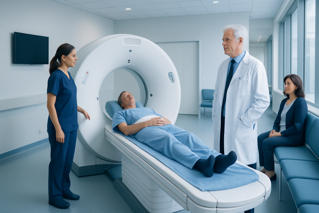
CT stands for computed tomography, a scan that combines multiple X-ray images into detailed cross-sectional views. Cardiac CT is often used to assess coronary artery calcium, coronary artery narrowing, heart structure, and some blood vessel conditions.
Key Takeaways
- CT stands for computed tomography, a scan that combines multiple X-ray images into detailed cross-sectional views.
- Cardiac CT is often used to assess coronary artery calcium, coronary artery narrowing, heart structure, and some blood vessel conditions.
- Preparation may include avoiding food or caffeine for a short period, depending on the type of scan and the imaging centre's instructions.
- Contrast dye is commonly used for coronary CT angiography and should be discussed in advance with the care team if there is kidney disease, an allergy history, or pregnancy.
- Most people return to usual activities immediately after a cardiac CT, unless they have received medication that requires short observation.
CT scan cardiac imaging is a non-invasive test that combines X-rays and computer processing to produce detailed images of the heart, its blood vessels, and nearby structures. It can help clinicians evaluate coronary artery narrowing, heart anatomy, calcium buildup, and selected causes of cardiac symptoms.
Overview: what CT scan cardiac imaging shows
CT scan cardiac imaging, also called cardiac computed tomography or cardiac CT, is an imaging examination that creates detailed pictures of the heart and blood vessels. It uses a rotating X-ray scanner and advanced computer processing to produce cross-sectional images. In many cases, the images are timed with the heartbeat so that the coronary arteries and moving heart structures can be seen clearly.
The test may be performed as a coronary artery calcium scan, which looks for calcified plaque in the coronary arteries, or as coronary CT angiography, which uses contrast dye to show the inside of the coronary arteries and other blood vessels. Clinicians may use the results alongside symptoms, medical history, examination findings, blood tests, and other cardiac investigations.
Cardiac CT does not replace every heart test. The best examination depends on the clinical question, the person’s symptoms, and factors such as heart rhythm, kidney function, and prior heart procedures. It is one of several tools used to investigate possible coronary artery disease and related heart concerns.
What does CT stand for?
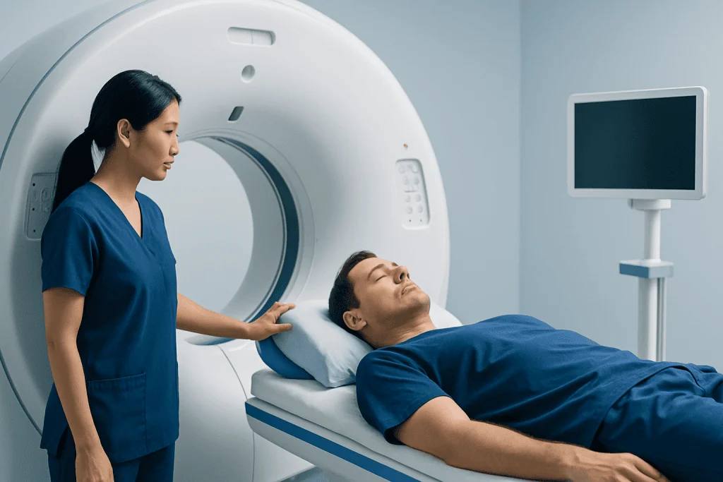
CT stands for computed tomography. “Computed” refers to the computer technology that processes many X-ray measurements, while “tomography” refers to creating images of thin slices or sections through the body.
Unlike a standard chest X-ray, which produces a single two-dimensional image, CT gathers images from many angles around the body. A computer then reconstructs these images into detailed views that can be examined one slice at a time or displayed as three-dimensional models.
For heart imaging, the scanner is coordinated with the cardiac cycle whenever possible. This reduces motion blur and helps the radiology and cardiology teams assess the coronary arteries, heart chambers, valves, aorta, and surrounding tissues.
What is a CT short for?

CT is short for computed tomography. It is sometimes also called a CAT scan, where CAT means computed axial tomography. Both terms describe the same general imaging technology, although CT is now the more commonly used name.
A CT scan uses ionising radiation in the form of X-rays. Modern scanners and imaging protocols are designed to obtain clinically useful images while keeping radiation exposure as low as reasonably achievable. The actual exposure varies according to the type of scan, the equipment used, and the individual imaging needs.
In cardiac CT, special techniques may be used to capture images during a particular phase of the heartbeat. This is why some people are given medication before the scan to slow a fast heart rate, when appropriate and safe.
What does a CT mean?
In a medical report, “CT” means computed tomography and identifies the type of imaging test performed. A CT result is not a diagnosis by itself; it is interpreted in the context of the reason for the scan and the person’s overall health.
A cardiac CT report may describe coronary calcium, narrowing or plaque in coronary arteries, the size and shape of heart chambers, the aorta, heart valves, or findings outside the heart that are visible in the chest. Some findings are common with ageing and may not require treatment, while others may lead to further evaluation.
The referring clinician explains what the result means for the individual. If a scan identifies coronary artery plaque or a possible narrowing, next steps may range from preventive lifestyle and medication discussions to additional testing or specialist assessment. A CT finding should not be interpreted without professional guidance.
How is CT different from MRI?
CT and MRI both create detailed images of the body, but they use different technology. CT uses X-rays, whereas magnetic resonance imaging (MRI) uses strong magnetic fields and radiofrequency waves. MRI does not use ionising radiation.
Cardiac CT is particularly useful for evaluating coronary artery calcium and for rapidly viewing coronary arteries with contrast dye. It is usually completed within a short time and is often more suitable for urgent or time-sensitive imaging questions. MRI can provide especially detailed information about heart muscle, heart function, inflammation, scarring, and certain structural conditions.
The choice between CT and MRI is based on what clinicians need to know. MRI may not be suitable for some people with certain implanted devices, severe claustrophobia, or other contraindications. CT may be less suitable when radiation or iodinated contrast poses a particular concern. The imaging team will recommend the most appropriate approach.
Candidacy and preparation for cardiac CT
A clinician may recommend cardiac CT for people with selected chest symptoms, an uncertain risk of coronary artery disease, suspected coronary artery abnormalities, known disease needing further anatomical assessment, or questions about the aorta or heart structure. A coronary calcium score is generally used for risk assessment in carefully selected people without symptoms, rather than as a test for acute chest pain.
Before the appointment, patients should tell the team about kidney disease, prior reactions to contrast dye, asthma, thyroid conditions, diabetes, pregnancy or possible pregnancy, breastfeeding, and all medications or supplements. These details help the team plan the scan safely. People should not stop prescribed medication unless specifically instructed to do so.
Preparation varies by examination. The centre may ask the person to avoid food for several hours before contrast CT and to avoid caffeine, nicotine, or exercise beforehand because these can raise the heart rate. Water may be encouraged. The care team may also ask for recent kidney blood test results when contrast dye is planned.
- Wear comfortable clothing and remove jewellery or metal objects near the chest where requested.
- Bring a list of medicines, allergies, prior imaging reports, and relevant medical records.
- Ask in advance whether someone should accompany the patient if heart-rate medication or a sedating medicine may be used.
Step-by-step: how the procedure works
On arrival, a radiographer or nurse confirms medical information, explains the examination, and may place an intravenous line in the arm if contrast dye is needed. Small adhesive electrodes are placed on the chest to monitor the heartbeat. The person lies on a padded table that moves through the circular opening of the CT scanner.
For coronary CT angiography, contrast dye is injected through the intravenous line during imaging. It may cause a brief warm sensation or a metallic taste, which usually passes quickly. The person may be asked to hold their breath for a few seconds at a time and to remain still. Clear communication from the team helps make the experience straightforward.
The scanning portion itself is usually brief, though the full visit can take longer because of preparation, heart-rate assessment, intravenous access, and observation when needed. In some cases, medication is given to reduce heart rate or widen the coronary arteries. This is considered individually, with monitoring for safety.
The images are reviewed by appropriately trained specialists. Results may be available promptly in some settings, but the timing of the formal report varies. If further evaluation is required, a clinician may discuss options such as medical management, functional heart testing, or coronary angiography.
Benefits, risks, recovery and when to seek medical care
The main benefit of CT scan cardiac imaging is detailed, non-invasive assessment of the heart and coronary arteries. It can help rule out significant coronary narrowing in selected patients and support decisions about prevention or further care. It may also identify structural and vascular findings that would be difficult to assess with standard X-rays alone.
Cardiac CT involves a small amount of radiation, and contrast CT carries a small risk of allergic-type reaction or effects on kidney function in susceptible people. Serious reactions are uncommon, and imaging teams have procedures to recognise and manage them. The scan may be less accurate when heart rhythm is irregular, heart rate is high, or there is extensive coronary calcium or prior coronary stenting.
Most people can return to normal activities after the scan. Drinking fluids after contrast, if medically appropriate, may help the body clear it. Those who receive certain medications before the test may be monitored briefly and should follow individual advice about driving, activity, and medicines.
When to seek medical care: New, severe, or persistent chest pressure; chest pain with shortness of breath, sweating, nausea, fainting, or pain spreading to the arm, jaw, back, or neck requires urgent medical assessment. After a contrast scan, urgent help is also needed for trouble breathing, swelling of the face or throat, widespread hives, or other signs of a serious allergic reaction. For planned cardiac evaluation, a qualified clinician can advise whether cardiology assessment or another test is appropriate.
Acibadem International’s multidisciplinary specialists and JCI-accredited hospitals support international patients who need cardiac imaging and follow-up care, with investigations planned according to individual clinical needs.
Frequently asked questions
Is CT scan cardiac imaging painful?
The scan itself is not painful. Patients need to lie still and may feel brief discomfort from intravenous line placement if contrast dye is used. Some people notice temporary warmth or a metallic taste during contrast injection.
How long does a cardiac CT scan take?
The actual image acquisition is often very short, sometimes only seconds. However, the complete appointment may take longer because it can include registration, screening questions, preparation, intravenous access, heart-rate checks, and post-scan observation.
Do patients need contrast dye for every cardiac CT?
No. A coronary artery calcium scan is generally performed without contrast dye. Coronary CT angiography usually requires iodinated contrast because it makes the inside of the coronary arteries visible.
Can someone eat before a cardiac CT scan?
Instructions depend on the exact examination. For some contrast cardiac CT scans, the imaging centre may ask patients not to eat for a few hours beforehand and to avoid caffeine or nicotine. Patients should follow the instructions provided by their own care team.
What can a cardiac CT scan detect?
It can help detect coronary artery calcium, plaque, possible coronary narrowing, some congenital coronary artery variations, and selected abnormalities of the heart, aorta, or surrounding chest structures. The test is chosen based on the clinical question and does not diagnose every heart condition.
When are cardiac CT results available?
A specialist must review the images and prepare a report, so timing varies by centre and clinical urgency. The referring clinician explains the findings and whether further testing, prevention measures, or treatment is needed.
References
- American College of Radiology
- Radiological Society of North America
- American Heart Association
- Society of Cardiovascular Computed Tomography
- National Heart, Lung, and Blood Institute
This article is for general information only and is not a substitute for professional medical advice. Please consult a qualified doctor about your individual situation.
Heart care in Turkey — expert evaluation and treatment
JCI-accredited · board-certified surgeons · reply within 24h
Add us as a Preferred Source to see more of our trusted health content across Google Search, AI Overviews and Discover.
Check your numbers in seconds
BMI, calories, due date, blood pressure and 30+ more clinical calculators — free, instant, doctor-reviewed ranges.

More from the Health Library



Related Specialists

Dr. Edip Yılmaz
Orthopedic Surgery & Traumatology
Prof. Dr. Soner Solmaz
Hematology
Dr. Müjgan Orman
Radiology
Embriyolog Seda Özkan
Vitro Fertilization and Reproductive Medicine Center