Microphthalmia Treatment: How It Works, Results and What to Expect

Microphthalmia is a congenital condition in which one or both eyes are abnormally small and incompletely developed. Treatment cannot usually make an underdeveloped eye develop normal vision, but it can protect existing sight and support socket and facial growth.
Key Takeaways
- Microphthalmia is a congenital condition in which one or both eyes are abnormally small and incompletely developed.
- Treatment cannot usually make an underdeveloped eye develop normal vision, but it can protect existing sight and support socket and facial growth.
- Children with little or no vision in the affected eye may use conformers, expanders and custom ocular prostheses.
- Care is often coordinated among pediatric ophthalmology, ocularistry, genetics, pediatrics and low-vision rehabilitation specialists.
- Prompt evaluation is especially important when a baby has a very small eye, an absent-looking eye, a cloudy cornea or other developmental concerns.
Microphthalmia treatment is individualized and usually focuses on protecting any remaining vision, supporting normal growth of the eye socket and surrounding face, and fitting expanders or a prosthetic eye when needed. Early assessment by pediatric eye specialists is important because the condition can range from a mildly small eye with useful sight to a severely underdeveloped eye with little or no vision.
Microphthalmia Treatment: How It Works
Microphthalmia treatment is planned around the individual child’s eye development, vision potential and overall health. The main goals are to identify and protect any usable vision, monitor for eye-related complications, encourage growth of the eye socket and eyelids, and provide practical and emotional support for the child and family. Treatment often begins in infancy and continues as the child grows.
Microphthalmia means that an eye is smaller than expected because it did not fully develop before birth. It may affect one eye or both. In some children, the eye is small but has structures that can support some vision; in others, the eye has very limited visual potential or is associated with anophthalmia, where eye tissue is absent or nearly absent.
Management is not one single operation or device. A pediatric ophthalmologist first assesses the eye carefully and then coordinates care with an ocularist, who makes custom prosthetic eyes, and other specialists when appropriate. When vision is present, treatment prioritizes preserving it. When sight is not possible in the small eye, care focuses on healthy socket development, comfort and appearance.
Assessment, Candidacy and Care Planning
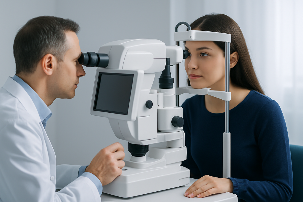
Every child with suspected microphthalmia should be assessed by an ophthalmologist experienced in childhood eye conditions. The examination may include measurement of the eye, assessment of the cornea, retina and optic nerve, and checks for pressure-related problems such as glaucoma. Infants and young children may need an examination under anesthesia so the eye can be evaluated safely and thoroughly.
Ultrasound and, in selected cases, MRI or other imaging can show the size and internal structures of the eye and orbit. Imaging may also help clinicians look for associated differences involving the optic nerves or brain. Genetic counseling and testing may be discussed, particularly when both eyes are affected, there is a family history, or the child has other physical or developmental findings.
Children may be candidates for socket expansion when the affected eye has little or no visual potential and the eye socket is too small to support balanced facial growth. A child with useful sight may instead need treatment directed at associated findings, such as cataract, refractive error, retinal changes or elevated eye pressure. The plan is reviewed regularly because a child’s needs change with growth.
- Vision assessment in each eye, using age-appropriate testing
- Evaluation of eyelids, eye socket size and facial symmetry
- Imaging when it will clarify anatomy or associated conditions
- Genetic and developmental assessment when clinically indicated
- Support for vision development, school access and family adjustment
Step-by-Step: Expanders, Prostheses and Surgery
For an eye with poor or absent visual potential, socket expansion usually starts with a small smooth device called a conformer. It sits behind the eyelids and is designed to gently maintain space in the socket. As the child grows, the ocularist or ophthalmology team replaces it with gradually larger conformers. This process may help the eyelids, orbit and surrounding soft tissues develop more evenly.
Once enough socket volume is present, a custom ocular prosthesis may be made. This is an artificial eye shell that is individually shaped and painted to match the other eye as closely as possible. It does not restore sight, but it can support the eyelids, improve symmetry and help a child feel comfortable in social settings. Prostheses need cleaning, regular reviews and periodic replacement or adjustment as the child grows.
Some children need surgery when conformers alone do not provide adequate expansion or when eyelid and socket anatomy requires correction. Procedures may involve orbital implants, tissue expanders, skin or mucosal grafts, or surgery to improve eyelid position. The exact approach depends on anatomy and is decided after detailed discussion of likely benefits, limitations and follow-up needs. Information about custom ocular prosthesis care may be helpful for families considering this part of treatment.
When a child has vision in one eye only, protecting that eye is especially important. The ophthalmology team may recommend protective eyewear for sports and activities with a risk of injury, regular eye examinations and prompt review of any new visual symptom. Low-vision services can also help children use their remaining sight as effectively as possible.
Benefits, Limits, Risks and Recovery Timeline
The potential benefits of microphthalmia treatment include preserving available vision, supporting growth of the eye socket, improving eyelid position and facial balance, and helping a child use a comfortable prosthesis. Families often find that early, planned care provides a clearer path through a complex condition. However, results vary with the degree of eye development and whether one or both eyes are affected.
It is important to understand that a prosthetic eye is cosmetic and supportive rather than visual: it cannot create sight. Similarly, socket expansion cannot make a severely underdeveloped eye function normally. If there is useful vision, the outlook depends on the structures within that eye and on related eye conditions that may be treatable.
After a conformer fitting, children may need a short adjustment period. Mild tearing, discharge or initial awareness of the device can occur, but persistent pain, marked redness, swelling or a device that repeatedly falls out should be reviewed. Following a surgical procedure, recovery time varies according to the operation; families receive specific instructions about wound care, medication, activity and follow-up.
Potential risks differ by treatment. Conformers and prostheses can cause irritation, mucus buildup or infection if fit or hygiene is poor. Surgery has general risks such as bleeding, infection, scarring, device exposure, asymmetry and the possible need for additional procedures as the child grows. Regular monitoring helps the team address problems early.
What Do People With Microphthalmia See?
Vision in microphthalmia varies widely. Some people have relatively useful vision in the affected eye, while others may perceive only light, shapes or movement, and some have no vision in that eye. The level of sight depends on how developed the retina, optic nerve, cornea, lens and other eye structures are.
When microphthalmia affects only one eye and the other eye is healthy, many children develop normal or near-normal vision overall by relying mainly on the unaffected eye. They may still have reduced depth perception and a smaller visual field on the affected side. Protecting the stronger eye and ensuring regular examinations are central parts of care.
When both eyes are affected, visual needs can be more significant. Early referral to low-vision and developmental services can help a child build mobility, learning and daily-living skills. Support should be tailored to the child’s actual functional vision rather than based only on the appearance or size of the eyes.
Does Microphthalmia Affect the Brain?
Microphthalmia itself is primarily an eye-development condition and does not automatically mean that the brain is affected. Many people with isolated microphthalmia have no brain abnormality. However, in some cases, especially when both eyes are affected or there are other congenital findings, microphthalmia can occur as part of a broader genetic or developmental condition.
For this reason, clinicians may recommend imaging, a pediatric assessment or genetic evaluation when the history or examination suggests associated concerns. These assessments are used to understand the full picture, guide support and identify any additional health needs early. They do not mean that an associated brain condition is expected in every child.
Development should be followed routinely by the child’s usual healthcare team. If developmental delays, seizures, feeding difficulties, unusual head growth or other neurological symptoms are present, the appropriate pediatric specialists can advise on further evaluation and support.
Can Microphthalmia Be Cured? Is It a Disability?
Microphthalmia cannot usually be cured in the sense of making an eye that did not fully form before birth develop normally. Nevertheless, treatment can make a meaningful difference. It may preserve usable vision, manage associated eye conditions, support normal socket growth and provide prosthetic rehabilitation where vision cannot be restored.
Whether microphthalmia is considered a disability depends on the degree of vision loss and how it affects daily functioning. A person with microphthalmia in one eye and normal vision in the other may have relatively few day-to-day limitations, although depth perception and side vision may be reduced. Bilateral microphthalmia may cause visual impairment that requires educational accommodations, rehabilitation services and accessibility support.
Disability status and available services vary by country and local regulations. Families can speak with the healthcare team, school professionals and social support services about an individual child’s functional needs. The emphasis should be on access, independence and participation rather than on a label alone.
When to Seek Medical Care
A newborn or child should be assessed promptly if an eye appears unusually small, absent, sunken, cloudy or significantly different in size from the other eye. Early assessment is also important if the eyelids do not open normally, the child does not seem to respond to light or faces, or there is a known family history of congenital eye conditions.
Urgent medical review is needed for eye redness with pain, marked swelling, pus-like discharge, fever with eye symptoms, a sudden change in the appearance of the eye, or any concern about vision in the stronger eye. A child using a conformer or prosthesis should be reviewed if there is persistent irritation, bleeding, recurrent device displacement or concern for infection.
Ongoing care commonly involves pediatric ophthalmology, ocularistry, genetics and rehabilitation professionals. Acibadem International’s multidisciplinary specialists and JCI-accredited hospitals diagnose and treat congenital eye conditions for international patients, with care plans based on the child’s anatomy, vision and developmental needs.
Frequently asked questions
What is the main goal of microphthalmia treatment?
The main goals are to protect any vision that is present and support normal growth of the eye socket and surrounding face. For an eye without useful vision, conformers, expanders or a custom prosthesis may help with comfort, eyelid support and appearance.
At what age can microphthalmia treatment begin?
Assessment should begin as soon as microphthalmia is suspected, often shortly after birth. Socket expansion may be started in infancy when appropriate because the orbit and facial tissues grow rapidly during early childhood.
Can an artificial eye restore vision in microphthalmia?
No. An ocular prosthesis does not restore vision because it is designed to replace visible eye volume and support the eyelids and socket. It can be an important part of rehabilitation when the eye has little or no visual potential.
Will a child need more than one prosthetic eye?
Usually, yes. A child’s face and eye socket change as they grow, so a prosthesis often needs adjustments, polishing and replacement over time. The ocularist and ophthalmology team advise on an individualized review schedule.
Is microphthalmia inherited?
Some cases are related to genetic changes, while others occur without a known inherited cause. Genetic counseling may help explain possible causes, recurrence considerations and whether further testing would be useful for the family.
Can children with microphthalmia attend regular school?
Many children can attend regular school, particularly if one eye has normal vision. Children with reduced vision in both eyes may benefit from individualized educational planning, low-vision aids and accessible learning materials.
References
- American Academy of Ophthalmology
- National Eye Institute
- Orphanet
- American Association for Pediatric Ophthalmology and Strabismus
- MedlinePlus Genetics
This article is for general information only and is not a substitute for professional medical advice. Please consult a qualified doctor about your individual situation.
Explore treatments in Turkey — costs, top hospitals & a free quote
JCI-accredited · board-certified surgeons · reply within 24h
Add us as a Preferred Source to see more of our trusted health content across Google Search, AI Overviews and Discover.