Radiation Plaque: How It Works, Results and What to Expect

Radiation plaque therapy delivers focused radiation from a small temporary device attached to the outer surface of the eye. It is commonly used for selected intraocular tumors, especially uveal melanoma, and is planned by an ophthalmic oncology and radiation oncology team.
Key Takeaways
- Radiation plaque therapy delivers focused radiation from a small temporary device attached to the outer surface of the eye.
- It is commonly used for selected intraocular tumors, especially uveal melanoma, and is planned by an ophthalmic oncology and radiation oncology team.
- The plaque is usually placed and removed during two separate operations, with treatment commonly lasting several days.
- Vision changes and delayed radiation-related effects are possible, so long-term eye examinations remain essential.
- The radioactive source is removed before the patient leaves treatment, so radiation does not remain in the body afterward.
Radiation plaque, also called plaque brachytherapy, is a localized form of radiation treatment most often used for uveal melanoma and certain other tumors inside the eye. A small radioactive plaque is temporarily placed on the outside of the eye to deliver radiation directly to the tumor while limiting exposure to nearby tissues.
Radiation Plaque Overview
Radiation plaque is a type of internal radiation treatment, also known as plaque brachytherapy. It is most often used to treat tumors within the eye, particularly uveal melanoma. The treatment uses a thin, curved metal disc containing radioactive seeds. During a short, planned treatment period, the disc is attached to the outside wall of the eye directly over the tumor.
Because the radioactive source is positioned close to the tumor, radiation plaque treatment can deliver a concentrated dose to the target area while reducing radiation exposure to the rest of the body. It is not the same as external-beam radiation, which sends radiation from a machine outside the body.
The goals are usually to control the tumor, preserve the eye where appropriate, and maintain as much useful vision as possible. Outcomes depend on factors such as the tumor’s type, size, location, genetic features, and whether it has spread. A specialist team can explain what the treatment is intended to achieve in an individual situation.
How Radiation Plaque Works
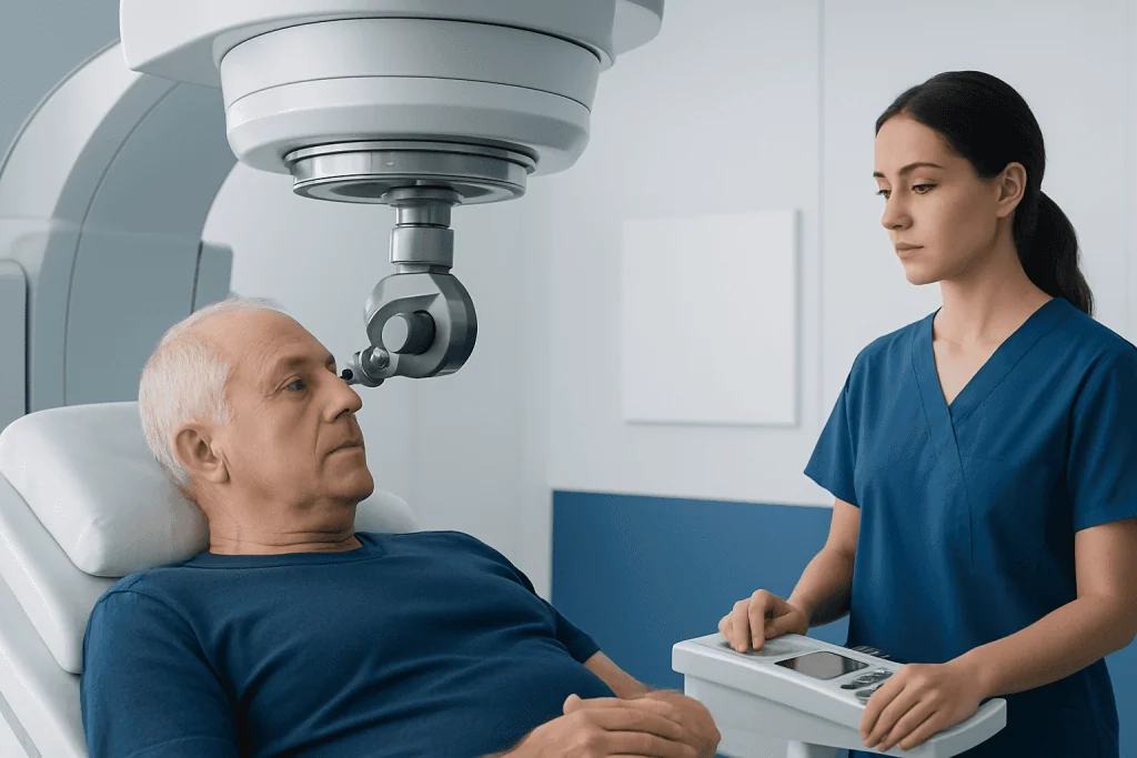
The plaque is designed to fit the curve of the eye. It may contain iodine-125, ruthenium-106, palladium-103, or another radioisotope, depending on local practice and the tumor being treated. Its radioactive seeds emit radiation over a limited distance, allowing doctors to direct treatment toward the tumor and a carefully calculated margin around it.
Before treatment, the team performs detailed eye imaging and measurements. These help determine the appropriate plaque size, its exact position, the radiation source, and how long it should remain in place. This planning is important because tumors near structures responsible for central vision, eye pressure control, or the optic nerve may carry different risks.
People sometimes ask, “How do you check for radiation in your body?” In plaque therapy, the radioactive device stays outside the eyeball and is removed at the end of treatment. It does not circulate through the bloodstream or remain in the body. Hospital staff follow radiation-safety procedures while the plaque is in place, and the care team provides instructions about visitors and activity during that time.
Who May Be a Candidate for Radiation Plaque Therapy

Radiation plaque therapy may be considered for people with a tumor inside the eye that is suitable in size and location for local treatment. It is frequently used for uveal melanoma, including choroidal and ciliary body melanoma. Some other selected eye tumors may also be evaluated for plaque therapy, although treatment choices vary by diagnosis.
Candidacy is determined through a specialist assessment rather than a single test. The evaluation may include a dilated eye examination, ocular ultrasound, retinal imaging, fluorescein or other angiography when needed, and scans to assess for disease elsewhere in the body. In some cases, a biopsy or tumor sample may be discussed for diagnostic or prognostic testing.
Radiation plaque may not be the best option when a tumor is very large, has extensively involved certain parts of the eye, or has features that require another approach. Alternatives can include laser-based treatments, external radiation approaches, surgery, medicines in selected situations, or removal of the eye. The most appropriate option should be decided with an ocular oncologist and radiation oncologist.
- Eligibility depends on tumor size, position, thickness, and diagnosis.
- Baseline vision and the condition of the retina and optic nerve are considered.
- General health, anesthesia suitability, and prior eye treatments may affect planning.
What Happens During the Procedure
Radiation plaque therapy generally involves two operations. During the first procedure, performed under local anesthesia with sedation or general anesthesia, the surgeon identifies the tumor location using preoperative planning and attaches the plaque to the sclera, the white outer layer of the eye. The plaque is sewn into position on the outside of the eye; it does not enter the eye itself.
The patient remains under medical supervision while the plaque delivers radiation. The treatment period commonly lasts several days, but the exact duration is calculated individually. Depending on the hospital’s safety policies and the type of radiation source used, the patient may stay in hospital and may have restrictions on close contact with visitors, particularly children and pregnant people.
At the end of the prescribed treatment time, the surgeon removes the plaque in a second procedure. The eye is then covered and protected as it heals. The treatment team will arrange follow-up visits to monitor the eye, measure tumor response, manage symptoms, and perform ongoing surveillance appropriate for the original diagnosis.
Radiation Plaque Therapy Recovery and Timeline
Radiation plaque therapy recovery begins after the plaque-removal procedure. In the first days, the eye may feel sore, scratchy, watery, or sensitive to light. Some people have eyelid swelling, redness, mild blurred vision, or a temporary feeling that something is in the eye. These symptoms are often related to surgery and usually improve as the eye heals.
The hardest days after radiation treatment are often the first few days after placement or removal, when discomfort, fatigue, disrupted sleep, and practical restrictions can be most noticeable. However, experiences differ. Some people feel relatively comfortable, while others need more time before they can resume normal routines. The care team may prescribe eye drops, pain relief, and protective instructions tailored to the procedure.
Most people need to avoid rubbing the eye, heavy lifting, strenuous activity, swimming, and activities that could expose the healing eye to injury for a period advised by the surgeon. Vision may be blurred initially, and driving should be avoided until vision is safe and the doctor confirms it is appropriate. Follow-up appointments are an important part of recovery because certain radiation effects can emerge months or years later.
How long before someone knows if radiation therapy is working depends on the tumor and its appearance on imaging. The tumor may not disappear immediately; instead, specialists commonly look for stabilization, reduced thickness, and changes in blood flow over months. Regular examinations and ocular ultrasound provide a more reliable picture of response than day-to-day symptoms alone.
Potential Benefits and Side Effects of Radiation Plaque Therapy
A key potential benefit of radiation plaque is that it treats the tumor locally while preserving the eye in many suitable cases. It can provide effective local tumor control and may preserve useful vision, especially when the tumor is away from the macula and optic nerve. Still, preserving the eye does not always mean vision will remain unchanged.
What are the potential side effects of radiation plaque therapy? Short-term effects can include eye pain or irritation, swelling, redness, dry eye symptoms, blurred vision, double vision, light sensitivity, and temporary changes in eye pressure. Surgical complications, though uncommon, can include infection, bleeding, wound problems, retinal detachment, cataract, or changes in eye movement.
Delayed effects are often related to radiation exposure of sensitive eye structures. They may include cataract, radiation retinopathy, radiation-related optic nerve damage, macular swelling, glaucoma, dry eye, retinal detachment, and gradual loss of vision. The likelihood depends strongly on where the tumor sits and how much radiation reaches nearby structures. Some complications can be monitored or treated, which is why long-term follow-up is essential.
There is also a risk that the tumor may not respond fully, may recur locally, or may spread depending on its biology. Plaque treatment is planned carefully, but no cancer treatment can guarantee a particular outcome. The oncology team will explain the expected benefits and limitations in the context of the individual diagnosis.
What I Wish I Knew Before Radiation
People preparing for plaque rads often find it helpful to know that treatment is a process rather than a single event. The procedure may be short, but follow-up continues for years. Tumor response is assessed gradually, and vision may fluctuate during healing or change later because of the tumor’s original location or radiation-related effects.
It is also useful to plan for temporary practical support. A patient may need help with transport, medications, household tasks, and attending follow-up appointments, particularly in the first week after surgery. Asking the care team about hospital stay rules, visitor limitations, work leave, eye drops, activity restrictions, and expected visual changes can make the experience more manageable.
Before treatment, patients should share any history of eye disease, glaucoma, diabetes, prior eye operations, medication allergies, or blood-thinning medicines. They should also ask who to contact outside clinic hours and what symptoms require urgent assessment. Keeping a written list of questions and bringing a trusted support person to appointments can help patients absorb complex information.
At Acibadem International, multidisciplinary ophthalmology, radiation oncology, and oncology specialists at JCI-accredited hospitals can assess and treat eligible international patients with eye tumors, with care plans based on the individual diagnosis and treatment needs.
When to Seek Medical Care
Patients should contact their eye surgeon or treatment team promptly if they develop worsening pain, increasing redness or swelling, pus-like discharge, a sudden decline in vision, new flashes or a curtain-like shadow in vision, persistent nausea or vomiting, severe headache, or fever. These symptoms do not always indicate a serious problem, but they need timely assessment after eye surgery or radiation treatment.
Urgent medical care is especially important for sudden or severe vision loss, significant eye injury, rapidly increasing eye pain, or symptoms of an allergic reaction to a prescribed medication, such as trouble breathing or facial swelling. Patients should follow the emergency contact instructions provided by their hospital rather than waiting for a routine follow-up visit.
Even if recovery feels uncomplicated, all scheduled reviews should be attended. Eye examinations, scans, and any recommended whole-body surveillance help clinicians evaluate treatment response, identify manageable complications early, and support long-term health after radiation plaque therapy.
Frequently asked questions
Is radiation plaque painful?
The plaque placement and removal procedures are performed with anesthesia, so pain should be controlled during surgery. Afterward, mild to moderate soreness, irritation, tearing, or light sensitivity can occur. The treating team can recommend appropriate pain relief and eye care instructions.
How long does a radiation plaque stay in the eye?
The plaque is attached to the outside of the eye for a calculated period, commonly several days. The exact duration depends on the radioactive source, the prescribed radiation dose, and the tumor's measurements. It is removed in a planned second procedure.
Does radiation plaque make a person radioactive?
While the plaque is attached, it contains the radiation source, so hospitals use specific safety measures and may limit close contact. Once the plaque is removed, the patient is not radioactive. Radiation does not stay in the body after the device has been taken out.
Will vision return after radiation plaque therapy?
Vision may be temporarily blurred after surgery and can improve as the eye heals. Long-term vision depends on the tumor's location, the vision present before treatment, and whether delayed radiation effects affect the retina, macula, optic nerve, or lens. The specialist can give the most individualized outlook after reviewing imaging and follow-up results.
How long before you know if radiation therapy is working?
The treatment response is usually evaluated over months rather than days or weeks. Specialists use eye examinations and ultrasound to look for tumor stabilization or shrinkage. Continued follow-up is needed because tumor control and radiation-related changes can develop over time.
What are the hardest days after radiation treatment?
For many people, the most difficult period is the first few days after plaque placement or removal because of eye discomfort, fatigue, sleep disruption, and activity restrictions. Symptoms vary, and some patients recover more quickly than others. New severe pain, worsening redness, or sudden vision changes should be reported promptly.
References
- American Academy of Ophthalmology
- National Cancer Institute
- American Brachytherapy Society
- National Eye Institute
This article is for general information only and is not a substitute for professional medical advice. Please consult a qualified doctor about your individual situation.
Oncology care in Turkey — second opinion and treatment plan
JCI-accredited · board-certified surgeons · reply within 24h
Add us as a Preferred Source to see more of our trusted health content across Google Search, AI Overviews and Discover.

More from the Health Library



Related Specialists

Dr. Hüseyin Çelik
Nephrology
Dt. Bilge Baş
Oral & Dental Health
Dr. Kübra Nursel Bölük
Dermatology
Assoc. Prof. Dr. Bedriye Koyuncu Sökmen
Gastrointestinal Oncology Unit Clinical Service