Plaque Radiotherapy: How It Works, Results and What to Expect

Plaque radiotherapy is most often used for uveal melanoma and selected other eye tumors. The radioactive plaque is placed during surgery and usually removed after several days.
Key Takeaways
- Plaque radiotherapy is most often used for uveal melanoma and selected other eye tumors.
- The radioactive plaque is placed during surgery and usually removed after several days.
- Treatment planning aims to control the tumor while preserving the eye when possible.
- Vision changes and radiation-related eye complications can develop gradually, sometimes months or years later.
- Follow-up with an ocular oncology team is essential for monitoring tumor response, vision and overall health.
Plaque radiotherapy, also called eye plaque brachytherapy, is a focused radiation treatment for certain tumors inside or around the eye. A small radioactive plaque is temporarily attached to the outside of the eye, allowing radiation to target the tumor while limiting exposure to surrounding tissues.
Overview: What Is Plaque Radiotherapy?
Plaque radiotherapy is a type of internal radiation treatment, also known as brachytherapy, used mainly for tumors inside the eye. It works by placing a small, disc-shaped device called a plaque on the outer wall of the eye, directly over the tumor. The plaque contains tiny radioactive sources that deliver a carefully planned dose of radiation to the tumor over a limited period, commonly several days.
This approach is most often used to treat uveal melanoma, the most common primary cancer inside the adult eye. It may also be considered for selected cases of retinoblastoma, conjunctival tumors, or tumors that have spread to the eye, depending on their size, location and tissue type. The goal is usually local tumor control and eye preservation when this can be achieved safely.
Unlike external-beam radiotherapy, plaque radiotherapy delivers radiation from a source positioned very close to the tumor. This close placement allows the care team to focus treatment and reduce radiation reaching more distant tissues. However, because delicate eye structures may still receive radiation, vision outcomes depend strongly on where the tumor is located and how close it is to the optic nerve, macula, lens and other important structures.
How Plaque Radiotherapy Works
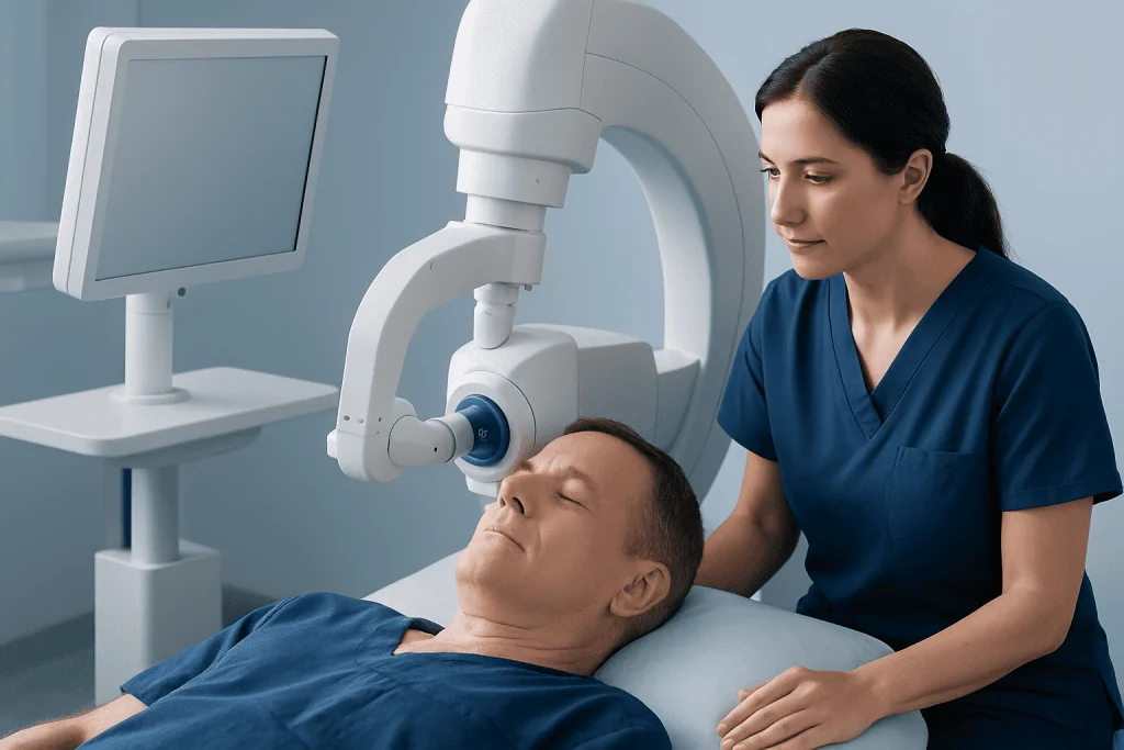
Before treatment, an ocular oncologist and radiation oncology team assess the tumor using a detailed eye examination and imaging. Ultrasound is commonly used to measure tumor thickness and its position inside the eye. Additional imaging may include retinal photography, optical coherence tomography, fluorescein angiography, magnetic resonance imaging or scans to evaluate whether disease has spread, depending on the diagnosis.
The treatment team uses these measurements to select a plaque size, radioactive source and placement plan. Iodine-125 is commonly used in eye plaque treatment, although other isotopes may be chosen in particular settings. The plaque is designed so that the radiation dose reaches the tumor and a small safety margin while limiting exposure to other parts of the eye as much as possible.
The plaque does not usually require radiation to be given in repeated daily appointments. Instead, radiation is delivered continuously while the plaque remains in place. The exact duration varies with the treatment plan, often ranging from a few days to about one week. Once the prescribed dose has been delivered, the plaque is removed in a second procedure.
Who May Be a Candidate for Radiation Plaque Therapy?

Candidacy for plaque radiotherapy is individualized. It may be an option for people with a small to medium-sized uveal melanoma and, in some circumstances, selected larger tumors. Tumor thickness, width, location, growth pattern and involvement of nearby eye structures all influence whether plaque treatment is appropriate.
The team also considers the person’s current vision, the condition of the other eye, general health and whether imaging shows disease outside the eye. Some tumors may be better treated with surgery, external-beam radiation such as proton therapy, laser-based approaches, systemic treatment, or a combination of therapies. In certain situations, removal of the eye may offer the safest or most effective local treatment.
A discussion with an ocular oncology specialist is important because no treatment can guarantee preservation of vision. For example, a tumor near the central retina or optic nerve can be controlled successfully but still carry a substantial risk of later vision loss from the tumor itself or radiation-related changes. Treatment planning therefore balances tumor control, eye safety and expected visual function.
- Typical considerations include tumor type, dimensions and location.
- Specialists assess whether the tumor can be covered safely by a plaque.
- Baseline vision and the health of the retina, optic nerve and other eye are considered.
- Staging tests may be needed to evaluate the rest of the body, especially for uveal melanoma.
Plaque Radiotherapy Procedure: Step by Step
The treatment process starts with consultation, eye imaging and radiation planning. The care team explains the expected treatment duration, practical safety instructions and the follow-up schedule. A medical or surgical evaluation may also be performed before anesthesia. Patients should tell the team about all medicines, allergies, blood-thinning medicines and relevant health conditions.
During the placement procedure, the patient receives anesthesia. The eye surgeon identifies the external location corresponding to the internal tumor and temporarily attaches the plaque to the sclera, the white outer layer of the eye. The plaque remains outside the eyeball; it is not inserted into the tumor. In some cases, the surgeon may place small markers or perform other planned procedures to help ensure precise positioning.
After placement, the patient stays in hospital or follows local radiation-safety procedures until removal. The exact arrangements vary according to the isotope used, local regulations and individual clinical needs. The plaque emits radiation only while it is in place, so staff will provide clear guidance about visitors, distance and contact precautions if needed.
At the end of the planned treatment interval, the plaque is removed during a second short surgical procedure. The eye is checked, and the patient receives instructions about eye drops, pain relief, activity limits and follow-up. For specialist assessment and coordinated care, patients may discuss plaque radiotherapy treatment with an experienced ocular oncology team.
Recovery Timeline, Benefits and Expected Results
After plaque removal, mild soreness, redness, tearing, light sensitivity or a scratchy sensation may occur for several days. Some people have swelling of the eyelids or temporary blurred vision. These symptoms often improve as the eye heals, although recovery is individual and the treating team should be informed of symptoms that worsen rather than improve.
The first follow-up visit is commonly scheduled soon after treatment, followed by regular examinations and eye imaging. Tumor regression is gradual. It may take months for the tumor to become flatter or show stable treatment-related changes. The care team monitors the tumor, retinal circulation, eye pressure and visual function over time.
A major benefit of plaque radiotherapy is that it can provide local treatment while preserving the eye in many suitable cases. It has a long-established role in ocular oncology. However, keeping the eye does not always mean keeping the same level of sight. Long-term visual outcome depends on the original tumor location, tumor response and whether radiation affects sensitive parts of the eye.
People treated for uveal melanoma generally need ongoing systemic surveillance as well as eye follow-up. This is because monitoring for spread to other organs is an important part of care, even when the eye tumor has been treated locally.
What Are the Potential Side Effects of Radiation Plaque Therapy?
Potential side effects of radiation plaque therapy include temporary eye irritation, pain, redness, swelling, blurred vision and dry eye symptoms after surgery. These early effects are often manageable with prescribed eye drops and pain relief recommended by the treating team. Infection, bleeding, wound problems and anesthesia-related complications are uncommon but possible surgical risks.
Some radiation-related effects develop later, often months or years after treatment. These may include radiation retinopathy, macular swelling, optic nerve damage, cataract, raised eye pressure or glaucoma, retinal detachment, chronic dry eye and changes to the appearance or movement of the eye. The chance of each complication varies according to the dose and the tumor’s proximity to sensitive structures.
Radiation retinopathy can affect the blood vessels of the retina and may reduce vision. Depending on the findings, treatment may involve eye injections, laser therapy or other retinal care. Cataracts and glaucoma can often be treated, but severe radiation-related damage may sometimes lead to substantial visual loss. Regular follow-up helps identify complications early, when management may be more effective.
It is also possible for a tumor not to respond fully or to recur locally. The team will discuss what further options may be appropriate if this occurs. Patients should seek prompt medical advice for sudden vision loss, severe pain, increasing redness, flashes, a new shower of floaters or a curtain-like shadow in vision.
What Are the Hardest Days After Radiation Treatment?
For plaque radiotherapy, the most challenging period is often the first few days after plaque placement or removal rather than the radiation delivery itself. The eye may feel irritated, swollen or sensitive to light, and vision can be temporarily blurred. Some people also find the hospital stay, limits on activity and uncertainty while awaiting follow-up emotionally tiring.
After removal, discomfort generally settles over days to a few weeks, but the timeline differs between individuals. The eye may look red or feel gritty while surgical tissues heal. Using prescribed drops correctly, avoiding rubbing the eye and attending scheduled reviews can support recovery.
Delayed side effects are different from the immediate recovery period. Radiation-related changes may appear gradually, and they do not necessarily cause symptoms at first. This is why long-term monitoring remains important even when the eye feels comfortable and vision seems stable.
What I Wish I Knew Before Radiation
Before plaque radiotherapy, it can be helpful to understand that treatment is a process rather than a single event. There are usually two procedures: one to place the plaque and another to remove it. The radiation is delivered continuously between these procedures, and the treatment schedule may require a short hospital stay or temporary changes to normal routines.
It is also useful to ask how the tumor’s location may affect vision. Tumors near the macula or optic nerve may carry a higher risk of future visual changes, even when local tumor control is achieved. Asking the care team about the purpose of treatment, the expected follow-up plan and possible options if complications arise can help patients make informed decisions.
Practical preparation can reduce stress. Patients may arrange help with transport, work responsibilities and daily tasks, particularly if vision is blurred after surgery. They should ask when it is safe to drive, exercise, travel, wear contact lenses or return to work, since recommendations depend on the procedure and recovery progress.
How Will I Feel After 5 Sessions of Radiotherapy?
Traditional plaque radiotherapy is not usually delivered as five separate radiation sessions. Instead, the plaque remains in position and provides continuous radiation over a planned number of days. Therefore, how a person feels after “five sessions” depends on whether they are receiving a different type of radiotherapy, such as external-beam treatment, or referring to five days with a plaque in place.
During the days after plaque treatment, many people feel tired from surgery, disrupted sleep, travel, stress or anesthesia rather than from widespread radiation effects. Local eye symptoms such as redness, tearing, light sensitivity and blurred vision are more typical than the whole-body effects associated with radiation to larger body areas.
If a person is receiving external-beam radiation in five fractions, the expected effects depend on the body area treated. Their radiation oncologist can explain what is most likely in their individual plan. New severe pain, sudden vision changes, fever, vomiting or worsening swelling should be reported promptly to the treatment team.
When to Seek Medical Care
Patients should contact their eye care or oncology team promptly if they develop severe or increasing eye pain, marked redness, pus-like discharge, fever, sudden worsening of vision, a dark curtain in the field of view, new flashes of light or many new floaters. These symptoms do not always indicate a serious problem, but they need timely assessment after eye surgery or radiation treatment.
Regular appointments are equally important when there are no new symptoms. Follow-up helps the team assess tumor response and check for complications that may be treatable before they noticeably affect vision. Patients should also attend recommended systemic surveillance appointments when treatment is for uveal melanoma.
Acibadem International’s multidisciplinary specialists and JCI-accredited hospitals provide evaluation and treatment planning for international patients with eye tumors, including coordinated ocular oncology and radiation care. A qualified specialist can explain whether plaque radiotherapy or another approach is suitable for the individual diagnosis.
Frequently asked questions
Is plaque radiotherapy painful?
The plaque placement and removal procedures are performed with anesthesia, so pain is not expected during surgery. Mild soreness, redness, a gritty feeling or light sensitivity can occur afterward and is usually managed with the care team’s recommended medicines and eye drops. Severe or worsening pain should be reported promptly.
How long does a radiation plaque stay in the eye?
The plaque is attached to the outside of the eye and commonly remains in place for several days. The precise duration depends on the radioactive source, tumor measurements and prescribed radiation dose. The treatment team schedules a second procedure to remove it once treatment is complete.
Will plaque radiotherapy save my vision?
Plaque radiotherapy can preserve the eye and may preserve useful vision in many appropriate cases, but visual outcomes vary. Tumor location is especially important because tumors near the macula or optic nerve can affect sight before or after treatment. The ocular oncology team can discuss the likely visual risks based on individual imaging.
Can a tumor come back after plaque radiotherapy?
A treated eye tumor can sometimes persist, regrow or recur locally, so regular imaging and eye examinations are necessary. If this happens, the team may consider additional local treatment, surgery, external-beam radiation or other options depending on the diagnosis. Ongoing surveillance also checks for treatment-related eye changes.
Are there radiation safety restrictions with an eye plaque?
Safety instructions vary according to the radioactive source, hospital policies and local regulations. Patients may need to stay in hospital or follow guidance about visitors and close contact while the plaque is in place. The radiation safety and treatment teams provide personalized instructions before treatment.
When can I return to normal activities after plaque radiotherapy?
Return to daily activities depends on healing after placement and removal, vision quality and the surgeon’s instructions. Many people need to avoid rubbing the eye, strenuous activity and driving until cleared by their team. Follow-up appointments help determine when it is safe to resume work, exercise and travel.
References
- American Cancer Society
- National Cancer Institute
- American Academy of Ophthalmology
- International Atomic Energy Agency
- National Comprehensive Cancer Network
This article is for general information only and is not a substitute for professional medical advice. Please consult a qualified doctor about your individual situation.
Oncology care in Turkey — second opinion and treatment plan
JCI-accredited · board-certified surgeons · reply within 24h
Add us as a Preferred Source to see more of our trusted health content across Google Search, AI Overviews and Discover.