

Quick answer
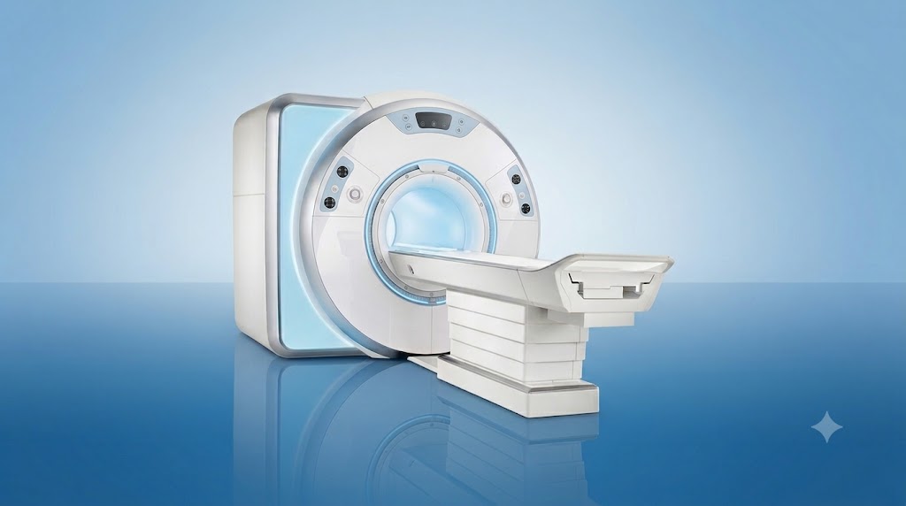
3 Tesla Prism MR is an advanced magnetic resonance imaging system that produces detailed anatomical and functional images, especially for neurological assessment, without using ionising radiation. At Acibadem in Turkey, it supports non-invasive evaluation of the brain, spine and other body structures to help clinicians diagnose conditions and plan care.
3 Tesla Prism MR: Advanced MRI for Detailed Neurological and Functional Imaging
3 Tesla Prism MR is an advanced magnetic resonance imaging (MRI) platform designed to produce highly detailed images of the body, with particular value in neurological and functional imaging. It helps specialists see anatomy, tissue characteristics and certain aspects of brain function with exceptional clarity, supporting earlier detection, accurate diagnosis and personalised treatment planning.
At Acibadem International, our multidisciplinary teams use advanced imaging technologies to understand each patient’s condition as clearly as possible before recommending care. For many patients, MRI can provide important diagnostic information without surgery, needles or ionising radiation.
The 3 Tesla Prism MR platform is especially useful when specialists need high-resolution images of the brain, spinal cord, nerves and other complex structures. Depending on the clinical question, it may also support detailed imaging of joints, blood vessels, internal organs and soft tissues.
What Is 3 Tesla Prism MR?
MRI is a medical imaging method that uses a strong magnetic field and radiofrequency energy to create detailed images of structures inside the body. Unlike X-ray-based imaging techniques, MRI does not use ionising radiation.
The term “3 Tesla” refers to the strength of the magnetic field used by the MRI system. A stronger magnetic field can help produce more detailed images and may allow specialists to examine small or complex structures with greater confidence. “Prism” refers to the advanced imaging platform and its capabilities for high-quality anatomical, neurological and functional assessment.
For patients with symptoms affecting the nervous system, imaging quality can make a meaningful difference. Small changes in brain tissue, blood vessels, nerves or the spine may be easier to assess when high-resolution images are available. This can help the clinical team determine the most appropriate next step, whether that involves monitoring, medication, rehabilitation, further testing or a procedure.
Every scan is planned according to the patient’s symptoms, medical history and the clinical question being investigated. A specialist will confirm whether 3 Tesla Prism MR is suitable for your individual situation.
How Does 3 Tesla Prism MR Work?
During an MRI examination, the patient lies on a movable table that enters the scanner. The system uses a powerful magnetic field to align naturally occurring particles in the body. Carefully controlled radiofrequency pulses then interact with these particles. As the body responds, the MRI system collects signals and a computer converts them into detailed cross-sectional images.
Different MRI sequences can highlight different types of tissue. For example, one sequence may be particularly useful for assessing fluid, while another may provide more detail about blood flow, nerve pathways, inflammation or tissue changes. This flexibility allows radiologists and referring specialists to tailor the examination to the patient’s needs.
For neurological care, the system may be used for specialised imaging approaches such as high-resolution brain imaging, spinal imaging, vascular MRI, diffusion imaging or functional imaging. Functional MRI can help identify areas of the brain associated with specific functions, such as movement or language, in selected clinical situations.
Some examinations may require a contrast agent. MRI contrast is administered only when clinically appropriate and after reviewing the patient’s health history, including kidney function, allergies and previous imaging experiences. Your care team will explain why contrast may be recommended and answer any questions before the scan.
Clinical Applications
3 Tesla Prism MR supports the work of radiologists, neurologists, neurosurgeons and other specialists by providing high-quality images for diagnosis, treatment planning and follow-up. The exact examination depends on your symptoms, referral reason and medical assessment.
| Clinical applications | How 3 Tesla Prism MR may help | Examples of conditions or concerns assessed |
|---|---|---|
| Brain and neurological imaging | Provides detailed images of brain structures, tissue changes and selected vascular features. | Headaches, seizures, dizziness, weakness, sensory changes, suspected stroke, brain lesions and neurological symptoms. |
| Functional brain imaging | May help map selected brain functions in carefully chosen cases, supporting clinical and surgical planning. | Pre-operative evaluation for selected brain conditions, assessment of areas related to language, movement or other functions. |
| Spine and spinal cord imaging | Helps assess discs, vertebrae, nerves, the spinal cord and surrounding soft tissues. | Back or neck pain, disc problems, spinal stenosis, nerve compression, spinal lesions and unexplained neurological symptoms. |
| Blood vessel imaging | MR angiography may visualise selected blood vessels without the use of ionising radiation. | Possible vascular narrowing, aneurysm assessment, vascular malformations and selected circulation-related concerns. |
| Musculoskeletal imaging | Offers high soft-tissue detail for joints, ligaments, tendons, cartilage and muscles. | Sports injuries, joint pain, ligament tears, tendon conditions, cartilage changes and soft-tissue injuries. |
| Follow-up and treatment planning | Supports comparison over time and helps specialists plan or evaluate treatment when MRI is clinically indicated. | Monitoring selected neurological, spinal, musculoskeletal or soft-tissue conditions. |
MRI findings are always interpreted in context. Imaging is one important part of a complete medical assessment, alongside your symptoms, examination findings, laboratory results and previous medical records.
Why High-Resolution Neurological Imaging Matters
The brain and nervous system are complex, and many neurological symptoms can have more than one possible cause. Detailed imaging can help specialists investigate the source of symptoms and distinguish between conditions that may appear similar clinically.
For example, a patient may be referred for MRI because of persistent headaches, changes in vision, numbness, balance problems, memory concerns, seizures or unexplained weakness. In other cases, MRI is requested after an emergency event, following an abnormal screening result or as part of preparation for a planned procedure.
High-quality MRI images can help the clinical team identify findings that require attention, clarify whether further tests are needed and support a more informed conversation about treatment options. Not every scan identifies a serious condition, and a normal result can also provide valuable reassurance when interpreted by the appropriate specialist.
Benefits and Advantages for Patients
3 Tesla Prism MR is designed to support precise, patient-centred imaging. The principal benefit is the ability to obtain detailed diagnostic information in a largely non-invasive way.
| Patient benefits at a glance | What this can mean for your care |
|---|---|
| High-resolution imaging | Detailed images may help specialists assess small or complex structures with greater clarity. |
| No ionising radiation | MRI uses magnetic fields and radiofrequency energy rather than X-rays, making it an important imaging option when clinically appropriate. |
| Largely non-invasive assessment | Many examinations can be completed without surgery or invasive diagnostic procedures. |
| Personalised examination protocols | The scan can be tailored to the body area, symptoms and clinical question identified by your specialist. |
| Support for treatment planning | Accurate imaging can help guide decisions about medication, monitoring, rehabilitation, surgery or further investigation. |
| Useful for follow-up care | When repeated imaging is needed, MRI can help specialists compare findings over time. |
While advanced MRI can provide valuable information, it is not the right test for every condition or every patient. Your doctor will recommend the most suitable imaging method based on your individual health needs.
What to Expect During a 3 Tesla Prism MR Examination
For international patients, the experience begins before travel whenever possible. Our international patient team can help coordinate the review of your available reports, imaging studies and medical history. This allows the relevant specialist to understand the reason for the scan and advise on the most appropriate next steps.
Before Your Appointment
You will be asked about your medical history and any previous imaging examinations. It is important to tell the team if you have a pacemaker, implantable device, cochlear implant, metal fragments, clips, prostheses, medication pump, pregnancy or possible pregnancy, kidney concerns or a history of reactions to contrast agents.
Many modern implants may be compatible with MRI under specific conditions, but this must always be checked carefully. Do not assume that an implant is safe for MRI without confirmation from the clinical team.
You may be asked to remove jewellery, watches, hearing aids, hair accessories, removable dental items, bank cards and other metal or electronic objects. Depending on the examination, you may change into a gown.
During the Scan
You will lie comfortably on the MRI table, and the radiology team will help position you for the area being examined. For brain imaging, a specialised coil may be placed around the head. This does not cause pain; it helps the scanner receive detailed signals.
The table moves into the scanner, which is open at both ends. MRI scans can be noisy, with tapping or knocking sounds during image acquisition. Ear protection is provided, and in many cases the team can maintain communication with you throughout the examination.
Remaining still is important because movement can reduce image quality. The scan itself is painless, although some patients may find the enclosed environment uncomfortable. If you are concerned about claustrophobia, tell the team in advance. They can discuss available support and determine the most appropriate approach for your situation.
If contrast is needed, it is usually administered through a small intravenous line. The radiology team will monitor you and provide instructions throughout the process.
After the Scan
Most patients can return to their usual activities after a standard MRI examination, unless they have received medication or have been given specific instructions by their doctor. A radiologist reviews the images, and the findings are shared with the referring specialist to support your care plan.
For patients travelling from abroad, our coordination team can help arrange appointments, facilitate communication between departments and support the organisation of follow-up discussions. If further care is recommended, we can assist with planning the next stages of your visit.
Safety, Clinical Standards and Accreditation
MRI is widely used in modern medicine and is considered safe for most appropriately screened patients. Because it uses a strong magnetic field, safety screening is essential. The imaging team follows established protocols to identify any implants, devices, materials or health factors that may affect whether MRI can be performed safely.
Acibadem International supports care in JCI-accredited hospital environments, where patient safety, quality standards and coordinated clinical practice are central to the patient experience. MRI examinations are performed by trained radiology professionals, with images interpreted by experienced radiologists and discussed with relevant specialists when needed.
Safety is individual. A scan may need to be modified, delayed or replaced with another imaging method if there are contraindications or if MRI is not the best option for your condition. Your team will explain recommendations clearly and ensure that you understand the purpose of the examination.
Preparing for Your International Visit
Travelling for diagnostic imaging can feel overwhelming, especially when you are managing symptoms or seeking answers after an uncertain diagnosis. Acibadem International aims to make the process more coordinated and understandable for patients and families coming from abroad.
Before your visit, you can share available medical reports, prior MRI or CT images, laboratory results and referral notes for preliminary review. Our team can coordinate the relevant specialties, help arrange imaging appointments and provide language support throughout your care journey.
Bringing previous imaging studies is particularly helpful. Comparing new images with earlier scans may give specialists a clearer understanding of whether a finding is new, stable or changing over time.
After your assessment, the care team can explain the findings, outline recommended next steps and help coordinate further consultations if needed. If you require ongoing care after returning home, you may also receive documentation to support communication with your local physician.
When Might a Specialist Recommend 3 Tesla Prism MR?
A specialist may recommend this advanced MRI platform when detailed neurological or functional imaging is clinically important. This may include investigating symptoms, evaluating an abnormality seen on another test, planning treatment or monitoring a known condition.
Examples include persistent or unexplained neurological symptoms, suspected spinal or nerve involvement, assessment before selected neurosurgical procedures, follow-up of certain conditions, and detailed evaluation of joints or soft tissues. The final decision is always based on an individual medical review.
If you already have a diagnosis, MRI may be used to provide additional information about the location, extent or activity of a condition. If you do not yet have a diagnosis, it may help narrow down possible causes and guide the next stage of evaluation.
Frequently Asked Questions
Is 3 Tesla Prism MR safe?
For appropriately screened patients, MRI is routinely used in clinical practice. It does not use ionising radiation. However, the strong magnetic field means that implants, devices, metal fragments and other safety considerations must be reviewed before the examination. Your clinical team will explain any specific considerations for your case.
Will I need contrast?
Not every MRI examination requires contrast. If contrast may improve the diagnostic value of your scan, the specialist will review your medical history and explain the reason for its use. You will have the opportunity to ask questions before the examination.
How long does the examination take?
The duration depends on the body area being studied and the imaging protocol needed. More detailed neurological or functional examinations may require additional sequences. Your team will provide practical guidance when your appointment is planned.
Can I have an MRI if I have an implant?
Some implants can be scanned safely under specific conditions, while others may not be compatible. Always inform the team about any implant, device, surgery or possible metal exposure before scheduling the scan. The radiology team will verify the relevant information before proceeding.
Will my insurance cover the examination?
Coverage depends on your individual insurance policy and clinical indication. You can share your insurer information with our international patient team, who can help clarify available options and provide guidance before your visit.
How do I get started?
You can submit your available medical reports and imaging records through a consultation request. A relevant specialist can review your case and advise whether 3 Tesla Prism MR may be suitable as part of your diagnostic or treatment plan.
Advanced Imaging with a Personalised Approach
3 Tesla Prism MR combines advanced MRI capability with a patient-centred diagnostic approach. For patients with neurological symptoms, complex imaging needs or treatment planning requirements, detailed and carefully interpreted images can provide an important foundation for confident clinical decisions.
At Acibadem International, radiology, neurology and related specialties work together to ensure that imaging is connected to the broader care plan. From report review and appointment coordination to language support and follow-up guidance, our international patient team is here to help make your experience clear, organised and reassuring.
Available at these Hospitals


















Speak with our medical team
Find out how this technology could support your diagnosis or treatment.