

Digital Mammography with 3D Tomosynthesis
Diagnosis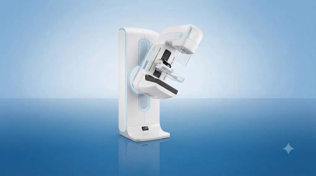
Quick answer
Digital mammography with 3D tomosynthesis creates layered breast images from multiple low-dose X-ray views, helping radiologists assess tissue that may be obscured on standard mammography. At Acibadem in Turkey, it is used for breast cancer screening and diagnostic evaluation when clinically appropriate.
Digital Mammography with 3D Tomosynthesis
Digital Mammography with 3D Tomosynthesis is an advanced breast imaging technology designed to provide a clearer, layer-by-layer view of breast tissue. It supports earlier detection of breast changes and helps radiologists assess areas that may be difficult to see on conventional two-dimensional mammography.
At Acibadem International, this technology is used by experienced radiology teams as part of a personalised approach to breast health. High-quality imaging can help specialists make more confident diagnostic decisions, recommend appropriate next steps and, when possible, avoid unnecessary invasive procedures.
For international patients, our care coordination teams can help organise imaging appointments, collect previous reports and images, arrange language support and guide you through each stage of your visit in Türkiye.
What Is Digital Mammography with 3D Tomosynthesis?
Digital mammography with 3D tomosynthesis, often called 3D mammography, is a breast X-ray examination that takes multiple low-dose images from different angles. Advanced software reconstructs these images into thin layers, allowing the radiologist to review the breast tissue slice by slice.
In a standard 2D mammogram, breast tissue is viewed as a single flattened image. When normal tissue overlaps, it can sometimes hide small abnormalities or create an appearance that needs further assessment. Tomosynthesis helps reduce the effect of tissue overlap by giving the radiologist a more detailed view of individual tissue layers.
This technology may be used for routine breast cancer screening and for diagnostic assessment when a patient has symptoms, an abnormal prior mammogram, a personal history of breast disease or another clinical reason for closer evaluation.
How 3D Tomosynthesis Works
During the examination, the breast is positioned and gently compressed between the mammography plates. Compression is important because it helps keep the breast still, spreads the tissue for clearer imaging and can reduce the amount of radiation needed for the examination.
The mammography system then moves in a short arc over the breast, capturing a series of low-dose X-ray images from different angles. These images are processed digitally to create a set of thin breast-tissue layers. The radiologist can scroll through these layers and examine the breast in greater detail.
Depending on the clinical purpose of the examination and the imaging protocol, the radiologist may review 3D images alone or together with reconstructed 2D images. The imaging plan is tailored to the patient’s needs and follows clinical and safety standards.
Why Layer-by-Layer Imaging Is Valuable
Breast tissue can appear dense or overlapping on a conventional mammogram. This does not necessarily mean there is a problem, but it can make interpretation more challenging. With tomosynthesis, specialists can examine the tissue in smaller sections, helping them distinguish between overlapping normal tissue and areas that may require further investigation.
This detailed imaging can support the identification of findings such as masses, architectural distortion, asymmetry and some calcifications. If an area needs more evaluation, the radiologist may recommend additional mammographic views, breast ultrasound, breast MRI or a biopsy, depending on the individual situation.
Clinical Applications
Digital Mammography with 3D Tomosynthesis is used in breast imaging when clinically appropriate. It is not a substitute for a specialist consultation, physical examination or other imaging studies when these are needed. Your radiologist and breast care team will consider your symptoms, age, breast density, personal and family history, previous imaging and other relevant factors.
| Clinical application | How 3D tomosynthesis may help | Possible next step |
|---|---|---|
| Routine breast cancer screening | Provides detailed breast imaging to support the early identification of suspicious changes before symptoms are present. | Return to the recommended screening schedule or arrange further assessment if an area requires review. |
| Assessment of an abnormal mammogram | Helps clarify whether a finding may be related to tissue overlap or needs closer investigation. | Additional mammographic views, ultrasound, MRI or biopsy may be considered. |
| Investigation of breast symptoms | May support assessment of concerns such as a new lump, focal thickening, nipple changes or unexplained breast discomfort. | A breast specialist may combine the imaging findings with clinical examination and other tests. |
| Dense breast tissue assessment | Can improve visualisation where overlapping dense tissue may make standard mammography more difficult to interpret. | Your doctor may discuss whether supplementary imaging is appropriate for your individual risk profile. |
| Follow-up after previous breast findings or treatment | Supports comparison with prior images and assessment of areas that require ongoing imaging surveillance. | Follow-up timing and imaging type are determined by the treating breast care team. |
| Pre-treatment breast evaluation | May help define the imaging appearance and location of a breast finding when planning further care. | Results can be discussed within a multidisciplinary breast care pathway when needed. |
Conditions and Breast Changes It Can Help Assess
3D mammography is an imaging tool rather than a treatment. It helps radiologists assess breast tissue and identify changes that may need monitoring, further imaging or tissue sampling. A result does not confirm a diagnosis on its own; findings are interpreted alongside your medical history, symptoms, physical examination and previous studies.
The examination may help assess suspicious breast masses, focal asymmetries, tissue distortion, certain patterns of calcification and other changes seen on prior imaging. It may also be used when a patient has symptoms that require a careful breast imaging evaluation.
Importantly, many breast imaging findings are benign. Cysts, fibroadenomas, benign calcifications and normal tissue overlap can all create appearances that require clarification. The purpose of detailed imaging is to help the care team identify which findings can be safely monitored and which need further investigation.
Benefits and Advantages for Patients
Digital Mammography with 3D Tomosynthesis combines the established value of mammography with a more detailed method of image review. It can support a more informed diagnostic pathway, particularly when overlapping tissue could make a 2D image harder to interpret.
Every patient’s experience and clinical outcome are different. Your care team will explain what the technology can contribute in your case and whether another examination may also be needed.
| Patient benefit | What it may mean for your care |
|---|---|
| Layer-by-layer breast imaging | Radiologists can examine breast tissue in thin sections rather than relying only on a single flat image. |
| Improved evaluation of overlapping tissue | Areas that appear unclear on conventional imaging may be assessed with greater detail. |
| Support for earlier detection | Detailed imaging may help identify suspicious changes at an earlier stage, when clinically detectable. |
| Digital image quality | Images can be reviewed, compared with prior studies and shared securely within the care team when appropriate. |
| Comfortable, non-invasive examination | The scan does not require injections or surgery, although brief breast compression is necessary for image quality. |
| Personalised diagnostic planning | Results can help your specialist decide whether routine follow-up, additional imaging or a biopsy is appropriate. |
| Coordinated international care | Our international patient teams can assist with scheduling, interpretation support and planning the next stage of care. |
What to Expect During Your Procedure
Before your appointment, the Acibadem International patient team can explain the preparation process in your preferred language and help you understand what documents or prior imaging may be useful. If you have had mammograms, breast ultrasound, MRI scans, biopsy reports or breast surgery in the past, please share this information whenever possible. Comparing new images with previous studies can be highly valuable.
Before the Examination
Your radiology team will review relevant medical information and confirm the purpose of the examination. Tell your team if you are pregnant, think you may be pregnant, are breastfeeding, have breast implants, have had breast surgery or have noticed new symptoms.
You may be asked not to apply deodorant, talcum powder, lotion or cream around the underarms or breasts on the day of your mammogram, as some products can affect the images. It is often helpful to wear a two-piece outfit so that you only need to remove clothing from the waist up.
During the Examination
A trained mammography technologist will guide you through the positioning. Each breast is placed on the imaging platform and gently compressed for a few seconds while images are taken. You will usually need several positions to capture the views required for a complete examination.
You may feel pressure during compression, but the process is brief. Let the technologist know if you experience significant discomfort, have limited shoulder movement or need assistance with positioning. The team will work carefully to make the examination as comfortable as possible while obtaining accurate images.
The image acquisition itself is quick. The overall appointment time can vary depending on whether previous images need review, whether additional views are required and whether the examination is being performed for screening or diagnostic assessment.
After the Examination
You can usually return to normal activities immediately after the mammogram. A radiologist will review the images and prepare a report for your doctor or breast care team. If the imaging raises a question that needs clarification, this does not necessarily mean cancer is present. Further assessment is common and may be recommended to obtain a more complete understanding of the finding.
For patients travelling from abroad, our coordination team can help arrange appropriate follow-up appointments during your stay whenever possible. If further tests are needed, the team can explain the recommended sequence of care and support communication with your doctor at home.
Safety, Radiation and Quality Standards
Digital Mammography with 3D Tomosynthesis uses X-rays, which means there is exposure to a small amount of ionising radiation. Modern mammography systems are designed to use low-dose imaging protocols while maintaining the image quality required for reliable interpretation. The exact dose and imaging approach depend on the system, breast characteristics, the number of views required and the clinical reason for the examination.
Your radiology team follows the principle of using the lowest radiation exposure reasonably achievable for the information needed. The expected benefit of accurate breast assessment is weighed carefully against radiation exposure, and imaging is recommended only when clinically appropriate.
Tell your physician and radiology team if there is any possibility of pregnancy. They will discuss whether mammography should be postponed, modified or performed with additional precautions according to your clinical needs.
Acibadem hospitals serving international patients include JCI-accredited facilities, where clinical processes, patient safety practices and quality standards are supported by structured care pathways. Mammography examinations are performed by trained imaging professionals, with results interpreted by specialist radiologists. Where needed, cases can be reviewed within a multidisciplinary breast care setting involving radiology, breast surgery, medical oncology, radiation oncology, pathology and other relevant specialties.
How Results Support Your Personalised Treatment Plan
Accurate imaging is an important foundation for breast care. Your results may support reassurance and routine screening, or they may indicate that further assessment is advisable. The next step is based on the imaging findings and your individual clinical profile.
If a suspicious area is identified, your specialist may recommend targeted ultrasound, breast MRI, a short-interval imaging follow-up or image-guided biopsy. If breast cancer is diagnosed, imaging findings can help the team understand the location and extent of the disease and develop an individual treatment plan.
At Acibadem International, patients can be supported through a coordinated pathway that brings together diagnostic imaging, pathology, surgery, oncology and supportive care when required. Your team will explain recommendations clearly, discuss available options and help you make informed decisions.
Why This Technology Matters for International Patients
Travelling for breast assessment can feel stressful, especially when you are waiting for answers. Our international patient services are designed to make the process more organised and understandable from the first contact through follow-up planning.
Before travel, you can share available medical reports, imaging summaries and pathology results for preliminary review. This helps the team understand your medical background and advise whether Digital Mammography with 3D Tomosynthesis may be appropriate as part of your assessment. Suitability is always confirmed by the responsible specialist after reviewing your individual case.
During your visit, patient coordinators can help with appointment planning, interpreter support, communication between departments and practical guidance throughout the care journey. If additional imaging, consultation or treatment is recommended, the team can work to coordinate the necessary steps efficiently.
After your visit, you can receive guidance on obtaining your medical documentation and sharing relevant information with your healthcare professionals in your home country. Continuity of care is particularly important in breast health, and clear documentation supports appropriate follow-up wherever you live.
Frequently Asked Questions
Is Digital Mammography with 3D Tomosynthesis safe?
Yes. It is routinely used in breast imaging and performed by trained professionals using established safety protocols. Because it uses X-rays, there is a low level of radiation exposure. Your team will explain the benefit and any individual considerations, including pregnancy or prior breast treatment.
Is 3D mammography better than standard mammography?
3D tomosynthesis provides a layer-by-layer view that can offer additional detail, particularly when breast tissue overlaps. Whether it is the best option for you depends on your symptoms, risk factors, breast density, prior imaging and the reason for the examination. Your radiologist will recommend the most appropriate imaging approach.
Does a 3D mammogram hurt?
Most patients experience pressure or temporary discomfort during breast compression. Compression lasts only a short time and is necessary for clear images. Tell the technologist if you are uncomfortable so they can support you with positioning and communication throughout the examination.
Can I have a mammogram if I have breast implants?
In many cases, yes. Please inform the radiology team before your appointment. Special positioning and additional views may be used to examine as much breast tissue as possible while taking care around the implants.
Will I need other tests after 3D mammography?
Not always. Many patients do not need additional tests. If the radiologist sees an area that requires clarification, further imaging such as ultrasound or MRI may be recommended. In some cases, a biopsy is needed to determine the nature of a finding.
Will my insurance cover the examination?
Insurance coverage depends on your policy, insurer and the reason for the examination. You may share your insurance details with our international patient team, who can help check available options and explain the expected administrative process before treatment or imaging is arranged.
How can I get started?
You may submit a consultation request and share your available reports, previous mammography images, ultrasound or MRI results and any pathology documentation. A specialist can review the information and advise whether Digital Mammography with 3D Tomosynthesis may be suitable within your individual breast assessment plan.
Related Medical Units
Available at these Hospitals


















Speak with our medical team
Find out how this technology could support your diagnosis or treatment.