

Quick answer
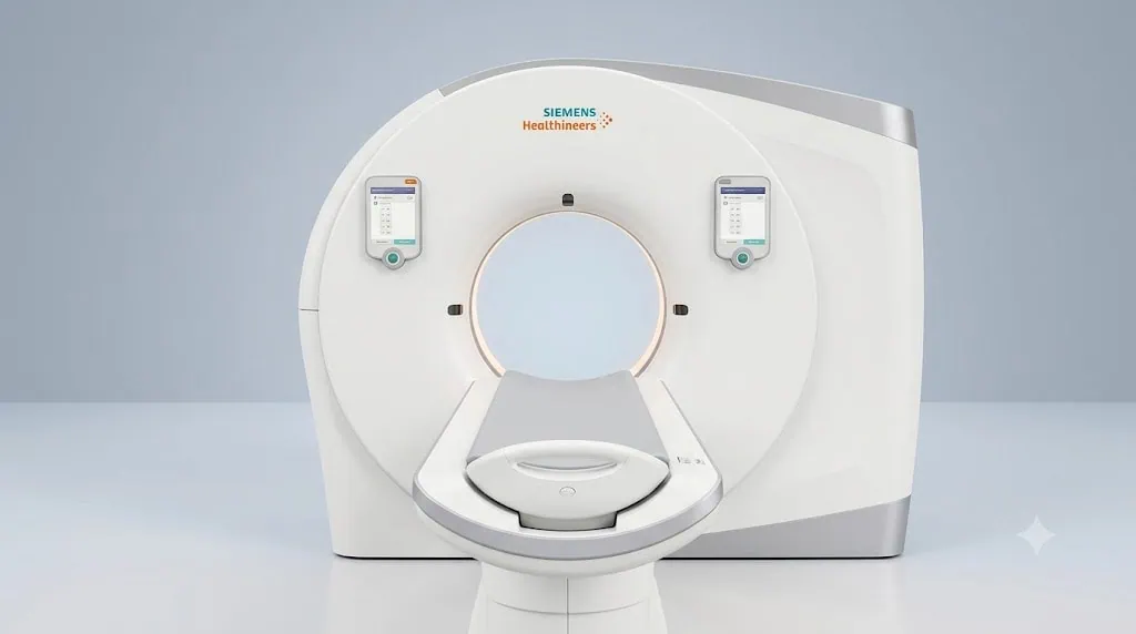
Somatom Drive is a dual-source, dual-energy CT system that produces detailed cross-sectional images rapidly for diagnostic assessment, treatment planning and follow-up. At Acibadem hospitals in Turkey, it is used by radiology teams to support clinically appropriate imaging protocols, including dose-optimization approaches when suitable.
Somatom Drive: Fast, Detailed Dual-Energy CT Imaging
Somatom Drive is an advanced computed tomography (CT) technology designed to create highly detailed images of the body quickly. It uses dual-source, dual-energy CT imaging to help specialists assess organs, blood vessels, bones and other structures with a high level of precision.
At Acibadem International, our radiology teams use advanced imaging technologies such as Somatom Drive to support accurate diagnosis, personalised treatment planning and follow-up care. Clear imaging can help doctors understand a condition earlier, evaluate its extent and make more confident clinical decisions.
CT imaging is a largely non-invasive examination. During the scan, the patient lies on a moving table while the scanner takes detailed cross-sectional images. Depending on the clinical need, the examination may be performed with or without contrast material.
Somatom Drive is particularly valuable when fast imaging, high-resolution detail and dose-optimisation approaches are important. Your radiologist and treating specialist will determine whether this technology is appropriate based on your symptoms, medical history, previous imaging and the clinical question being investigated.
What Is Somatom Drive?
Somatom Drive is a dual-source CT scanner. Unlike conventional CT systems that use a single X-ray source, dual-source technology uses two X-ray tubes and two detectors. This allows the system to capture imaging data rapidly and can improve image quality in situations where movement may otherwise affect the scan.
The system also supports dual-energy imaging. In simple terms, it can scan the body using two different X-ray energy levels. This gives radiologists additional information about the composition of tissues and materials within the body. It may help distinguish between different types of tissue, identify certain deposits, assess contrast flow or provide more detailed information for selected clinical conditions.
Somatom Drive does not replace the expertise of the medical team. Its value comes from the combination of advanced imaging, experienced radiologists and coordinated care across specialties. Imaging findings are reviewed in the context of each patient’s symptoms, examination results, laboratory data and wider treatment plan.
How Dual-Energy CT Imaging Works
CT scans use X-rays and computer processing to create detailed images in thin layers, often called slices. These slices can then be reviewed individually or reconstructed into three-dimensional views of the body.
With Somatom Drive, two X-ray sources work at the same time. This high-speed acquisition can be helpful when imaging areas affected by natural movement, such as the heart, lungs or blood vessels. Faster scanning may also support comfortable examinations for patients who find it difficult to hold their breath or remain still for a prolonged period.
Dual-energy imaging adds another layer of information. Different materials absorb X-ray energy differently. By comparing information obtained at two energy levels, the system can help specialists analyse certain tissues more closely. Depending on the examination, this may support assessment of calcium, iodine contrast, uric acid deposits, lung tissue changes or other clinically relevant features.
The imaging protocol is tailored to the patient and the medical question. The radiology team considers factors such as the body area being scanned, the reason for the examination, age, body size, previous imaging, kidney function where contrast is planned and any known allergies.
Clinical Applications
Somatom Drive may support diagnostic assessment, treatment planning and follow-up across a wide range of specialties. The precise use of the scanner depends on the individual patient and the decision of the treating physician.
| Clinical applications | How Somatom Drive may help | Relevant specialties |
|---|---|---|
| Cardiac and coronary artery assessment | Fast image acquisition may help evaluate the heart and coronary arteries, particularly when motion control is important. | Cardiology, Radiology, Cardiovascular Surgery |
| Chest and lung imaging | Detailed imaging can support assessment of the lungs, airways, chest structures and blood vessels. | Pulmonology, Thoracic Surgery, Oncology, Radiology |
| Vascular imaging | CT angiography may help visualise blood vessels and support planning for vascular, cardiac or interventional procedures. | Cardiology, Vascular Surgery, Interventional Radiology |
| Abdominal and pelvic assessment | Detailed cross-sectional imaging may assist in evaluating organs, blood vessels, inflammatory conditions and abdominal symptoms. | Gastroenterology, General Surgery, Urology, Radiology |
| Kidney stones and urinary tract conditions | Dual-energy imaging may help provide additional information about certain urinary stones and support treatment planning. | Urology, Nephrology, Radiology |
| Musculoskeletal conditions | CT may be used to evaluate bones, joints, fractures, complex anatomy and selected crystal-related joint conditions. | Orthopaedics, Rheumatology, Physical Medicine and Rehabilitation |
| Oncology imaging | Detailed imaging can support staging, treatment planning, response assessment and follow-up when clinically indicated. | Medical Oncology, Radiation Oncology, Surgical Oncology, Radiology |
| Emergency and trauma assessment | Fast scanning may help doctors assess injuries or acute conditions when timely diagnostic information is required. | Emergency Medicine, Trauma Surgery, Neurosurgery, Radiology |
Conditions It May Help Assess
Somatom Drive may be used as part of the assessment of many different health concerns. It does not diagnose every condition on its own, but it can provide important information to guide the next clinical step.
Examples may include coronary artery disease, vascular narrowing or aneurysms, pulmonary conditions, suspected blood clots, abdominal pain, kidney stones, inflammatory bowel conditions, liver and pancreatic findings, urinary tract disorders, fractures, joint abnormalities and selected cancer-related concerns.
For patients undergoing cancer care, CT imaging may be used before treatment, during planning or after treatment to evaluate response and monitor for recurrence when appropriate. For surgical patients, detailed CT images can assist specialists in understanding anatomy and preparing a tailored approach.
Your results are interpreted by a radiologist and shared with the relevant specialist. If further tests are needed, your care team will explain why they are recommended and how they fit into your overall treatment plan.
Benefits and Advantages for Patients
Advanced CT technology is designed to support clear imaging while keeping the patient experience as efficient and comfortable as possible. The benefits vary by examination and clinical indication, but Somatom Drive can offer important practical advantages.
| Patient benefits at a glance | What this can mean for your care |
|---|---|
| Fast image acquisition | The examination may be completed quickly, which can be helpful for patients with pain, breathlessness, anxiety or difficulty remaining still. |
| High-resolution imaging | Detailed images can help specialists evaluate small structures, complex anatomy and areas that require careful assessment. |
| Dual-energy information | Two energy levels can provide additional tissue-characterisation information in selected examinations. |
| Motion-sensitive imaging support | High-speed scanning may be particularly useful for areas affected by heartbeat, breathing or other natural movement. |
| Dose-optimisation approaches | Protocols can be adapted to the clinical need, with the goal of obtaining medically useful images while avoiding unnecessary radiation exposure where possible. |
| Largely non-invasive experience | Most CT examinations do not require surgery or instruments inside the body. Some scans may require intravenous contrast material. |
| Support for personalised care | Accurate imaging can help your physicians select appropriate next steps, whether this involves monitoring, medication, intervention, surgery or further investigation. |
What to Expect During a Somatom Drive CT Procedure
Before your scan, a member of the Acibadem International team will help coordinate your appointment and explain the preparation instructions. We understand that travelling for medical care can involve many questions, especially when tests are scheduled close together. Our teams aim to make the process clear, organised and easy to follow.
Your radiology team will review your referral, medical history and previous test results. Please tell the team about pregnancy or a possible pregnancy, allergies, kidney disease, thyroid conditions, diabetes, medications, previous reactions to contrast material and any recent imaging examinations.
Before the scan
Preparation depends on the type of CT examination. For some scans, you may be asked not to eat for a period before your appointment. You may also be asked to drink water or take specific instructions regarding medication. If contrast material is planned, the team may assess relevant blood test results and ask further questions about kidney function or allergies.
You will usually be asked to remove jewellery, metal accessories and other items that could affect image quality. Depending on the body area being examined, you may change into a hospital gown.
During the scan
You will lie on a padded examination table that moves smoothly through the CT scanner. The scanner is a wide, open ring rather than a long enclosed tunnel. The radiology team can see and speak with you throughout the examination.
You may be asked to hold your breath briefly while images are taken. It is important to remain as still as possible for a few seconds at a time. The scanning itself is generally painless.
If contrast material is used, it is commonly given through a small intravenous line. Some patients notice a temporary warm sensation, a metallic taste or a feeling of warmth spreading through the body. These sensations usually pass quickly. Inform the team immediately if you feel unwell, itchy, short of breath or experience any unexpected symptom.
After the scan
Most patients can return to their normal activities shortly after the examination. If contrast has been used, your team may advise you to drink fluids unless you have been given different instructions for a medical reason.
A radiologist reviews the images and prepares a report. Your treating physician discusses the findings with you, explains what they mean and recommends the next steps. For international patients, our coordination teams can support the sharing of reports and images with relevant specialists, subject to medical and privacy processes.
Safety, Radiation Awareness and Clinical Oversight
CT uses ionising radiation, which is why every examination should have a clear medical reason. At Acibadem International, imaging protocols are selected and adjusted by qualified professionals according to the patient’s clinical needs. The aim is to obtain diagnostic-quality images using an approach appropriate to the indication.
Somatom Drive supports dose-optimisation strategies when appropriate. However, the exact radiation dose depends on many factors, including the body region, the type of examination, the patient’s body characteristics, whether contrast is needed and the diagnostic objective. Your radiology team can discuss any relevant considerations for your individual scan.
Contrast-enhanced CT is safe for many patients, but contrast material is not suitable for everyone. The team will review your history and take precautions when there is a known allergy, reduced kidney function or another relevant medical concern. If a different imaging method is more appropriate, your physician may recommend alternatives such as MRI, ultrasound or another test.
Somatom Drive imaging is performed by trained radiology professionals and interpreted by specialist radiologists. Our JCI-accredited hospitals follow structured quality and patient-safety standards designed to support safe, coordinated care.
Why Accurate Imaging Matters in Treatment Planning
Good clinical decisions begin with reliable information. Detailed CT imaging can help a medical team identify the location, size and characteristics of a finding, evaluate how a condition may affect surrounding structures and determine whether further assessment is needed.
For some patients, a clear scan can help avoid unnecessary invasive procedures. For others, it can support more precise planning for surgery, interventional radiology, radiation therapy or medical treatment. In follow-up care, imaging may help the team monitor a known condition or assess response to treatment.
No single scan should be viewed in isolation. Your specialists bring together imaging, pathology where relevant, laboratory results, physical examination and your personal health history. This multidisciplinary approach helps ensure that recommendations are clinically appropriate and tailored to you.
Support for International Patients
When you are travelling abroad for diagnostic imaging or treatment, coordination matters. Acibadem International provides support throughout the process, from the first review of your medical documents to appointment planning and communication with the relevant clinical team.
Before travel, you can share available reports, imaging summaries and laboratory results through a consultation request. A specialist can review the information and advise whether Somatom Drive CT imaging may be relevant to your assessment or treatment plan. Final suitability is always confirmed after medical evaluation.
Our international patient coordinators can help organise the sequence of consultations, imaging and follow-up appointments so that your visit is as efficient as possible. Language support is available to help you understand the process, preparation requirements and discussions with your healthcare team.
If you have previous CT, MRI, ultrasound or X-ray images, bring them with you or send them in advance where possible. Previous imaging can help the team compare changes over time and may reduce the need for repeat examinations when the existing studies are adequate for clinical review.
After your assessment, your coordinator can assist with the practical aspects of continuing your care plan, including arranging follow-up discussions, sharing medical documentation and helping you understand the recommended next steps before returning home.
Frequently Asked Questions
Is Somatom Drive safe?
Somatom Drive is used routinely for clinically indicated CT imaging in our JCI-accredited hospitals. The examination is performed by trained radiology professionals using protocols selected for the patient and the diagnostic question. Because CT involves radiation, your doctor recommends it only when the expected clinical benefit is considered appropriate.
Is the CT scan painful?
The scan itself is usually painless. You will need to lie still and may be asked to hold your breath briefly. If contrast material is used, you may feel a short-lived warmth or metallic taste. Tell the team if you feel discomfort or have any concerns.
Do I need contrast material?
Not every CT examination requires contrast material. Whether it is needed depends on the part of the body being examined and the information your doctors need. Your team will explain the recommendation and review any relevant safety considerations.
How long does the examination take?
The actual scan is often very quick, but the total appointment includes registration, preparation, screening questions and, if needed, contrast administration. Your coordinator or radiology team will provide a clearer estimate based on your planned examination.
Will my insurance cover Somatom Drive CT imaging?
Coverage depends on your insurance policy, medical indication and authorisation requirements. You may share your insurer details with our international patient team so they can help review available options and explain the process before treatment.
How can I get started?
You can submit a consultation request with your available medical reports and imaging information. A specialist will review your case and advise whether Somatom Drive CT imaging may be suitable as part of your diagnostic assessment, treatment planning or follow-up care.
Related Medical Units
Available at these Hospitals


















Related Treatments
Speak with our medical team
Find out how this technology could support your diagnosis or treatment.