Melanoma: ABCDE Warning Signs, Biopsy, and Treatment Planning

The ABCDE rule helps identify moles or spots that may need medical evaluation: asymmetry, border, color, diameter, and evolution. A skin biopsy is the only reliable way to confirm whether a suspicious lesion is melanoma.
Key Takeaways
- The ABCDE rule helps identify moles or spots that may need medical evaluation: asymmetry, border, color, diameter, and evolution.
- A skin biopsy is the only reliable way to confirm whether a suspicious lesion is melanoma.
- Treatment planning depends on melanoma thickness, ulceration, lymph node involvement, spread to other organs, and molecular test results when appropriate.
- Surgery is the main treatment for early melanoma, while advanced melanoma may involve immunotherapy, targeted therapy, radiation therapy, or combined approaches.
- Regular skin self-checks, sun protection, and dermatologist visits are important for prevention and early detection.
Melanoma is a serious but often treatable form of skin cancer, especially when found early through careful attention to changing moles or new skin spots. Understanding the ABCDE warning signs, what happens during a biopsy, and how treatment is planned can help patients seek care promptly and make informed decisions.
Overview
Melanoma is a type of skin cancer that begins in melanocytes, the cells that produce melanin, the pigment that gives skin, hair, and eyes their color. It can develop on skin that is frequently exposed to the sun, such as the face, arms, back, and legs, but it may also appear in less exposed areas, including the soles of the feet, under the nails, or in the genital area. Although melanoma is less common than some other skin cancers, it deserves careful attention because it has a greater ability to spread if not detected and treated early.
Many melanomas are first noticed as a new spot on the skin or a change in an existing mole. Not every changing mole is cancer, and many skin growths are harmless, but a suspicious lesion should be checked by a qualified doctor rather than watched indefinitely. Early diagnosis usually allows for simpler treatment and a better chance of complete removal.
Melanoma care is individualized. Doctors consider the appearance of the lesion, biopsy findings, depth of growth, whether lymph nodes are involved, and the patient’s overall health. The goal is to remove or control the cancer while choosing the most appropriate treatment intensity for each stage.
ABCDE Warning Signs and Other Symptoms
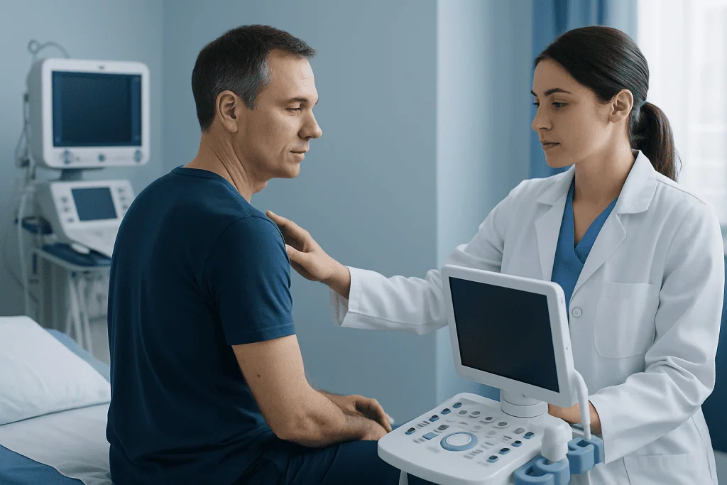
The ABCDE rule is a practical guide for recognizing mole changes that may need medical evaluation. It does not diagnose melanoma by itself, but it helps patients know when to schedule a skin examination. Any spot that looks different from the person’s other moles, sometimes called an “ugly duckling” lesion, also deserves attention.
- A – Asymmetry: one half of the mole or spot does not match the other half.
- B – Border: edges are irregular, blurred, notched, or uneven.
- C – Color: color is not uniform, with shades of brown, black, tan, red, white, gray, or blue.
- D – Diameter: the spot is larger than about 6 millimeters, although melanomas can be smaller.
- E – Evolution: the spot changes in size, shape, color, height, sensation, or begins to bleed or crust.
Other possible signs include a sore that does not heal, a mole that becomes itchy or tender, bleeding without obvious injury, or a streak of pigment under a fingernail or toenail. In people with darker skin tones, melanoma may be more likely to appear on the palms, soles, nail beds, or mucous membranes, so these areas should not be overlooked.
Some melanomas do not fit the classic pattern. A lesion may be pink, skin-colored, or only mildly pigmented. Because visual checks have limits, a dermatologist may use dermoscopy, a handheld magnifying device with light, to examine structures beneath the skin surface and decide whether biopsy is needed.
Causes and Risk Factors
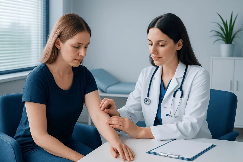
Melanoma develops when DNA changes in melanocytes allow cells to grow in an uncontrolled way. Ultraviolet radiation from sunlight and tanning beds is a major contributor because it can damage skin cell DNA. However, melanoma can also occur in areas with little sun exposure, which means genetics and other biological factors may also play a role.
Risk factors do not mean a person will definitely develop melanoma, and people without obvious risk factors can still be diagnosed. Knowing personal risk can guide how often skin checks are needed and how careful sun protection should be. A dermatologist can help tailor recommendations for people with many moles, atypical moles, or a personal history of skin cancer.
- History of intense sunburns, especially blistering sunburns
- Use of indoor tanning beds or sunlamps
- Fair skin, light eyes, red or blond hair, or skin that burns easily
- Many moles or atypical-looking moles
- Personal or family history of melanoma
- Weakened immune system, including after certain organ transplants or medical treatments
- Living at high altitude or in regions with strong ultraviolet exposure
Inherited mutations are responsible for a minority of melanomas, but family history is still important. Patients with several close relatives affected by melanoma, multiple primary melanomas, or unusual patterns of cancer in the family may be referred for genetic counseling or more intensive surveillance.
Diagnosis and Skin Biopsy
The diagnosis of melanoma begins with a medical history and full skin examination. The doctor may ask when the lesion appeared, how it has changed, whether it bleeds or itches, and whether there is a personal or family history of melanoma. A full-body skin check is often recommended because patients with one suspicious lesion may have other spots that also need evaluation.
If melanoma is suspected, a biopsy is needed. During a biopsy, the doctor removes all or part of the suspicious spot so a pathologist can examine the tissue under a microscope. When possible, an excisional biopsy that removes the entire lesion with a small rim of normal skin is commonly preferred because it allows accurate measurement of depth. In some locations, such as the face, fingers, toes, or very large lesions, another biopsy technique may be chosen to balance diagnostic accuracy with function and appearance.
The pathology report provides essential details, including whether melanoma is present, the Breslow thickness, ulceration status, mitotic activity, margins, and sometimes subtype. Breslow thickness describes how deeply the melanoma has grown into the skin and is one of the most important factors in staging and treatment planning. If the biopsy shows melanoma, the patient may be referred to a dermatologist, surgical oncologist, medical oncologist, or a multidisciplinary cancer team.
Patients should avoid trying to remove or treat suspicious moles at home. Creams, home cautery, or cosmetic removal without pathology can delay diagnosis and make staging more difficult. A medically performed biopsy is typically brief, done with local anesthesia, and provides information that cannot be obtained from visual examination alone.
Staging and Treatment Planning
Staging describes how far melanoma has developed and whether it has spread beyond the original skin site. For very thin melanomas, surgery may be the only treatment needed. For thicker melanomas or those with certain high-risk features, doctors may recommend additional evaluation, such as sentinel lymph node biopsy, imaging, or blood tests depending on the situation.
A sentinel lymph node biopsy is a procedure that checks the first lymph node or nodes most likely to receive drainage from the melanoma area. It is not necessary for every patient, but it may be discussed when melanoma thickness or other pathology features suggest a higher chance of microscopic lymph node involvement. The result helps refine staging and guide decisions about follow-up and additional therapies.
Treatment planning may also include molecular testing in more advanced melanoma. Some melanomas have changes in genes such as BRAF, which may make targeted therapy an option. The best plan depends on tumor stage, test results, melanoma location, patient age, other medical conditions, personal preferences, and the expected benefits and side effects of treatment.
Clear communication is important. Patients can ask what stage the melanoma is, whether lymph node testing is recommended, whether the biopsy completely removed the lesion, what the next surgery involves, and whether oncology referral is needed. Understanding these steps can make the care pathway feel more manageable.
Treatment Options
Surgery is the main treatment for most early melanomas. After the diagnostic biopsy, a wider excision is usually performed to remove additional normal-looking skin around the original site. This helps reduce the risk that microscopic melanoma cells remain. The recommended surgical margin depends on the thickness and location of the melanoma, and the wound may be closed directly or repaired with a skin flap or graft if needed.
For melanoma that has reached lymph nodes or has a higher risk of returning, additional treatments may be considered. Immunotherapy helps the immune system recognize and attack cancer cells. Targeted therapy may be used when the melanoma has specific mutations, such as certain BRAF mutations. These medicines are selected carefully because they can have meaningful benefits but also require monitoring for side effects.
Radiation therapy is not usually the primary treatment for early melanoma, but it may be useful in selected cases, such as treatment of certain lymph node areas, symptom control, or treatment of melanoma that has spread to specific sites. Chemotherapy is used less often than in the past because newer immunotherapy and targeted therapy approaches have changed the treatment landscape for many patients with advanced melanoma.
Follow-up care is part of treatment. The schedule depends on melanoma stage and individual risk. Visits may include skin examinations, lymph node checks, discussion of symptoms, imaging in selected cases, and education about self-examination. Patients who have had melanoma have an increased risk of another melanoma, so ongoing surveillance is important even after successful treatment.
Prevention, Self-Care, and When to See a Doctor
Not all melanomas can be prevented, but ultraviolet protection can reduce risk and also helps protect against other skin cancers and premature skin aging. Practical sun safety includes seeking shade, wearing broad-brimmed hats and UV-protective clothing, using broad-spectrum sunscreen on exposed skin, and reapplying it as directed, especially after swimming or sweating. Indoor tanning should be avoided because it exposes skin to concentrated ultraviolet radiation.
Monthly self-skin checks can help people become familiar with their normal pattern of moles and spots. A mirror, good lighting, and help from a partner or family member can make it easier to check the scalp, back, soles, and between toes. Photos may be useful for tracking changes, but they should not replace medical assessment of a concerning lesion.
A doctor or dermatologist should evaluate any new, changing, bleeding, non-healing, or unusual-looking skin spot. People with a history of melanoma, many atypical moles, strong family history, or immune suppression may need regular professional skin examinations. It is reasonable to seek prompt assessment rather than waiting for a routine visit if a lesion is evolving quickly or looks distinctly different from others.
For international patients who need coordinated evaluation or treatment planning, Acibadem International’s multidisciplinary specialists and JCI-accredited hospitals diagnose and treat melanoma using dermatology, pathology, surgery, oncology, imaging, and follow-up care when appropriate. Patients should consult a qualified doctor for individualized recommendations based on their diagnosis and overall health.
Frequently asked questions
Can melanoma look like a normal mole?
Yes. Some melanomas can resemble ordinary moles in the early stages, which is why change over time is so important. A mole that grows, changes color, becomes irregular, or looks different from a person’s other moles should be examined by a dermatologist.
Does every suspicious mole need to be removed?
Not every mole needs removal, but a suspicious lesion may need biopsy to confirm the diagnosis. A dermatologist may first examine it with dermoscopy and compare it with other moles. If melanoma cannot be ruled out, biopsy is the safest way to obtain a clear answer.
Is a skin biopsy painful?
A skin biopsy is usually performed with local anesthesia, so patients may feel a brief injection and pressure but should not feel sharp pain during the procedure. Mild soreness, bruising, or a small scar can occur afterward. The doctor will give instructions on wound care and when to expect results.
What does melanoma thickness mean?
Melanoma thickness, often called Breslow thickness, measures how deeply the melanoma has grown into the skin. It is one of the key factors used to estimate risk and decide whether additional surgery, sentinel lymph node biopsy, or oncology consultation is needed. Thinner melanomas are generally treated with less extensive approaches than thicker ones.
Can melanoma be treated successfully?
Many melanomas are treated successfully, especially when found early and removed completely with surgery. If melanoma is more advanced, modern treatments such as immunotherapy and targeted therapy may be considered. The outlook depends on the stage, biology of the tumor, response to treatment, and the patient’s overall health.
How often should someone check their skin?
Many people benefit from checking their skin about once a month so they can notice new or changing spots. The recommended schedule for professional skin examinations varies based on risk factors such as previous melanoma, many atypical moles, family history, or immune suppression. A dermatologist can suggest a personalized follow-up plan.
References
- American Academy of Dermatology
- National Cancer Institute
- American Cancer Society
- European Society for Medical Oncology
- World Health Organization
This article is for general information only and is not a substitute for professional medical advice. Please consult a qualified doctor about your individual situation.
Melanoma in Turkey — costs, top hospitals & a free quote
JCI-accredited · board-certified surgeons · reply within 24h
Add us as a Preferred Source to see more of our trusted health content across Google Search, AI Overviews and Discover.

Medical Oncology Department
Medical treatment of cancer with chemotherapy, immunotherapy and targeted therapies under a multidisciplinary tumor board.
60 specialists in this unitMore from the Health Library



Related Specialists

Assoc. Prof. Dr. Deniz Atasoy
General Surgery
Dr. Mustafa Kemal Gürsel
Otorhinolaryngology
Dr. Taylan Özgür Çetin
Intensive Care
Prof. Dr. İkbal Esen Aydingöz
Dermatology