Dural Arteriovenous Fistula: Pulsatile Tinnitus, MRI, and Endovascular Treatment

A dural arteriovenous fistula, or DAVF, is an abnormal blood vessel connection in the dura, the protective covering around the brain or spinal cord. Pulsatile tinnitus, often described as a heartbeat-like whooshing in one ear, is a common symptom when the fistula is near a venous sinus by the ear.
Key Takeaways
- A dural arteriovenous fistula, or DAVF, is an abnormal blood vessel connection in the dura, the protective covering around the brain or spinal cord.
- Pulsatile tinnitus, often described as a heartbeat-like whooshing in one ear, is a common symptom when the fistula is near a venous sinus by the ear.
- MRI and MR angiography can suggest the diagnosis, but catheter angiography is usually the most detailed test for mapping blood flow and planning treatment.
- The main factor guiding risk and treatment is whether blood drains backward into cortical veins, called cortical venous reflux.
- Endovascular embolization through a small catheter is often the first-line treatment, while surgery or radiosurgery may be considered in selected cases.
- New or worsening neurological symptoms, vision changes, severe headache, seizures, or sudden weakness require prompt medical assessment.
A dural arteriovenous fistula is an abnormal connection between arteries and veins in the tough outer covering of the brain or spinal cord. Many people first notice a rhythmic whooshing sound called pulsatile tinnitus, and modern imaging and endovascular techniques can often diagnose and treat the condition effectively.
Overview
A dural arteriovenous fistula, often shortened to DAVF, is an abnormal connection between arteries and veins located in the dura mater. The dura is the firm outer membrane that covers and protects the brain and spinal cord. In a DAVF, blood bypasses the usual tiny capillary network and flows directly from higher-pressure arteries into lower-pressure veins or venous sinuses.
This abnormal flow can change the pressure and direction of venous drainage. Some DAVFs are low risk and cause mainly bothersome symptoms such as pulsatile tinnitus. Others are more complex, especially when blood drains backward into veins on the surface of the brain, a pattern known as cortical venous reflux. This feature is important because it influences the likelihood of neurological symptoms and the need for treatment.
DAVFs are different from arteriovenous malformations, or AVMs, which are usually located within the brain tissue itself and often include a tangled central nidus. A DAVF is based in the dura and is typically acquired rather than present from birth. With careful evaluation by neurology, neurosurgery, and interventional neuroradiology specialists, treatment can be planned according to the exact anatomy and risk profile.
Symptoms: Why Pulsatile Tinnitus Matters
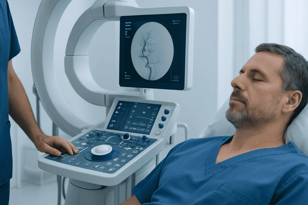
One of the most recognized symptoms of a dural arteriovenous fistula is pulsatile tinnitus. This is a rhythmic sound, usually in one ear, that may feel synchronized with the heartbeat. People often describe it as whooshing, pulsing, rushing, or a machinery-like sound. It may be more noticeable at night, when lying down, or in quiet environments.
Pulsatile tinnitus happens because turbulent blood flow near the ear or skull base can transmit sound through nearby bone and tissues. It is not the same as the more common continuous ringing tinnitus, which is often related to hearing loss or inner ear conditions. While many causes of pulsatile tinnitus are not dangerous, this symptom deserves medical evaluation because some vascular causes, including DAVF, are treatable.
Other symptoms depend on the location and drainage pattern of the fistula. Possible symptoms include headaches, pressure behind the eye, double vision, eye redness or swelling, reduced vision, dizziness, imbalance, seizures, speech difficulty, weakness, numbness, or memory and concentration changes. A DAVF near the cavernous sinus may cause prominent eye symptoms because it affects veins around the eye and nerves that control eye movement.
Symptoms can be mild, intermittent, or slowly progressive. In some cases, a DAVF is found incidentally during imaging for another reason. The presence of neurological changes, visual symptoms, or signs of increased venous pressure usually leads doctors to investigate more urgently and to consider active treatment.
Causes and Risk Factors

Most dural arteriovenous fistulas are considered acquired conditions. They may develop after changes in venous drainage or pressure within the dural venous sinuses, which are large channels that carry blood away from the brain. In some people, the exact trigger is never identified, and this is common.
One important association is venous sinus thrombosis, meaning a clot in a venous sinus. When normal venous drainage is narrowed or blocked, the body may form new small vessel connections as part of a repair response, and some of these connections can become abnormal fistulas. Prior head injury, surgery, infection, inflammation, or procedures involving the skull or venous sinuses may also be associated in selected cases.
DAVFs are more often diagnosed in adults, especially middle-aged and older adults, but they can occur at different ages. They are not usually inherited in a simple family pattern. Having high blood pressure, clotting tendency, smoking history, or conditions that affect blood vessels may influence overall vascular health, but a person with a DAVF should not assume they caused it through lifestyle choices.
The most important medical question is not only why the fistula formed, but how it drains. Classification systems such as Borden and Cognard describe venous drainage patterns. DAVFs without cortical venous reflux are generally lower risk, while those with reflux into cortical veins need closer attention because the veins may be exposed to higher pressure.
Diagnosis: MRI, Angiography, and Specialist Assessment
Diagnosis begins with a detailed history and examination. A doctor will ask about the sound of tinnitus, whether it follows the heartbeat, whether it changes with head position or gentle pressure around the neck, and whether there are headaches, eye changes, neurological symptoms, or previous clotting or head injury. Examination may include listening around the skull and neck with a stethoscope for a bruit, checking cranial nerves, eye movements, vision, strength, sensation, and coordination.
MRI of the brain is often an important first imaging test. It can show indirect signs such as enlarged vessels, abnormal flow-related changes, swelling from venous congestion, or previous bleeding. MR angiography and MR venography can provide additional information about arteries and veins. CT angiography may also be used, especially when fast assessment of blood vessels and bone structures is helpful. These tests are commonly performed and interpreted by specialists in neuroradiology.
However, the most precise test for a suspected dural arteriovenous fistula is usually digital subtraction angiography, also called catheter cerebral angiography. During this procedure, a thin catheter is guided through an artery, commonly from the groin or wrist, to inject contrast dye while detailed X-ray images are taken. This test shows which arteries feed the fistula, where the abnormal connection is located, and how the veins drain.
Catheter angiography is especially useful when treatment is being considered because it allows the team to create a detailed map before any intervention. In some cases, diagnosis and treatment may be performed in the same overall session, depending on safety, anatomy, and planning. The decision is individualized after reviewing imaging, symptoms, and the patient’s general health.
Treatment Options
Treatment depends on symptoms, fistula location, venous drainage, and overall risk. Some low-risk DAVFs without cortical venous reflux may be observed with regular follow-up, particularly if symptoms are mild. Observation does not mean ignoring the condition; it means monitoring symptoms and imaging while avoiding unnecessary procedures when the expected benefit is low.
For many symptomatic or higher-risk DAVFs, endovascular treatment is the preferred first approach. In endovascular embolization, a specialist guides a microcatheter through blood vessels to reach the arteries or veins connected to the fistula. Materials such as liquid embolic agents or coils are then used to close the abnormal connection while preserving normal circulation. This is a key area of interventional neurology and interventional neuroradiology.
The endovascular route may be transarterial, through the feeding artery, or transvenous, through the draining vein or venous sinus. The best route depends on the anatomy. Some fistulas can be completely closed in one procedure, while others require staged treatment or a combination of approaches. Patients are monitored closely during and after treatment, and follow-up imaging is often needed to confirm durable closure.
If endovascular treatment is not suitable or does not fully close the fistula, microsurgical disconnection may be recommended. In selected cases, stereotactic radiosurgery may be considered, particularly for DAVFs that are difficult to reach by catheter or surgery, although its effect may take time. The goal of all treatments is to stop abnormal shunting, reduce symptoms, and protect the brain or spinal cord from harmful venous pressure.
Recovery, Follow-Up, and Daily Life
Recovery varies depending on the fistula, the treatment performed, and the person’s baseline health. After endovascular treatment, patients are usually observed for access-site healing, neurological status, headache, and blood pressure control. Some people notice rapid improvement in pulsatile tinnitus once the abnormal flow is closed, while others improve gradually as venous pressure settles.
Follow-up is an essential part of care. Doctors may recommend MRI, MR angiography, CT angiography, or repeat catheter angiography to confirm that the fistula remains closed and that no new abnormal drainage is present. Follow-up timing is individualized, especially for complex DAVFs, partially treated lesions, or cases treated with radiosurgery.
Daily life guidance should be personalized. In general, patients should follow instructions about activity restrictions after angiography or surgery, take prescribed medications as directed, attend all follow-up visits, and report any new neurological or visual symptoms. Good vascular health habits may also support recovery and long-term wellbeing.
- Manage blood pressure, diabetes, cholesterol, and other vascular risk factors with medical guidance.
- Avoid smoking and seek support for quitting if needed.
- Discuss blood thinners, hormone therapy, clotting disorders, or a history of venous thrombosis with the treating doctor.
- Return gradually to exercise, work, and travel according to the care team’s advice.
When to See a Doctor
A person should arrange medical evaluation for pulsatile tinnitus, especially if it is new, persistent, one-sided, or clearly synchronized with the heartbeat. Evaluation is also important when tinnitus is accompanied by headaches, dizziness, vision changes, eye redness or swelling, or symptoms that change with position. An ear, nose, and throat assessment may be part of the workup, but vascular imaging may be needed when a blood-flow cause is suspected.
Urgent medical care is appropriate for sudden weakness or numbness on one side of the body, difficulty speaking, new seizure, sudden severe headache, sudden vision loss, confusion, fainting, or rapidly worsening neurological symptoms. These symptoms can occur for many reasons, including stroke-like conditions, and should be assessed promptly.
Patients with eye movement problems, double vision, bulging or painful eye, or unexplained vision changes may also need assessment by specialists in neuro-ophthalmology, because some DAVFs affect venous drainage around the eye. Coordinated care helps ensure that both the vascular cause and vision-related effects are addressed.
Acibadem International’s multidisciplinary teams and JCI-accredited hospitals evaluate and treat conditions such as dural arteriovenous fistula for international patients, including advanced imaging, neurological assessment, and endovascular treatment when appropriate. Any treatment decision should be made after individualized consultation with qualified specialists.
Frequently asked questions
What is a dural arteriovenous fistula?
A dural arteriovenous fistula is an abnormal connection between arteries and veins in the dura, the tough membrane covering the brain or spinal cord. Blood flows directly from an artery into a vein or venous sinus, bypassing the normal capillary network. The significance depends largely on where it is located and how the blood drains.
Why does a dural arteriovenous fistula cause pulsatile tinnitus?
A DAVF can create turbulent, high-flow blood movement near the ear or skull base. This turbulence may be transmitted as a heartbeat-like whooshing sound, called pulsatile tinnitus. Because some vascular causes of pulsatile tinnitus are treatable, persistent one-sided pulsatile tinnitus should be evaluated.
Can MRI detect a dural arteriovenous fistula?
MRI and MR angiography can show signs that suggest a dural arteriovenous fistula, such as enlarged vessels, abnormal flow, or venous congestion. However, small or complex fistulas may be difficult to fully define on MRI alone. Catheter angiography is often needed to confirm the diagnosis and plan treatment.
Is a dural arteriovenous fistula dangerous?
Some DAVFs are low risk and may cause mainly symptoms such as pulsatile tinnitus. Others are higher risk, especially if blood drains backward into cortical veins on the brain surface. Doctors assess the drainage pattern, symptoms, and imaging findings to decide whether observation or active treatment is safest.
What is endovascular treatment for a DAVF?
Endovascular treatment is a minimally invasive procedure performed through a small catheter placed inside the blood vessels. The specialist guides the catheter to the fistula and uses embolic materials or coils to close the abnormal connection. The approach may be through an artery, a vein, or both, depending on the anatomy.
Will pulsatile tinnitus go away after treatment?
Pulsatile tinnitus may improve quickly if the DAVF causing the turbulent flow is completely closed. In some people, improvement is gradual, and other ear or vascular factors may also contribute to symptoms. Follow-up with the treating team helps confirm whether the fistula is closed and whether further evaluation is needed.
Can a dural arteriovenous fistula come back?
Recurrence is possible but not inevitable. The likelihood depends on the fistula type, treatment method, completeness of closure, and underlying venous drainage factors. This is why follow-up imaging and specialist visits are important even after successful treatment.
References
- American Heart Association
- American Stroke Association
- Mayo Clinic
- Cleveland Clinic
- Radiological Society of North America
This article is for general information only and is not a substitute for professional medical advice. Please consult a qualified doctor about your individual situation.
Joint and spine care in Turkey — expert assessment & treatment
JCI-accredited · board-certified surgeons · reply within 24h
Check your numbers in seconds
BMI, calories, due date, blood pressure and 30+ more clinical calculators — free, instant, doctor-reviewed ranges.