Neurointerventional Radiology: Conditions Commonly Treated Without Open Surgery

Neurointerventional radiology uses image-guided catheters inserted through blood vessels to reach the brain or spine. Commonly treated conditions include stroke caused by blocked arteries, brain aneurysms, arteriovenous malformations, carotid artery disease, and some vascular tumors or fistulas.
Key Takeaways
- Neurointerventional radiology uses image-guided catheters inserted through blood vessels to reach the brain or spine.
- Commonly treated conditions include stroke caused by blocked arteries, brain aneurysms, arteriovenous malformations, carotid artery disease, and some vascular tumors or fistulas.
- Treatment is highly individualized and depends on the patient’s symptoms, anatomy, overall health, and imaging findings.
- Diagnosis usually involves CT, MRI, ultrasound, and catheter angiography to map blood vessels in detail.
- These procedures may help avoid open surgery in selected cases, but they still require expert evaluation and follow-up.
- Urgent symptoms such as sudden weakness, trouble speaking, severe headache, or vision loss need immediate medical attention.
Neurointerventional radiology is a subspecialty that uses tiny catheters and real-time imaging to diagnose and treat certain brain, spine, head, and neck conditions without traditional open surgery. It can be an important option for selected vascular disorders, stroke, and other problems where a minimally invasive approach may reduce recovery time and tissue disruption.
Overview
Neurointerventional radiology, also called endovascular or interventional neuroradiology, is a medical specialty that treats problems of the brain, spinal cord, head, and neck from inside the blood vessels. Instead of making a large incision, the specialist guides a thin catheter through an artery or vein, usually starting in the wrist or groin, and navigates it to the area that needs treatment using advanced imaging.
This approach is most often used for vascular conditions, meaning disorders that involve arteries, veins, or abnormal connections between them. Because the treatment is targeted, many patients benefit from less disruption to surrounding tissues, shorter hospital stays, and faster recovery than with open surgery. However, minimally invasive does not mean minor; these are specialized procedures that require careful planning and experienced teams.
Neurointerventional radiology may be used on its own or together with neurology, neurosurgery, vascular surgery, stroke medicine, oncology, and rehabilitation. The goal is to restore blood flow, prevent bleeding, close abnormal vessels, or reduce the risk of future neurological injury while preserving as much healthy brain and spinal tissue as possible.
Conditions Commonly Treated Without Open Surgery
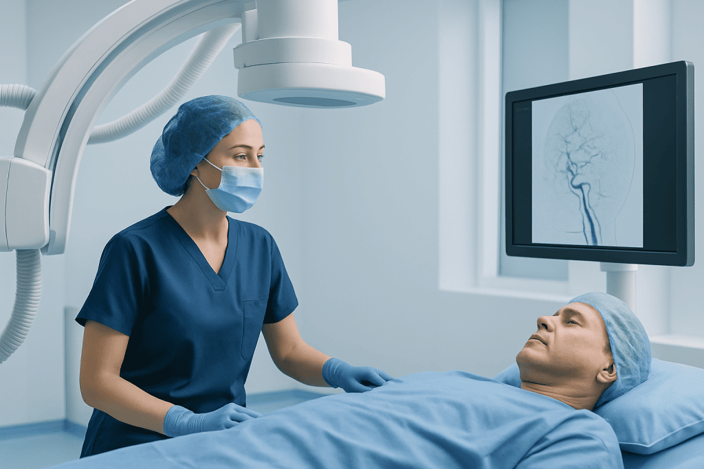
One of the best-known uses of neurointerventional radiology is emergency treatment for ischemic stroke, which happens when a blood clot blocks an artery supplying the brain. In selected patients, doctors can remove the clot with a catheter-based procedure called mechanical thrombectomy, helping restore circulation and limit brain damage. Time matters greatly, so rapid assessment is essential.
Another common group of conditions includes brain aneurysms and other weak or ballooned areas in blood vessel walls. Depending on the aneurysm’s size, shape, and location, specialists may seal it from the inside using tiny coils, flow-diverting devices, or stents. This can lower the risk of rupture or rebleeding in carefully chosen patients with brain aneurysm disease.
Neurointerventional methods are also used for arteriovenous malformations (AVMs), dural arteriovenous fistulas, carotid or intracranial artery narrowing, and some causes of pulsatile tinnitus. In AVMs or fistulas, the aim may be to close abnormal vessel connections using an embolic material delivered through a catheter. For narrowed arteries, balloon angioplasty or stenting may sometimes be considered, especially when symptoms continue despite medical therapy.
In addition, certain head, neck, or spine tumors with a rich blood supply may be treated with preoperative embolization to reduce bleeding before surgery. Some patients with nosebleeds that do not stop, spinal vascular malformations, or cerebrospinal venous abnormalities may also benefit from image-guided endovascular procedures after specialist evaluation.
Symptoms and Situations That May Lead to Evaluation

The symptoms that lead to neurointerventional evaluation depend on the underlying condition. In stroke, warning signs often appear suddenly and may include weakness on one side of the body, facial drooping, numbness, difficulty speaking, confusion, loss of balance, or sudden vision changes. These symptoms should always be treated as a medical emergency.
Brain aneurysms do not always cause symptoms, and some are found incidentally during imaging for another reason. When symptoms do occur, they may include headache, eye pain, double vision, drooping eyelid, or nerve-related symptoms caused by pressure on nearby structures. A ruptured aneurysm can cause a sudden, severe headache and requires urgent care.
AVMs and fistulas may cause headaches, seizures, neurological symptoms, pulsating noises in the ear, or bleeding. Carotid artery disease may be discovered after a transient ischemic attack, stroke, or an ultrasound done for circulation concerns. Symptoms of reduced brain blood flow can overlap with other disorders, so imaging is often needed to clarify the cause.
- Sudden weakness, numbness, or speech difficulty
- Severe or unusual headache, especially if abrupt
- Sudden vision loss or double vision
- Seizures or unexplained neurological changes
- Pulsatile tinnitus or symptoms linked to abnormal blood flow
How Diagnosis Is Made
Diagnosis usually begins with a medical history, neurological examination, and imaging studies. CT and MRI can show bleeding, stroke, vessel abnormalities, and surrounding brain tissue changes. CT angiography and MR angiography provide additional detail about blood vessels and are commonly used to screen for aneurysms, narrowing, or vascular malformations.
Ultrasound, especially carotid Doppler ultrasound, may help evaluate blood flow in the neck arteries. In emergency stroke care, imaging also helps determine whether brain tissue may still be saved and whether catheter-based treatment is likely to help. This decision is based on the timing of symptoms, the location of the blockage, and the patient’s overall condition.
The most detailed test is catheter angiography, also called cerebral angiography. During this procedure, contrast dye is injected through a catheter while X-ray images are taken. It can define the exact anatomy of an aneurysm, AVM, fistula, or narrowed vessel and often serves as the roadmap for treatment planning. Because it is invasive, it is used when the information is likely to affect management.
Diagnosis is rarely based on one test alone. Specialists usually combine imaging findings with symptoms, age, medical history, bleeding risk, prior stroke history, and the expected natural course of the condition. This helps determine whether observation, medication, or an intervention is the safest and most appropriate next step.
Treatment Options in Neurointerventional Radiology
Treatment depends on the condition being addressed. In ischemic stroke, neurointerventional specialists may perform clot removal procedures to reopen a blocked artery. For aneurysms, options can include endovascular coiling, stent-assisted coiling, or flow diversion, each chosen according to the vessel anatomy and rupture risk. Some patients with stroke or aneurysm need treatment urgently, while others can be planned electively.
For AVMs, fistulas, and some tumors, embolization is a common approach. This means that the doctor delivers tiny particles, glue-like agents, coils, or other materials to block abnormal blood vessels. Embolization may be used as a definitive treatment or as one step before surgery or radiosurgery, depending on the lesion’s size and location. In selected aneurysm cases, endovascular aneurysm treatment may reduce the need for open procedures.
For narrowed arteries, treatment may involve medications alone, or in carefully selected cases, angioplasty and stenting. These are not appropriate for every patient, and the expected benefit must be balanced against possible risks such as bleeding, vessel injury, or stroke during the procedure. Careful patient selection is one of the most important parts of safe neurointerventional treatment.
Most procedures are performed under local anesthesia with sedation or under general anesthesia, depending on urgency and complexity. After treatment, patients are monitored closely for blood pressure control, neurological changes, and access-site complications. Follow-up imaging is often needed because some treated vessels or lesions can change over time and may require ongoing observation.
Benefits, Risks, and Recovery
The main benefit of neurointerventional radiology is that it can treat certain complex conditions without opening the skull or performing large surgical exposures. This may mean smaller puncture sites, less postoperative pain, shorter hospitalization, and an earlier return to usual activities for many patients. It can also offer a treatment option for lesions that are difficult to reach with conventional surgery.
Even so, every procedure has risks. Possible complications include bleeding, infection, allergic reaction to contrast dye, blood vessel injury, stroke, kidney strain from contrast, or recurrence of the treated problem. Some risks are related to the condition itself rather than the procedure, which is why a personalized discussion with the care team is so important.
Recovery varies widely. A patient treated for uncomplicated aneurysm coiling may recover differently from someone treated for a major stroke or a complex AVM. Many people need observation in the hospital for at least a short period, and some require rehabilitation afterward to address speech, movement, balance, or memory changes.
Long-term follow-up matters. Patients may need repeat scans to confirm that an aneurysm remains sealed, that a stent is open, or that an AVM has been completely treated. Medicines such as blood thinners or antiplatelet therapy may be prescribed in some situations, but the exact plan should always come from the treating physician.
Prevention, Self-care, and Living With Vascular Risk
Not every neurovascular condition can be prevented, but many vascular risks can be reduced. Good control of blood pressure, diabetes, and cholesterol helps protect blood vessels throughout the body, including those that supply the brain. Avoiding tobacco, limiting excess alcohol, staying physically active, and following a balanced eating pattern also support long-term brain and vascular health.
People who have had a stroke, transient ischemic attack, aneurysm treatment, or carotid disease often need structured follow-up. This may include prescribed medication, repeat imaging, and lifestyle adjustments. Taking medicines exactly as directed is especially important after stent placement, because stopping some medications too early can increase the risk of clot formation.
Self-care also includes knowing the warning signs of stroke and seeking help immediately if they appear. Family members can play an important role by recognizing sudden facial droop, arm weakness, or speech trouble and calling emergency services without delay. Rapid treatment can improve outcomes and may expand the options available.
For patients who need specialized evaluation, centers with coordinated neurology, neurosurgery, neuroradiology, and intensive care support can be valuable. Acibadem International’s multidisciplinary specialists and JCI-accredited hospitals evaluate and treat complex neurovascular conditions for international patients, including advanced image-guided options such as embolization procedures when appropriate.
When to See a Doctor
A doctor should be consulted promptly for any new neurological symptom, especially sudden weakness, numbness, trouble speaking, severe dizziness, or vision changes. Immediate emergency care is essential if stroke is suspected or if there is a sudden severe headache unlike previous headaches. Waiting to see if symptoms improve can reduce treatment options.
Non-emergency consultation is also important for ongoing headaches with unusual features, pulsatile tinnitus, seizures, unexplained fainting, transient neurological episodes, or imaging findings such as an incidental aneurysm or vascular malformation. A specialist can help explain the significance of the finding and whether monitoring or treatment is recommended.
People with strong vascular risk factors, prior stroke, or known carotid disease should keep regular follow-up appointments and ask about warning signs that require urgent attention. Because treatment decisions are highly individualized, patients are encouraged to discuss the risks, benefits, alternatives, and expected follow-up of any proposed neurointerventional procedure with a qualified doctor.
Frequently asked questions
What is neurointerventional radiology?
Neurointerventional radiology is a specialty that treats certain brain, spine, head, and neck conditions from inside the blood vessels using catheters and imaging guidance. It is often used for vascular problems such as stroke, aneurysms, and abnormal vessel connections.
Is neurointerventional radiology the same as open brain surgery?
No. These procedures are minimally invasive and usually involve a small puncture in the wrist or groin rather than a large surgical opening. However, they are still advanced medical procedures that require expert teams, hospital monitoring, and careful follow-up.
Which conditions are commonly treated this way?
Common examples include ischemic stroke caused by a large vessel blockage, brain aneurysms, AVMs, dural arteriovenous fistulas, and some cases of carotid or intracranial artery narrowing. It may also be used before surgery for certain highly vascular tumors to reduce bleeding.
How is a patient evaluated for treatment?
Evaluation usually includes a neurological examination and imaging such as CT, MRI, angiography, or ultrasound. Doctors consider the exact anatomy of the problem, the patient’s symptoms, the urgency of treatment, and overall health before recommending a plan.
What are the advantages of a minimally invasive approach?
A minimally invasive approach may reduce tissue disruption, shorten hospital stay, and support faster recovery in selected patients. It can also allow treatment of deep or delicate vascular areas that may be difficult to reach with conventional surgery.
Are there risks?
Yes. Risks can include bleeding, blood vessel injury, stroke, contrast reaction, infection, or recurrence of the treated condition. The exact risk depends on the procedure, the anatomy being treated, and the patient’s general health.
When should someone seek emergency help?
Emergency care is needed right away for sudden weakness, facial drooping, speech difficulty, confusion, vision loss, or a sudden severe headache. These symptoms can signal stroke or bleeding and should never be ignored.
References
- World Health Organization
- American Stroke Association
- National Institute of Neurological Disorders and Stroke
- Society of NeuroInterventional Surgery
- Radiological Society of North America
This article is for general information only and is not a substitute for professional medical advice. Please consult a qualified doctor about your individual situation.
Joint and spine care in Turkey — expert assessment & treatment
JCI-accredited · board-certified surgeons · reply within 24h
Add us as a Preferred Source to see more of our trusted health content across Google Search, AI Overviews and Discover.