Endovascular Treatment for Brain AVM: Options, Risks, and Treatment Planning

A brain AVM is an abnormal tangle of blood vessels connecting arteries and veins. Endovascular treatment usually works by embolization, which reduces or blocks blood flow within the AVM.
Key Takeaways
- A brain AVM is an abnormal tangle of blood vessels connecting arteries and veins.
- Endovascular treatment usually works by embolization, which reduces or blocks blood flow within the AVM.
- This approach may be used alone or before surgery or radiosurgery, depending on the AVM’s size, location, and structure.
- Treatment planning is individualized and balances the benefits of reducing bleeding risk against procedure-related risks.
- Care is best provided by a multidisciplinary team with expertise in interventional neurology, neurosurgery, and imaging.
Endovascular treatment for brain AVM is a minimally invasive procedure that closes off abnormal blood vessels in the brain using a catheter and special materials. It may be used on its own in selected cases, or as part of a broader treatment plan with surgery or radiosurgery.
Overview
Endovascular treatment for brain AVM is a minimally invasive method used to treat a brain arteriovenous malformation, or AVM. A brain AVM is a tangle of abnormal blood vessels where arteries connect directly to veins without the usual tiny capillaries between them. Because of this unusual blood flow, the vessels can be fragile and may lead to bleeding, seizures, headaches, or other neurological symptoms.
In endovascular treatment, a doctor guides a thin tube called a catheter through the blood vessels, usually starting from the groin or wrist, and advances it to the arteries feeding the AVM. Through this catheter, the specialist places embolic material such as medical glue, liquid agents, or tiny particles or coils to reduce or stop blood flow into the malformation. This process is often called embolization.
Endovascular therapy is not the right choice for every AVM, and it is not always meant to completely cure the problem by itself. In many people, it is one part of a larger plan that may also include microsurgery or stereotactic radiosurgery. The main goal is to make treatment safer and more effective while protecting healthy brain tissue as much as possible.
When Endovascular Treatment Is Considered
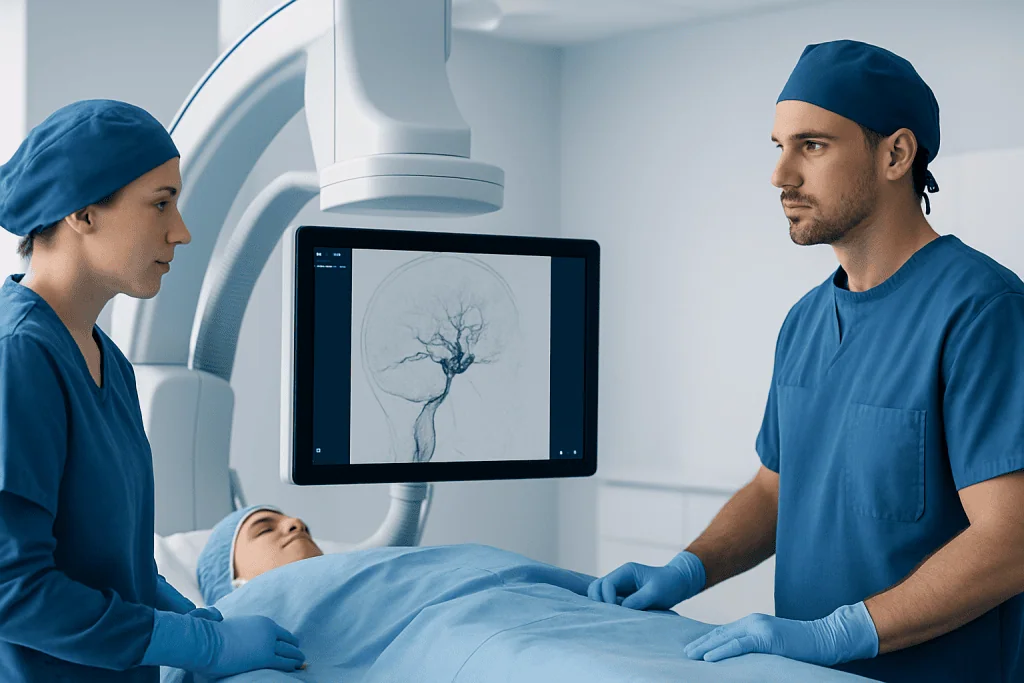
Doctors consider endovascular treatment based on several factors, including whether the AVM has bled, its size, shape, and location, and which arteries supply it. AVMs that are deep in the brain, close to important functional areas, or supplied by multiple vessels may require especially careful planning. A person’s age, general health, symptoms, and previous treatments also matter.
In some cases, embolization is used as the main treatment when the AVM anatomy is favorable. More often, it is used to reduce the size or blood flow of the AVM before another treatment. For example, reducing blood flow first can make surgery technically easier or lower the risk of bleeding during an operation. It may also help prepare selected AVMs for focused radiation treatment.
Endovascular treatment can also be considered urgently if there is bleeding from a weak point related to the AVM, such as an associated aneurysm or a high-risk vessel. In that setting, the aim may be to stabilize the immediate source of danger rather than treat the entire malformation at once. A multidisciplinary review helps determine the safest and most effective strategy.
How the Procedure Works

Before the procedure, the care team reviews detailed brain imaging to understand the AVM’s structure. The patient may need blood tests and an anesthesia assessment, and medications are reviewed carefully. The procedure is commonly performed under general anesthesia so the patient remains still and comfortable, although practice may vary depending on the situation.
During embolization, the interventional specialist inserts a catheter into a blood vessel and navigates it under X-ray guidance toward the arteries feeding the AVM. Contrast dye is used to map blood flow in real time. Once the correct position is confirmed, the specialist injects embolic material to block the abnormal channels. The material used depends on the AVM anatomy and the treatment goal.
The procedure may treat only part of the AVM in one session, especially if the lesion is complex. Some people need staged treatments over time to reduce risk. After the procedure, patients are monitored closely for blood pressure control, neurological changes, headache, or nausea. A short hospital stay is common, though this depends on the complexity of treatment and recovery.
Because treatment decisions often overlap with other specialties, endovascular therapy may be discussed alongside Gamma Knife radiosurgery or brain AVM surgery when building a complete care plan.
Benefits, Limits, and Risks
The main benefit of endovascular treatment for brain AVM is that it is less invasive than open brain surgery. It may reduce blood flow to the AVM, lower the chance of bleeding from high-risk parts of the malformation, and improve the safety of additional treatment. In carefully selected cases, embolization may achieve complete closure of the AVM.
However, embolization also has limits. Some AVMs have a complex structure that makes full closure difficult or unsafe through a catheter alone. Even when blood flow is reduced significantly, a partially treated AVM may still require follow-up and further therapy. Long-term imaging is often needed to confirm the result.
As with any procedure involving brain blood vessels, there are risks. Possible complications include stroke, bleeding, vessel injury, swelling in the brain, seizures, contrast-related problems, infection, and neurological deficits such as weakness, numbness, speech difficulty, or vision changes. The exact risk varies based on the AVM’s anatomy, whether it has already bled, and how extensive treatment needs to be.
Patients often feel reassured by knowing that treatment planning is designed to weigh these risks carefully against the natural risks of the AVM itself. The goal is not simply to intervene, but to choose the approach with the best balance of safety and benefit for that individual person.
Diagnosis and Treatment Planning
Accurate diagnosis is essential before deciding on treatment. Many AVMs are first detected after symptoms such as headache, seizure, or bleeding, while others are found incidentally on brain imaging. Doctors may begin with CT or MRI, but detailed vascular imaging is usually needed to map the AVM fully.
The most important planning test is often cerebral angiography, also called a catheter angiogram. This test gives a highly detailed picture of the feeding arteries, the central tangle of vessels, and the draining veins. It helps doctors understand how quickly blood moves through the AVM and whether there are fragile associated aneurysms or weak points.
Treatment planning typically involves specialists from interventional neuroradiology, neurosurgery, neurology, and radiation oncology. Together, they consider whether observation, embolization, surgery, radiosurgery, or a combination would be most appropriate. Factors such as AVM grade, brain location, prior hemorrhage, and expected effect on quality of life are all part of the discussion.
Patients may also hear the AVM described in relation to broader cerebrovascular care, including evaluation of conditions such as brain aneurysm or other abnormal vessel findings that can affect treatment choices.
Recovery, Follow-Up, and Self-Care
Recovery after embolization varies. Some people return home within a day or two, while others need longer monitoring, especially after treatment of a large or complex AVM. Temporary tiredness, headache, or mild soreness at the catheter insertion site can occur. The care team gives instructions on activity, medicines, hydration, and when normal routines can be resumed.
Follow-up is a key part of care. Even when the procedure goes well, repeat imaging may be needed to check how much of the AVM has been closed and whether more treatment is necessary. Depending on the overall plan, this may involve MRI, CT angiography, or repeat catheter angiography after a period of recovery.
Patients are usually advised to manage general vascular health, take prescribed medications exactly as directed, and avoid stopping seizure medicines or other treatments without medical advice. Keeping follow-up appointments is important because treatment results can evolve over time. Family members may also be asked to watch for new neurological symptoms during recovery.
At experienced centers, treatment may be coordinated with other advanced procedures such as interventional neuroradiology procedures when the anatomy is especially complex. Acibadem International’s multidisciplinary specialists and JCI-accredited hospitals diagnose and treat brain AVMs for international patients as part of individualized care planning.
When to See a Doctor
Anyone with symptoms that could suggest a brain AVM should seek medical evaluation. These symptoms can include seizures, unexplained severe headaches, weakness, numbness, speech changes, balance problems, vision changes, or sudden confusion. Not every headache or seizure is caused by an AVM, but prompt assessment is important.
Emergency care is needed for sudden severe headache, loss of consciousness, sudden neurological deficit, or any signs of stroke or bleeding in the brain. These symptoms require urgent medical attention rather than watchful waiting at home. Fast treatment can be important for both diagnosis and stabilization.
People who already know they have a brain AVM should contact their doctor if symptoms change, new seizures occur, or recovery after treatment does not seem to follow expectations. Questions about activity, travel, pregnancy, medicines, or future procedures are also worth discussing with a qualified specialist. Personalized guidance is especially important because AVMs vary so much from one person to another.
Frequently asked questions
What is endovascular treatment for brain AVM?
It is a minimally invasive procedure used to reduce or block blood flow in a brain arteriovenous malformation. A specialist passes a catheter through the blood vessels and delivers embolic material into the arteries feeding the AVM.
Can embolization cure a brain AVM by itself?
Sometimes, but not always. In selected AVMs, embolization may completely close the malformation, but in many cases it is used to prepare for surgery or radiosurgery rather than serve as the only treatment.
What are the risks of brain AVM embolization?
Possible risks include stroke, bleeding, vessel injury, seizures, brain swelling, and temporary or permanent neurological symptoms. The level of risk depends on the size, location, and complexity of the AVM and the overall health of the patient.
How do doctors decide whether I need treatment or monitoring?
The decision depends on whether the AVM has bled, what symptoms it causes, where it is located, and how risky treatment may be. Specialists compare the natural risk of leaving the AVM untreated with the potential benefits and risks of intervention.
How long is recovery after endovascular treatment?
Recovery is often shorter than recovery from open brain surgery, but it varies from person to person. Some patients go home within a day or two, while others need more monitoring and follow-up, especially after complex treatment.
Will I need more than one procedure?
Possibly. Complex AVMs are often treated in stages to improve safety, and some patients need additional surgery or radiosurgery after embolization. Follow-up imaging helps show whether further treatment is needed.
References
- World Health Organization
- National Institute of Neurological Disorders and Stroke
- American Heart Association
- American Stroke Association
- Society of NeuroInterventional Surgery
This article is for general information only and is not a substitute for professional medical advice. Please consult a qualified doctor about your individual situation.
Explore treatments in Turkey — costs, top hospitals & a free quote
JCI-accredited · board-certified surgeons · reply within 24h
Add us as a Preferred Source to see more of our trusted health content across Google Search, AI Overviews and Discover.
Check your numbers in seconds
BMI, calories, due date, blood pressure and 30+ more clinical calculators — free, instant, doctor-reviewed ranges.